Transcription
NeuroImage 185 (2019) 222–235Contents lists available at ScienceDirectNeuroImagejournal homepage: www.elsevier.com/locate/neuroimageFirst-year development of modules and hubs in infant brainfunctional networksXuyun Wen a, b, Han Zhang b, *, Gang Li b, Mingxia Liu b, Weiyan Yin b, Weili Lin b, Jun Zhang c, **,Dinggang Shen b, d, ***aSchool of Data and Computer Science, Sun-Yat Sen University, ChinaDepartment of Radiology and BRIC, University of North Carolina at Chapel Hill, Chapel Hill, NC, USASchool of Computer Science and Engineering, South China University of Technology, Guangzhou, ChinadDepartment of Brain and Cognitive Engineering, Korea University, Seoul, 02841, Republic of KoreabcA R T I C L E I N F OA B S T R A C TKeywords:Brain developmentModuleHubInfantFunctional brain networkThe human brain develops rapidly in the first postnatal year, in which rewired functional brain networks couldshape later behavioral and cognitive performance. Resting-state functional magnetic resonances imaging (rsfMRI) and complex network analysis have been widely used for characterizing the developmental brain functionalconnectome. Yet, such studies focusing on the first year of postnatal life are still very limited. Leveraging normallydeveloping longitudinal infant rs-fMRI scans from neonate to one year of age, we investigated how brain functional networks develop at a fine temporal scale (every 3 months). Considering challenges in the infant fMRIbased network analysis, we developed a novel algorithm to construct the robust, temporally consistent andmodular structure augmented group-level network based on which functional modules were detected at each age.Our study reveals that the brain functional network is gradually subdivided into an increasing number of functional modules accompanied by the strengthened intra- and inter-modular connectivities. Based on the developingmodules, we found connector hubs (the high-centrality regions connecting different modules) emerging andincreasing, while provincial hubs (the high-centrality regions connecting regions in the same module) diminishing. Further region-wise longitudinal analysis validates that different hubs have distinct developmental trajectories of the intra- and inter-modular connections suggesting different types of role transitions in network, suchas non-hubs to hubs or provincial hubs to connector hubs et al. All findings indicate that functional segregationand integration are both increased in the first year of postnatal life. The module reorganization and hub transitionlead to more efficient brain networks, featuring increasingly segregated modular structure and more connectorhubs. This study provides the first comprehensive report of the development of functional brain networks at a 3month interval throughout the first postnatal year of life, which provides essential information to the futureneurodevelopmental and developmental disorder studies.1. IntroductionIn the first postnatal year, the human brain undergoes the most dramatic growth with its total volume expanding up to double its size(Knickmeyer et al., 2008; Tau and Peterson, 2010; Gao et al., 2016; Liet al., 2018). Concurrently, remarkable functional brain milestones areachieved during this period, including not only the improved vision(Courage and Adams, 1990) and body manipulation, but also a number ofhigher-order cognitive functions, such as self-awareness (Amsterdam,1972), spatial attention (Haith et al., 1988), and working memory(Reznick, 2009). These dramatic changes greatly shape subsequentcognitive and behavioral development and lay foundations for theessential skills in later life. Thus, it is critical to improve our limitedunderstandings of developmental patterns and mechanisms of the humanbrain in the first postnatal year.In recent years, resting-state functional magnetic resonance imaging* Corresponding author. Department of Radiology and BRIC, University of North Carolina at Chapel Hill, NC, 27599, USA.** Corresponding author. School of Computer Science and Engineering, South China University of Technology, Guangzhou, 510006, China.*** Corresponding author. Department of Radiology and BRIC, University of North Carolina at Chapel Hill, Chapel Hill, NC, USA.E-mail addresses: hanzhang@med.unc.edu (H. Zhang), csjun@scut.edu.cn (J. Zhang), dgshen@med.unc.edu (D. .019Received 15 May 2018; Received in revised form 9 September 2018; Accepted 4 October 2018Available online 10 October 20181053-8119/ 2018 Elsevier Inc. All rights reserved.
X. Wen et al.NeuroImage 185 (2019) 222–235efficiencies (Fair et al., 2009; Supekar et al., 2009; Dosenbach et al.,2010; Wu et al., 2013; Cao et al., 2014). From neonate to early childhood,a similar developmental trend was suggested, but it came from only onestudy on the large-scale functional brain networks of the infants atthree-week, one-year and two-years of age (Gao et al., 2011), whichrevealed increasing local and global efficiency, as well as the increasinglyuniform spatial distribution of “hub” regions (the nodes with significantly more connections or higher degrees). While the study is stilllacking for the first postnatal year, a bold hypothesis has been proposed,which assumes more rapid changes in functional integration/segregation(Cao et al., 2016a, 2017). Therefore, it is highly important and necessaryto fill this gap and test such a hypothesis by characterizing fine-grained,month-to-month spatiotemporal developmental trajectories of the brainfunctional networks and its topological structures.In this paper, leveraging a large longitudinal rs-fMRI dataset fromnormally developing infants with each having multiple dense scans (atevery three months from birth), we aim to (1) delineate the fine-graineddevelopmental trajectories of the large-scale functional brain networks inthe first postnatal year, and (2) explore the possible driving forces of sucha dynamic evolution with explicit evidences from network topologicalanalysis. Instead of using global network metrics (e.g., local/global efficiency), we focus on a group of densely interconnected brain regions, i.e.,communities or modules (Rubinov and Sporns, 2010), which are suggested to be relevant to specific cognitive/behavioral functions (Bertolero et al., 2015). Based on the modular structure, we further investigatedthe longitudinal reconfiguration and rewiring involving a small set of“hub” regions. In this sense, the functional segregation and integrationcan be assessed by quantifying the modular structures and hubs (Hwanget al., 2012; Huang et al., 2013; Van den Heuvel and Sporns, 2013;Bertolero et al., 2015). Based on different topological roles of hubs, weinvestigate and identify the longitudinally sensitive spatial locationchanges of both provincial hubs (for integrating the nodes within amodule) and connector hubs (for integrating the nodes belonging todifferent modules) (Van den Heuvel and Sporns, 2013; Bertolero et al.,2015). We hypothesize that from neonate to one year of age, (1) thenumber of functional brain modules will gradually increase, driven bythe increasingly strengthened within-module FC (i.e., functional segregation); (2) the number of connector hubs will increase, driven bystrengthened inter-modular FC (i.e., functional integration); and (3) thespatial distribution of different types of hubs are continuously changing,which represents the different maturation orders for different functionalsystems.(rs-fMRI) has been emerging for probing functional brain development(Gao et al., 2009; Smyser et al., 2010; Fransson et al., 2011; Gao et al.,2011; Smyser et al., 2011; Barrett and Satpute, 2013; Alcauter et al.,2014; Damaraju et al., 2014; De Asis-Cruz et al., 2015; Gao et al., 2015a;Gao et al., 2015b; Van den Heuvel et al., 2015; Cao et al., 2016a).However, only a few studies have focused on the longitudinal aspects offunctional brain development in this most important period, i.e., the firststage of postnatal life. In a large-scale longitudinal study, Gao et al.delineate the developmental trajectories of nine brain functionalsub-networks every three months from neonate to one year of age. Itreveals that different brain sub-networks have distinct growth trajectories following a sequence from the primary to higher-order functionalsystems (Gao et al., 2015a). This finding investigates the developmentalfunctional segregation in the brain, but largely neglects the developmentof functional integration (i.e., the functional interactions among differentbrain regions or different functional systems). It is worth noting thatstudying the development of functional integration is essential in thedevelopmental neuroscience, due to the matter that the functional interactions are believed to mediate more complex cognitive functions(Barrett and Satpute, 2013; Behrmann and Plaut, 2013; Friederici andGierhan, 2013; Sporns, 2013; Gao et al., 2015a; Cohen and DEsposito,2016).The spatiotemporal characteristics of developing large-scale functional brain networks could deepen our understanding on how thehuman brain develops well-coordinated functional systems that mirrorcognitive milestones at such a pivotal age. To this end, it is highlyrequired to have more sophisticated and fine-grained network modelingand topological analyses. The most popular method is to represent theinter-regional functional interactions as a complex network that consistsof all brain regions as nodes linked by functional connectivity (FC) asedges. In the network, the edge strength is usually measured by thetemporal synchronization of regional blood-oxygen level-dependent(BOLD) rs-fMRI signals (Friston, 2011). Graph theory analyses can helpuncover the details of the topological properties of such a complexnetwork. Abundant evidence indicates that matured adult brain networksfollow a small-world property with both high local and global efficienciesto facilitate the efficient information exchange (Watts and Strogatz,1998; Liao et al., 2017). From another viewpoint, this efficient organization also suggests well-balanced functional segregation (supportingintra-functional-system information exchange to process nablinginter-functional-system collaboration to process multimodal information andsupport higher-level functions) with limited wiring resources (Sporns,2013). Such properties have been observed in many other age groups,such as infants (Gao et al., 2011; De Asis-Cruz et al., 2015; Van denHeuvel et al., 2015; Cao et al., 2016a), older children (Fair et al., 2009),adolescents (Smyser et al., 2011), and the elderly (Onoda and Yamaguchi, 2013). Despite the well-observed global network properties, thedetailed rewiring processes, i.e., the longitudinal development of thelocalized nodes and edges, have not currently been investigated,although they could be more important and helpful to understand thedeveloping brain.It has been hypothesized that the developmental brain networkcontinuously reconfigures with a small amount of connections rewired todrive the entire network toward a more efficient organization (bothglobally and locally), while keeping the whole system relatively stable(Fair et al., 2009; Supekar et al., 2009; Gao et al., 2011; Uddin et al.,2011; Chen et al., 2013; Huang et al., 2013; Damaraju et al., 2014;Thomason et al., 2015; Zhao et al., 2015; Cao et al., 2016a, 2017).However, most evidence comes from the age groups spanning from latechildhood ( five years of age) to early adolescence ( 12 years of age).Based on these studies, a few key connections that link different functional systems are prone to rewire in order to achieve enhanced functional integration/segregation, as well as increased local and global2. Materials and methods2.1. Subject informationImages were obtained from subjects enrolled in the “Multi-visitAdvanced Pediatric brain imaging study for characterizing structural andfunctional development (MAP Study)“. Fifty-one typically developinginfants with 158 longitudinal rs-fMRI scans, i.e., 0 month (n ¼ 33), 3months (n ¼ 29), 6 months (n ¼ 31), 9 months (n ¼ 30), and 12 months(n ¼ 35), in the first postnatal year were used in this study. Fig. S1 inSupplementary Materials presents a distribution of age for all the includedsubjects whose image quality passed the quality control (QC). The percentages of the subjects who completed each of the five scans are19.61%, 19.61%, 13.73%, 25.49%, and 21.57%, respectively. All subjects were in a natural sleeping state during rs-fMRI acquisition. For thedetailed inclusion and exclusion criteria for infants, please see our previous study (Gao et al., 2015a). Informed written consent was obtainedfrom the parents of all participants and all study protocols were approvedby the University of North Carolina at Chapel Hill Institutional ReviewBoard.223
X. Wen et al.NeuroImage 185 (2019) 222–235on the weighted networks.2.2. Data acquisitionAll images were acquired with a Siemens 3T MRI scanner. Rs-fMRIwas acquired using a T2-weighted EPI sequence. The imaging parameters were as follows: TR ¼ 2 s, TE ¼ 32 ms, 33 slices, voxel size ¼ 4 4 4 mm3, total volumes ¼ 150 (5 min). In order to provide an anatomicalreference, structural images were also acquired using a 3D MP-RAGEsequence with the following parameters: TR ¼ 1820 ms, TE ¼ 4.38 ms,inversion time ¼ 1100 ms, voxel size of 1 1 1 mm3.2.5. Module-guided group-level network constructionTo detect robust functional brain modules for each age group, onewidely-adopted strategy is to generate a group-level weighted FC matrixby averaging all individual-level FC matrices of the same age and detectthe modular structure based on it. However, this strategy bears twolimitations. First, the potential image noises could lead to spuriousindividual-level FC matrices that might dominate the group-level result.Second, the resultant averaged FC matrix is usually over-blurred, makingthe following module and hub detection difficult and biased. Suchproblems could be magnified in the neonate/infant study due to theincreased noise and artifact in infants’ fMRI data (Zhang et al., 2018b).To generate robust group-level networks, we proposed a novel methodcalled “Module-Guided Group-Level Network Construction”, which leverages the information of the modular structure detected at the individual level and utilizes it as the anatomical prior to guide the group-levelFC network construction.The flowchart of our method is illustrated in Fig. 1, consisting of threesteps. First, for each subject, we kept top 10% strongest positive connections to ensure the sparsity of FC networks and to remove weakconnections (Power et al., 2012; Cole et al., 2013; Yeo et al., 2014; Najafiet al., 2016). We then detected corresponding modular structure for eachindividual FC matrix by maximizing modularity (Q) with a widely-usedtoolbox (Radtools, deim.urv.cat/ sergio.gomez/radatools.php). It allows to use multiple searching algorithms in an iterative manner andoutput the best partition with the highest Q. We combined tabu search(Arenas et al., 2008), extremal optimization (Duch and Arenas, 2005),fast heuristics (Newman, 2004), and spectral optimization (Newman,2006) as recommended by Radtools developers. For each individual-levelnetwork, the module detection was repeated for 100 times and themodular structure obtained from each run was converted to a “modularpartition matrix”, where the element with two nodes assigned to thesame module has a value of one; otherwise, zero. We then averaged 100module partition matrices to obtain an individual-level “modular partition probability” (MPP) matrix. Second, by averaging individual-levelMPP matrices across all subjects of the same age group, we generated agroup-level MPP matrix, where each element represents the probabilityof assigning a pair of nodes to the same module. Third, we generated aMPP-guided group-level FC network. The edges with higher MPPs aremore likely to be intra-modular connections with strong FC, while thosewith lower MPPs are more probably inter-modular connections withweak FC. Therefore, we divided the MPPs of all edges into three categories using two cutoffs, i.e., thrl and thrh, which respectively representthe low and high thresholds for MPPs. We set the final weight of agroup-level FC edge to be the mean of the five highest individual-level FCof the same edge across all subjects, if the MPP of this edge is larger thanthrh (i.e., max Pool). Similarly, we set it to be the mean of the five weakestindividual-level FC across all subjects, if this edge has an MPP lower thanthrl (i.e., min Pool). For the edges in-between of thrl and thrh, we treatedthem as uncertain modular partition and set their final weights to themean FC of all subjects (i.e., mean Pool). The values of thrl and thrh wereset to 0.1 and 0.5 in the following experiments. Other settings for thrl andthrh are discussed in Section 4.5.3.Rather than calculating the MPP matrix independently for each agegroup, we added an extra step, i.e., temporal smoothness, during the MPPcalculation to adjust it and to ensure its temporal consistency across ages(Li et al., 2012; Wang et al., 2012; Yan et al., 2017). We assume that theneighboring age groups share similar brain network architectures tocertain extent. In light of this, we let each element in the adjusted MPPmatrix at each time point shares the information from its temporalneighborhood. Specifically, for an edge (i, j) in the r-th time point, theadjusted MPPr(i, j) was calculated as the weighted sum of the originalMPP t(i, j) (t ¼ 1, , T) as follows:2.3. Imaging preprocessingAllowing for the equilibration of the magnetic field, the first 10 volumes were discarded. The remaining data were pre-processed using FSL(http://www.fmrib.ox.ac.uk/fsl). It includes the following procedures:slice-timing correction, head motion correction, spatial smoothing (6mm full width at half maximum Gaussian kernel), low-pass temporalfiltering ( 0.08 Hz), and mean signal removal (including white matter,cerebrospinal fluid (CSF), whole-brain averaged signal and six headmotion parameters) by using a linear regression model. To further reducehead motion effects, we conducted data “scrubbing”, which removed onevolume before and two volumes after each bad frame to control theglobal measure of signal change (DVARS 0.5%) and frame-wisedisplacement (FD 0.5 mm) (Power et al., 2012). After scrubbing, subjects with less than 90 volumes were excluded, and twenty rs-fMRI scanswere removed from further analysis. No significant differences werefound in terms of mean FD (p ¼ 0.538) and the number of censoredframes (p ¼ 0.923) among all the age groups by using Kruskal-Wallis test.No correlations were observed between the mean FD (r ¼ 0.112,p ¼ 0.164) and the number of censored frames (r ¼ 0.144, p ¼ 0.072)with age. Additionally, three benchmark metrics, including QC-FC correlation, QC-FC distance dependence and the loss of temporal degrees offreedom (tDOF-loss), were reported in Fig. S2 in Supplementary Materialsto further evaluate the effectiveness of the head motion control in ourstudy (Parkes et al., 2018).To improve registration accuracy, we used structural tissue-labeledimages derived by a learning-based multi-source integration framework(LINKS) (Wang et al., 2015) for longitudinal and cross-sectional imageregistration. In the labeled images, the voxels were labeled as graymatter, white matter and CSF, rather than their original intensity. Specifically, for each subject at each age, the first volume of the rs-fMRI datawas affine registered to the corresponding structural images. We thenadopted a group-wise registration method using GLIRT (Wu et al., 2012)to implement within-subject longitudinal registration, thus aligningdifferent structural images of the same subject scanned at different ages.After that, we registered the mean image of each subject to a standardsymmetric Montreal Neurological Institute (MNI-152 adult) template byusing Demons (Thirion, 1998). By combing the linear transformationmatrix and the deformation fields from the above steps, we obtained afinal deformation field of each subject at each scan from its native spaceto the MNI space.2.4. Individual-level brain functional network constructionWe parcellated the whole brain into 200 regions by using the atlasprovided by Craddock et al. (2012). As cerebellum registration is difficultfor infants due to its small size and weak contrast, we excluded 20cerebellar regions and only used 180 regions. For each subject, wewarped the atlas back to each subject's native space by using the inverteddeformation field obtained from the registration, and then extracted theaveraged rs-fMRI time series in each brain region. We calculated pairwisePearson's correlation between each pair of the nodes (i.e., regions), thusgenerating a weighted brain functional network for each subject at eachage. Only the positive connections were kept and all negative values wereset as zeros, as the anti-correlations are still biologically unclear(Garrison et al., 2015). Of note, all the following experiments were based224
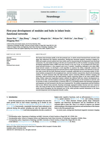
X. Wen et al.NeuroImage 185 (2019) 222–235Fig. 1. Flowchart of the proposed Module-Guided Group-Level Network Construction method. First, based on each subject-level FC network, we detected the subjectlevel modular structure of FC network for each subject and represented such modular structure as a matrix. In this matrix, if two nodes were assigned to the samemodule, the corresponding value was set to one; otherwise, zero. Second, we averaged modular partition matrices across all subjects in each age group to generate thegroup-level “probability matrix”, i.e., “Module Partition Probability” (MPP) matrix. In the MPP matrix, each element represents the probability of assigning a pair ofnodes to the same module. Third, we determined the final weights of edges in the group-level FC network guided by the MPP matrix. For each edge, if its MPP is largerthan thrh, the edge weight is set to the mean of the five highest individual-level FC of the same edge across all subjects (i.e., max Pool). If the MPP is smaller than thrl,the edge weight is the mean of the five weakest individual-level FC across all subjects (i.e., min Pool); otherwise, the weight is the average value across all subjects(i.e., mean Pool).MPPadjustði; jÞ ¼rXTt¼1wrt MPPorigt ði; jÞinter-modular connectivities were calculated using whole-brain FC links,they provide the direct measurement for the whole-brain functionalsegregation/integration changes.After obtaining the mean intra- and inter-modular connectivities ofeach subject at all age groups, we adopted a linear mixed-effect regression (LMER) model to delineate the developmental trajectories of intraand inter-modular connectivities. The LMER model was used due to itsability to handle missing data for the longitudinal study (Verbeke andMolenberghs, 2000). Specifically, both a linear model (using age as afixed-effect variable to test for a linear change) and a log-linear model(using log(age) as a fixed-effect variable to test for a nonlinear change)were built. The log-linear model has been widely used in previous braindevelopment studies (Gao et al., 2015a, 2015b). Random intercept andsubject effects were included as the random effects to characterize thetemporal correlation. The Akaike information criterion (AIC) was adopted for model selection between the linear and nonlinear models. For allmodels, the significance was defined as p 0.05. Next, we identified andcharacterized key brain regions (i.e., hubs) that contributed to thedeveloping functional segregation/integration.(1)where T is the total number of the age time points, and wrt is thecontribution ratio of the t-th time point (t ¼ 1, , T) to the r-th timepoint, as defined below:wrt1ðt rÞ2¼ pffiffiffiffiffi exp 2δ22π δ!,XTt¼1wrt(2)where δ is the standard deviation of the Gaussian function to control the“temporal smoothness”, which is set to 1.5 in this study (please see thefurther discussion on the choice of this parameter in Section 4.5.2). According to Sections 2.4-2.5, we generated “augmented” group-level FCnetworks for each age group by both enhancing the modular structure andmaking them temporally consistent for following longitudinal moduledetection (Section 2.6) and hub assessment (Sections 2.7-2.8).2.6. Module detection from augmented group-level FC networksBased on the “augmented” group-level FC networks, we detected themodular structure for each age separately using Radtools with the samesparsity (10%) as used for individual-level module detection (Poweret al., 2012; Cole et al., 2013; Yeo et al., 2014; Najafi et al., 2016). Foreach group-level network, we repeated the module detection for 100times and used a consensus clustering method (Bassett et al., 2013) toobtain the consistent modules. The modular structure for the age-specificFC networks was compared across different ages (0, 3, 6, 9, and 12months of age). Based on the detected modular structures, we furthercalculated mean intra- and inter-modular connectivities across all modules for each subject at all age groups for quantitative assessment of thedevelopmental changes in the FC network segregation and integration.The mean intra-modular connectivity (reflecting functional segregation)was defined as the averaged FC strength across all the within-modularconnections. The mean inter-modular connectivity (measuring functional integration) was calculated as the averaged FC strength across allthe edges that linked different modules. All the positive connections wereused, and the negative connections were not counted. As the intra- and2.7. Provincial and connector hub detectionInstead of investigating hubs as a whole, we step further to classifyhubs into provincial and connector hubs based on their topological rolesin the modular partitions measured by within-module degree (WD) andparticipation coefficient (P) (Guimera and Amaral, 2005; Meunier et al.,2009, 2010; Bertolero et al., 2015). WD measures how well-connected anode is in the same module that this node belongs to (Guimera andAmaral, 2005). P measures the distribution of nodes’ connections amongdifferent modules (Guimera and Amaral, 2005). In other words, WDevaluates the importance of a region to its module and P characterizes theimportance of a region in connecting with different modules. Specifically, suppose a modular partition is M ¼ {mc jc ¼ 1, , C}. For a node iin module mc, WDi and Pi were respectively calculated by:WDi ¼225κ imc κ mcδκ mc(3)
X. Wen et al.Pi ¼ 1 XC κim cc¼1kiNeuroImage 185 (2019) 222–235months of age compared to other ages (see the blue arrow in Fig. 2a). Incontrast, from our proposed method, the frontal area is consistently andsmoothly partitioned into three modules during the development in thefirst postnatal year. We also find that the introduction of “temporalsmoothness” in the MPP prior calculation could further improve thetemporal consistency of modular structures. As shown in Fig. 2b, without“temporal smoothness”, a small region in the inferior parietal cortex issingled out from the main parietal modules (see the red arrow in Fig. 2b),but such a result does not exist at neighboring ages. With the method ofintegrating “temporal smoothness”, such inconsistent results can beavoided. We also evaluated the effectiveness of our method on an adultdataset. For detailed information, please see Supplementary Materials.(4)where κimc is the total FC strength between node i and the other nodes inmodule mc. κ mc and σ κmc represent the average and standard deviation(SD) of κ imc across all nodes in module mc, respectively. ki is the sum of FCstrength connecting node i.Provincial hubs provide structures within the local module, andconnector hubs mediate connections between multiple modules (Guimera and Amaral, 2005; Van den Heuvel and Sporns, 2013). Thus, provincial hubs should have high WD but low P, while connector hubs haveboth high WD and P. By setting thresholds for within-module degree(thrWD) and participation coefficient (thrP), we detected provincial andconnector hubs from each age-specific group-level FC network. For nodei, it is categorized as a provincial hub if WDi thrWD and Pi thrP, or aconnector hub when WDi thrWD and Pi thrP. In the experimental results, we only showed the detected hubs when thrWD and thrP were set to1.0 and 0.6, respectively. The other settings for thrWD and thrP are discussed in Section 4.5.6.3.2. Development of modular structures in infant functional brain networksThe full views of the detected modules from neonate to twelvemonths of age are visualized in Fig. 3a. Asides from the temporallyconsistent modular patterns across ages, we find that the brain is gradually divided into more and more modules, from 4 modules at neonate to8 after 12 months of maturation (Fig. 3b). Table 1 summarizes how thesenew modules emerge by splitting from or merging to the existing modules at previous ages. Specifically, the quantity of modules increases withthe emergence of new modules in spatially-independent brain regions atdifferent ages, which seems to follow a particular order as describedbelow.In neonates, the brain is separated into four modules: neonatal-modulea (mainly covering the visual cortex in the occipital lobe), neonatalmodule b (encompassing the temporal lobe and subcortical regions),neonatal-module c (in the frontal area), and neonatal-module d (located atcentral areas for sensorimotor function, i.e., pre- and post-central gyrus).With development, all modules are gradually reshaped or divided andnew modules emerge. Compared to the other three modules, the spatialpattern of the neonatal-module a has less change from neonates to oneyear of age.At three months of age, the neonatal-module b is divided into twomodules: one at subcortical regions and the other covering the temporalarea.At six months of age, the neonatal-module c is divided into
First-year development of modules and hubs in infant brain functional networks Xuyun Wena,b, Han Zhangb,*, Gang Lib, Mingxia Liub, Weiyan Yinb, Weili Linb, Jun Zhangc,**, Dinggang Shenb ,d *** a School of Data and Computer Science, Sun-Yat Sen University, China b Department of Radiology and BRIC, University of North Carolina at Chapel Hill, Chapel Hill, NC, USA