Transcription
Rhythm & 12 Lead EKG ReviewMarch 2011 CECondell Medical CenterEMS SystemSite code # 107200E-1211Prepared by: FF/PMD Michael Mounts – Lake Forest FireRevised By: Sharon Hopkins, RN, BSN, EMT-P
ObjectivesUpon successful completion of this module,the EMS provider will be able to: Identify the components of a rhythm strip Identify what the components represent onthe rhythm strip Identify criteria for sinus rhythms Identify criteria for atrial rhythms Identify AV/junctional rhythms
Objectives cont. Identify ventricular rhythmsIdentify rhythms with AV blocksIdentify treatments for different rhythmsIdentify criteria for identification of ST elevationon 12 lead EKG’s Identify EMS treatment for patients with acutecoronary syndrome (ST elevation) Demonstrate standard & alternate placementof ECG electrodes for monitoring
Objectives cont. Demonstrate placement of electrodes forobtaining a 12 lead EKG Demonstrate the ability to identify avariety of static or dynamic EKG rhythmstrips Demonstrate the ability to identify thepresence or absence of ST elevation whenpresented with a 12 lead EKG Review department’s process to transmit12 lead EKG to hospital, if capable Successfully complete the post quiz with ascore of 80% or better.
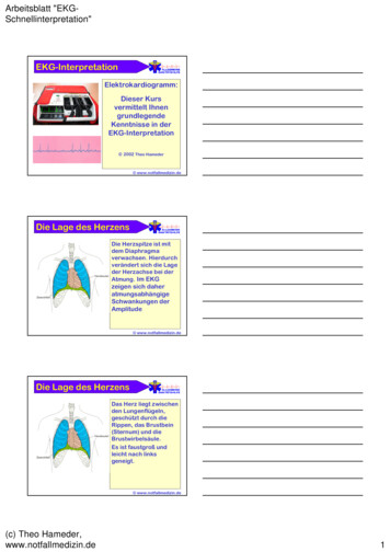
ECG Paper What do theboxesrepresent? How do youmeasure time& amplitude?
Components of the Rhythm Strip ECG Paper Wave forms Wavecomplexes Wavesegments Waveintervals
Wave Forms, Complexes,Segments & Intervals P wave – atrial depolarization QRS – Ventricular depolarization T wave – Ventricularrepolarization
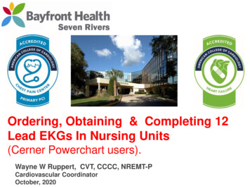
What’s a J point and where is it? J point – point tomark end of QRSand beginning of STsegment– Evaluate ST elevation0.4 seconds after Jpoint– Based on relationshipto the baseline
Intervals and Complexes PR interval – atrial and nodal activity– Includes atrial depolarization & delay in theAV node (PR segment) QRS complex– Corresponds to the patient’s palpated pulse– Large in size due to reflection of ventricularactivity
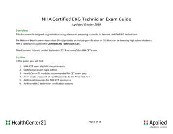
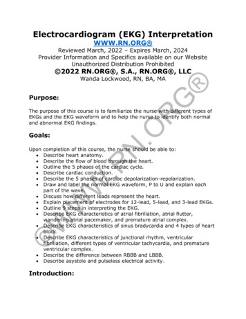
The Electrical Conduction System SA Node AV Node RightBundleBranch Bundle of HIS PurkinjeFibers Left BundleBranch
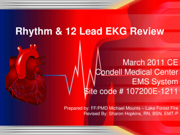
Correlation of ECG Wave Forms
Sinus Rhythms Originate in the SA node– Normal sinus rhythm (NSR)– Sinus bradycardia (SB)– Sinus tachycardia (ST)– Sinus arrhythmia Inherent rate of 60 – 100 Base all other rhythms on deviations fromsinus rhythm
Sinus Rhythm
Sinus Bradycardia
Sinus Tachycardia
Sinus Arrhythmia
Atrial Rhythms Originate in the atria– Atrial fibrillation (A Fib)– Atrial flutter– Wandering pacemaker– Multifocal atrial tachycardia (MAT)– Supraventricular tachycardia (SVT)– PAC’s– Wolff–Parkinson–White syndrome (WPW)
A - Fib
A - Flutter
Wandering Pacemaker
Multifocal Atrial Tachycardia (MAT)(Rapid Wandering Pacemaker) Similar to wandering pacemaker ( 100) MAT rate is 100 Usually due to pulmonary issue COPD Hypoxia, acidotic, intoxicated, etc. Often referred to as SVT by EMS Recognize it is a tachycardia and QRS is narrow
SVT
PAC’s
Wolff–Parkinson–White - WPW Caused by an abnormalaccessory pathway(bridge) in theconductive tissue Mainly nonsymptomatic withnormal heart rates If rate becomestachycardic (200-300)can be lethal– May be brought on bystress and/or exertion
Wolff–Parkinson–White(AKA - Preexcitation Syndrome)
AV/Junctional Rhythms Originate in the AV node– Junctional rhythm rate 40-60– Accelerated junctional rhythm rate 60-100– Junctional tachycardia rate over 100– PJC’s Inherent rate of 40 - 60
Junctional Rhythm
Accelerated Junctional
Junctional TachycardiaOften difficult to pick out so often identified as “SVT”
PJC’sFlat or inverted P Waveor P wave after the QRS
Ventricular Rhythms Originate in the ventricles / purkinje fibers– Ventricular escape rhythm (idioventricular) rate 20-40– Accelerated idioventricular rate 42 - 100– Ventricular tachycardia (VT) rate over 102 Monomorphic – regular, similar shaped wide QRS complexes Polymorphic (i.e. Torsades de Pointes) – life threatening ifsustained for more than a few seconds due to poor cardiacoutput from the tahchycardia)– Ventricular fibrillation (VF) Fine & coarse– PVC’s
Idioventricular
Accelerated Idioventricular
VT (Monomorphic)
VT (Polymorphic)Note the “twisting of the points”This rhythm pattern looks likeRibbon in it’s fluctuations
VF
PVC’s
R on T PVC’s
R on T PVC’s cont. Why is R on T so bad?– Downslope of T wave is the relative refractory period Some cells have repolarized and can be stimulatedagain to depolarize/discharge– Relatively strong impulse can stimulate cells toconduct electrical impulses but usually in aslower, abnormal manner» Can result in ventricular fibrillation Absolute refractory period is from the beginning ofthe QRS complex through approximately the firsthalf of the T wave– Cells not repolarized and therefore cannot bestimulated
Synchronized Cardioversion Cardioversion is synchronized to avoid the refractory periodof the T wave The monitor “plots” out the next refractory period in order toshock at the right moment – the safe R wave– With a QRS complex & T wave present, the R wave canbe predicted (cannot work in VF – no wave forms present)
A/V Heart Blocks 1st degree– A condition of a rhythm, not a true rhythm– Need to always state underlying rhythm 2nd degree– Type I - Wenckebach– Type II – Classic – dangerous to the patient–Can be variable (periodic) or have a setconduction ratio (ex. 2:1) 3rd degree (Complete) – dangerous to the patient
Atrioventricular (AV) Blocks Delay or interruption in impulseconduction in AV node, bundle of His,or His/Purkinje system Classified according to degree of blockand site of block– PR interval is key in determining type ofAV block– Width of QRS determines site of block
AV Blocks cont. Clinical significance dependent on: Degree or severity of the block Rate of the escape pacemaker site Ventricular pacemaker site will be aslower heart rate than a junctional site Patient’s response to that ventricular rate Evaluate level of consciousness /responsiveness & blood pressure
1st Degree Block
2nd Degree Type I
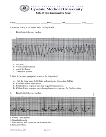
2nd Degree Type II (constant)P WavePR IntervalUniform.12 - .20QRSNarrow & UniformCharacteristicsMissing QRS afterevery other P wave(2:1 conduction)Note: Ratio can be 3:1, 4:1, etc. The higher the ratio, the “sicker” the heart.(Ratio is P:QRS)
2nd Degree Type II (periodic)P WavePR IntervalUniform.12 - .20QRSNarrow & UniformCharacteristicsMissing QRS aftersome P waves
3rd Degree (Complete)
How Can I Tell What Block It Is?49
Helpful Tips for AV Blocks Second degree Type I– Think Type “I” drops “one”– Wenckebach “winks” when it drops one Second degree Type II– Think 2:1 (knowing it can have variableblock like 3:1, etc.) Third degree - complete– Think completely no relationship betweenatria and ventricles
Implanted Pacemaker Most set on demand– When the heart rate falls below a preset rate,the heart “demands” the pacemaker to takeover51
Paced Rhythm - 100% Capture
Goal of Therapy Is rate too slow?– Speed it up (Atropine, TCP) Is rate too fast?– Slow it down (Vagal maneuvers, Adenosine, Verapamil) Blood pressure too low?– Is there enough fluid (blood) in the tank?– Improve contractility of the heart (dopamine,Epinephrine) Are the ventricles irritable?– Soothe with antidysrhythmic (Amiodarone, Lidocaine)
Treatments for Rhythms As always treat the patient NOT the monitor Obtain baseline vitals before and/or during ECGmonitoring Identify rhythm and determine correspondingSOP to follow– Helpful to have at least one more personverify strip Obtain patient history & OPQRST of currentcomplaint
Transcutaneous Pacing No response to doses of atropine Unstable patient with a wide QRS Set pacing at a rate of 80 beats per minute inthe demand mode Start output (mA) at lowest setting possible(0) and increase until capture noted– Spike followed by QRS complex Consider medications to help with the chestdiscomfort
Tachycardias Can be generally well tolerated rhythmsOR Can become lethal usually related to the heartrate and influence on cardiac output Ask 2 questions:– Is the patient stable or unstable? If unstable, needs cardioversion– If stable, determine if the QRS is narrow orwide QRS width drives decisions for therapy instable patient
12-lead ECG Review Lead placement– Lead II monitoring– Obtaining 12 lead EKG Lead / location correlation of ST elevation ST elevation criteria 12 – lead practice EKG’s
12 Lead & Monitoring Lead Placement Einthoven’s Triangle Arm and Leg leads Chest leads (V leads)
Where do those chest stickers go? Make sure to “feel” for intercostal space – don’t just use your eyes!
and the FEMALES Bras loosened,people, if in your way Not all nipple linesare created equal Cover the chest witha towel/sheet afterleads are placed topreserve modesty
Lead Placement in the Female Avoid placing electrodes on top of breasttissue Use the back of the hand to displacebreast tissue out of the way to placeelectrode– Avoids perception of “groping”– Can ask the patient to move left breast out ofway.
Lead Placement The more accurate the lead placement, themore accurate the 12-lead interpretation 12-leads are often evaluated on a sequentialbasis, each interpretation considering theprevious one V4 - V6 should be in a gentle upward curvefollowing the same 5th intercostal rib space CMC has done many trainings– We should be doing this right by now– Placement was standardized in 1938; this isproven science and placement must be accurate!
Heart & 12 – Lead StripCorrelation
12 – Lead StripsRemember: Every lead is like a “camera angle”
12 – Lead Strips cont.Imagine your strips broken into groups like this aVRIV4V1IIV5aVLIIIV2aVFV6V3
Remember You will see Lead II first! This is how you monitor patient’s rhythms May see “reciprocal changes” as ST depression
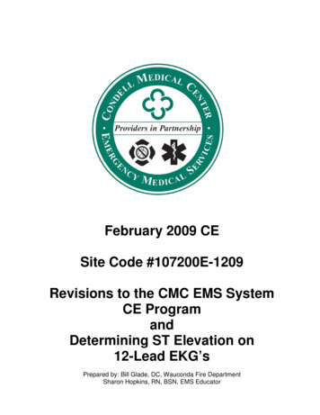
Value of Reciprocal Changes* ST elevation means acute transmuralinjury – injury across all 3 layers of theheart muscle Diagnosis often based on: Presence of ST elevation in 2 or morecontiguous leads Reciprocal changes*Confirms acute-injury patterns Patient history Presentation
12 – Lead Comparison Chart forReciprocal Change(Main ones are highlighted)
Contiguous ECG Leads EKG changes aresignificant when theyare seen in at leasttwo contiguous leads Two leads arecontiguous if theylook at the same areaof the heart or theyare numericallyconsecutive chestleads
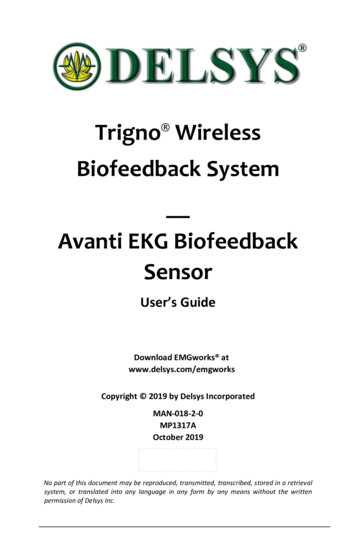
ST Elevation Evaluation Locate the J-point Identify/estimate where the isoelectric line isnoted to be– Check the standardized 2mm mark at the far left orbeginning of each row of the EKG strip Compare the level of the ST segment 0.4seconds after the J point to the isoelectric line Elevation (or depression) is significant if morethan 1 mm (one small box) is seen in 2 or moreleads facing the same anatomical area of theheart (ie: contiguous leads)
Measuring for ST Elevation Find the J point Is the ST segment 1mm above theisoelectric line in 2or more contiguousleads?
Complications of Lateral Wall MII, aVL, V5, V6 Monitor for lethal heart blocks– Second degree type II – classical– Third degree heart block – complete Treat with TCP–Consider sedation for patient comfort–Monitor for capture–Monitor for improvement by measuring levelof consciousness and blood pressure
Complications of Inferior Wall MIII, III, aVF May see Mobitz type I – Wenckebach– Due to parasympathetic stimulation & not injury to conductionsystem Hypotension– Right ventricle may lose some pumping ability Venous return exceeds output, blood accumulates in rightventricle– Less blood being pumped to lungs to left ventricle and outto body– Develop hypotension, JVD, with clear lung sounds Treated with additional fluid administered cautiously Contact Medical Control prior to NTG administration
Complications of Septal Wall MIV1 & V2 Monitor for lethal heart blocks– Second degree type II – classical– Third degree heart block – complete Treat with TCP Rare to have a septal wall MI alone– Often associated with anterior and/or lateralwall involvement
Complications of Anterior Wall MIV3 & V4 Occlusion of left main coronary artery – the “widowmaker”– Cardiogenic shock and death without prompt reperfusion Second degree AV block type II– Often symptomatic– Often progress to 3rd degree heart block– Prepare to initiate TCP Third degree heart block – complete– Rhythm usually unstable– Rate usually less than 40 beats per minute– Prepare to initiate TCP
Acute Coronary SyndromeRoutine Medical Care12 Lead ECG and transmit, if availableStablePatient AlertSkin warm and drySystolic BP 100 mmHg Aspirin 324 mg by mouth Nitroglycerine 0.4 mg SLMay be repeated every 5 minIf pain persists following 2 doses, advance toMorphine Sulfate Morphine Sulfate 2mg IVPSlowly over 2 minutesMay repeat every 2 minutes as needed, to amaximum total dose of 10 mg TransportUnstableAltered Mental StatusSystolic BP 100 mmHg Aspirin 324 mg by mouth, if pt cantolerate Contact Medical control Monitor and TransportNote: ASPIRIN my be withheld if patient is reliable and states has taken within 24 hours
Patient Presenting with Coronary ChestPain – AMI Until Proven Otherwise Oxygen– May limit ischemic injury– New trends/guidelines coming out in 2011 SOP’s Aspirin - 324 mg chewed (PO)– Blocks platelet aggregation (clumping) to keepclot from getting bigger– Chewing breaks medication down faster & allowsfor quicker absorption– Hold if patient allergic or for a reliable patient thatstates they have taken aspirin within last 24 hours
Acute Coronary SyndromeMedications cont. Nitroglycerin - 0.4 mg SL every5 minutes– Dilates coronary vessels to relieve vasospams– Increases collateral blood flow– Dilates veins to reduce preload to reduceworkload of heart Watch for hypotension If inferior wall MI (II, III, aVF), contact MedicalControl prior to administration– If pain persists after 2 doses, move to Morphine– Check for recent male enhancement drug use(ie: viagra, cialis, levitra) Side effect could be lethal hypotension
Acute Coronary SyndromeMedications cont. Morphine - 2 mg slow IVP– Decreases pain & apprehension– Mild venodilator & arterial dilator Reduces preload and afterload– Given if pain level not changed after the2nd dose of nitroglycerin– Give 2mg slow IVP repeated every 2 minutesas needed– Max total dose 10 mg
Practice Rhythms Break into groups of 2 or 3 for rhythm quizzing Instructor will use wristwatch or stopwatch togive each group 30 seconds to determine strip– You don’t have 5 min in the field, you don’t get 5 minin the classroom Message to Instructor:– Refer to slide notes for more information
Sinus w/ 1st degree BlockNo symptoms are due to the first degree heartblock; symptoms would be related to theunderlying rhythm
Junctional TachycardiaNote: Inverted P waves; heart rate 140How is the patient tolerating the heart rate?
2nd Degree Type 1 – WenckebachPR getting longer and finally 1 QRS drops;patient generally asymptomatic; can benormal rhythm for some patients
WPW SyndromeSlurring at beginning of QRS (delta wave)Better diagnosed on 12 lead EKG; patientasymptomatic unless heart rate becomestachycardic
2nd Degree Type II (2:1 conduction)Should be preparing the TCP for this patient
3rd degree heart block (complete)with narrow QRSSymptoms usually based on overall heart rate– the slower the heart rate the moresymptomatic the patient. Prepare the TCP.
NSR to Torsade des PointesIf torsades is long lasting, patient maybecome unresponsive and arrest. Prepare fordefibrillation followed immediately with CPR
Intermittent 2nd Degree Type II(Long PR intervals; periodic dropped beat)Consider need to apply TCP and then turn on if patientsymptomatic
Why would this patient havesymptoms of a stroke? Atrial fibrillation puts patient at risk from clots inthe atria breaking loose and lodging in a vesselin the brain Rhythm irregularly irregular Patient most likely on Coumadin and digoxin
Ventricular Tachycardia What 2 questions should you ask for alltachycardias?– Is the patient stable or unstable?– If stable, then you have time to determine ifthe QRS is narrow or wide What’s this strip?
Paroxysmal SupraventricularTachycardia (PSVT) into sinus rhythmEvidence of abrupt stopping of the SVT
Sinus ArrhythmiaCommon in the pediatric patient and influenced byrespirations. Treatment is not indicated
Sinus with unifocal PVC’sin trigeminyOften PVC’s go away after administration ofoxygen
Multifocal Atrial Tachycardia(MAT)Rapid Wandering PacemakerIdentification can be SVT and treatment wouldbe based on patient symptoms
12 – Lead Time! Same as Lead II strips– Identify St elevation and try to give anatomicallocations– Remember to be watchful for typicalcomplications based onlocation of infarct andblocked coronary vessel
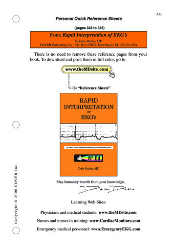
ST elevation in V2 – V5(Anterior wall)
No ST elevation but peaked Twaves (Hyperkalemia)
Hyperkalemia Can be caused by– Over medication of potassium supplement (ex. K-dur)– Excessive intake of foods (bananas are high in potassium)– Crush syndrome After pressure/crush is released, the heart is hit with thepotassium that built-up in the poorly perfused crushed area Too much potassium can lead to critical heartdysrhythmias; difficult to treat Other populations at risk Dialysis patient Patient in diabetic ketoacidosis
ST elevation in II, III & aVF(Inferior wall with LBBB)Watch for hypotension
ST ElevationInferior Wall – II, III, aVFWatch for hypotension
ST elevation in II, III, aVF(Inferior wall - note reciprocal changes)Watch for hypotension
ST elevation in V1 – V6, I & aVL(Anteroseptal with lateral extension)Extensive anteroseptalWatch for heart block
ST elevation V2-V5Watch for heart block
Documentation Rhythm strip interpretation If 12 lead EKG obtained: Note presence or absence of ST elevation If ST elevation noted, in which leads If EKG was transmitted to hospital
Hospital Notification Notify the receiving hospital as soon aspossible about a cardiac alert How did you determine this may be acardiac alert?– Your general impression was made based on: Gathering patient history Performing a cardiac assessment Obtaining a 12 lead EKG as quickly as possibleafter first patient contact 12 lead EKG evaluated for presence of STelevation
Field Trip If your department can obtain12 lead EKG’s review the processfor marking– Patient age and race– Your department name on the 12 lead If your department can fax, review theprocess– Go to the ambulance to review the equipmentand process
Questions?
Bibliography Atwood, S., Stanton, C., Storey-Davenport, J.Introduction to Basic Cardiac Dysrhythmia 3rd Edition.MosbyJems. 2003. Bledsoe, B., Porter, R., Cherry, R. Paramedic CarePrinciples & Practices Third Edition. Brady. 2009. Page, B. 12 Lead ECG for Acute and Critical CareProviders. Brady. 2005. Previous CMC Cardiac CEs Region X SOP March 2007; amended January 1, 2008 Various webpages– For pictures, rhythms, and graphs Walraven, G. Basic Arrhythmia 7th Edition. Brady. 2011. www.MikeCowley.co.uk/leads.htm
variety of static or dynamic EKG rhythm strips Demonstrate the ability to identify the presence or absence of ST elevation when presented with a 12 lead EKG Review department's process to transmit 12 lead EKG to hospital, if capable Successfully complete the post quiz with a score of 80% or better.