Transcription
Suez Canal University Medical JournalVol. 25 (1), 2022Pages 13-21Acute Meningitis in Mansoura Fever HospitalSameh M. Abdel Monem1, Aya M. Al Amely1, Naglaa A. Khalifa2,Omima T. Taha3*, Ahmed L. Sharaf11Department of Tropical Medicine, Faculty of Medicine, Zagazig University, 2Department of Clinical Pathology,Faculty of Medicine, Zagazig University, 3Department of Obstetrics and Gynecology, Faculty of Medicine, SuezCanal UniversityAbstractBackground: Meningitis is a serious health problem that results in grave morbidity and mortality. It is caused by several organisms and present with various presenting pictures impartingsome challenge. Aim: Highlighting the etiology and prognosis of acute meningitis in MansouraFever Hospital. Subjects and Methods: A prospective cross sectional observational study. Thestudy included 350 cases admitted with suspected meningitis. Lumbar puncture: for collectionof cerebrospinal fluid (CSF) samples. CSF analysis, microbiological examination of CSF, andblood culture and antibiotic sensitivity test were done. Brain magnetic resonant imaging (MRI)was performed before lumbar puncture in some patients. Detailed analysis of epidemiologiccharacteristics, clinical data and laboratory findings, the causative organisms, prognosis of allthe patients with acute bacterial meningitis (ABM) were studied. Results: Meningitis occurred atall age groups. A significant proportion of meningitis patients (60%) reported a positive historyof antibiotic intake in the few days (3-5) before admission to the hospital. 79.1% of ABM patientshad a CSF leukocyte count in the range of 100 up to 1,000 cell/mm3 and 80% had CSF neutrophilpercentage 50%. The pathogens were detected by direct Gram-stained smear in 86.4% of patients. Gram ve bacteria was detected in 55/110 (50%) of cases. Gram -ve bacteria detected in40/110 (36.4%) of cases. ABM carried a high mortality rate (22.7%). ABM had 22.7% mortality rate.Conclusions: Meningitis is endemic in Egypt with a significant mortality rate. Strept. pneumoniawas the most common offending organism.Keywords: Acute bacterial meningitis; CSF markers; epidemiology; mortality.IntroductionMeningitis is a drastic health concern,with multiple causative organisms thatdiffer according to age and distribution.The rout of infection might be from theears, nasopharynx, cranial injury, or congenital meningeal defect, or by bloodstream spread(1). Bacterial meningitis isassociated with significant morbidity andmortality due to severe meningeal in*Corresponding Author: omimatharwat@yahoo.comflammation(1). More than one million people are affected each year globally and it ismore common in developing countries asthe meningitis belt in Africa (2). This beltstretches from Senegal to Ethiopia wherethe highest disease burden occurs. Morethan 400 million people are in danger ofinfection in 26 countries(3). It might be attributed to Escherichia coli, Neisseria meningitis, Streptococcus pneumonia, Haemophilus influenza, Streptococcus Group
14B, Staphylococcus aureus, and Listeriamonocytogenes(4). On the contrary, viralmeningitis, also called aseptic meningitis,is considered a self-limited disease with abenign course. Offending viruses includehuman herpes virus 6, herpes simplex(HSV), varicella zoster virus (VZV), cytomegalovirus (CMV), Epstein- Barr virus, JCvirus and enterovirus(5). Non- infectiousetiologies include malignant cells, drugsand blood following subarachnoid hemorrhage(6). Although viral meningitis is morecommon, bacterial infection is more serious. Bacterial meningitis occurs at a rateof 40/100,000 in developing countries(7).The clinical picture may include definitivemeningitis, encephalitis or meningismwhere the CSF is normal(8). Bacterial meningitis is endemic in Egypt with StreptPneumonia considered the leading offending organism (42%)(9). The disease is associated with significant long-term complications as epilepsy, hearing loss, hydrocephalus, and cognitive defects(10). Significant morbidity and mortality occur in developing and developed countries(11) withmortality rates reaching 5% and long-termmorbidity of 15%(12). Earlier studies conducted in Egypt reported high rates ofmorbidity and mortality(9,13) highlightingthe importance of updating the epidemiological aspects of such endemic disease inEgypt.Patients and MethodsThis cross-sectional study was conductedin Mansoura fever hospital from April 2019to March 2020. Informed consent was obtained from all patients and the study protocol was approved by the ethical committee of Faculty of Medicine, ZagazigUniversity. The study included 350 patients admitted to Mansoura fever hospital with suspected meningitis. Bacterialmeningitis was confirmed when patientshad fever, meningeal symptoms, alteredMeningitis updates in Egyptmental status, and with an abnormalnumber of white blood cells in CSF(8). Viralmeningitis was considered in patientswith symptoms suggestive of meningitis,absence of an identifiable cause (bacterialinfection, fungal infection, autoimmunedisease, injury, cancer, or drugs), andcomplete recovery with only conservativemeasures(14). Patients were recruited according to inclusion and exclusion criteria.Inclusion criteria: a) any person with sudden onset of fever and one of the following signs: neck stiffness, altered consciousness, other meningeal signs or purpura fulminans, b) any child under 2 yearsof age with sudden onset of fever and oneof the following signs: neck stiffness, orflaccid neck, bulging fontanel, convulsion,other meningeal signs or purpura fulminans. Exclusion criteria: Cases admittedwith suspicious meningitis following headtrauma or neurosurgical procedure, orcerebral abscess. All patients underwenthistory taking and clinical examination.Lumbar punctureCSF was collected from the subarachnoidspace by a sterile spinal needle (25 or 27G) between the fourth and the fifth lumbar vertebrae. The CSF was collected in 2sterile, screw-capped tubes. Two ml ofCSF was collected in each tube. The firsttube was labeled as No 1 (for direct Gram'stain, standard bacteriological culturemethods and antibiotics sensitivity testing). The second tube was labeled as No 2(for physical (color, aspect), chemical(glucose level, protein concentration) andcytological examination). A strict aseptictechnique was used to collect the bloodfrom patients. The top of the culture bottle was wiped using an ethanol swab, and10-12 ml of blood was taken.CSF analysisThe specimen number one was centrifuged at 2000-3000 rpm for 20 minutes.
Abdel Monem SM et al.The supernatant was aspirated with asterile pipette, leaving approximately0.5ml of fluid in the specimen tube (supernatant was reserved for biochemicalstudies), the sediment shaken to resuspend---Physical characteristics of CSF: Colorand aspect.Cytological examination: The presenceof leukocytes was graded as: negative( 10), 1 (10 – 50), 2 (51 – 290), and 3 ( 291 cells/uL).Microbiological examination: Directsmears stained by Gram were doneaccording to microbiological standards. Microscopical examination ofGram-stained smears was performed.Culture of CSF: Inoculation was doneon blood, chocolate and Mac-Conkey'sagar plates and incubated aerobicallyand anaerobically in presence of 5-10%CO2 at 37C for 72h. Identification ofbacterial colony by colony morphology and biochemical reactions e.g.: Coagulase test, Catalase test, Optochintest and bile solubility test, Indoleproduction test and Oxidase test.Antibiotics sensitivity testingDisk diffusion (Kibry-Bauer) method wasused to determine the susceptibility of therecovered clinical isolates to antimicrobialagents. The diameters of inhibition zoneswere measured in mm using a ruler andclassified as either susceptible, intermediate, or resistant to the agents that wastested.Brain MRIMRI may be performed before the lumberpuncture in patients with neurological deficit, seizure, Glasgow score 11.Inpatient follow-up and outcome assessmentPatients were evaluated daily for symptom improvement or occurrence of newsymptoms. Vital signs were assessed every four hours for the first 48 hours and15based on the need thereafter. Daily follow-up with a neurosign chart that included the following variables: (Glasgow comascale (GCS), seizure, headache, and nuchalrigidity) was done during the inpatienttreatment. Patients were also assessed atdischarge for gross neurologic deficits(visual problems, hearing deficit, and bodyweakness) and mini-mental state examination. Detailed analysis of epidemiologiccharacteristics, clinical data and laboratory findings, the causative organisms,prognosis of all the patients with ABMwere studied.ResultsWe recruited 350 patients. One hundredand ten patients had confirmed diagnosisof acute bacterial meningitis (ABM) (Figure 1). ABM occurred in males (61.8%)more than females (38.2%). It affected patients from urban (70.0%) much morecommonly than patients from rural (30%)communities. Antecedent illnesses (i.e.,any disease or condition which has givenrise to another disease or condition in themorbid train of events leading to death)(13)were found in 74.5% of patients with ABM.Pneumonia was the most common predisposing condition (33.6%). 60 % of meningitis patients reported a positive historyof antibiotic intake in the few days beforeadmission to the hospital (Table 1). Theclinical presentation of ABM was characterized mainly by fever (90.9%) and headache (88%). Signs of meningeal irritationi.e., neck rigidity (90.9%), Kernig sign(72.7), and Brudziniski sign (69%) are significantly higher in patients with meningitis. Once neck rigidity, or headache, orboth are present with fever meningitis issuspected (Table 2). 79.1% of ABM patientshad a CSF leukocyte count in the range of 100 up to 1,000 cell/mm3 and 80% ofABM patients had CSF neutrophil percent-
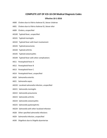
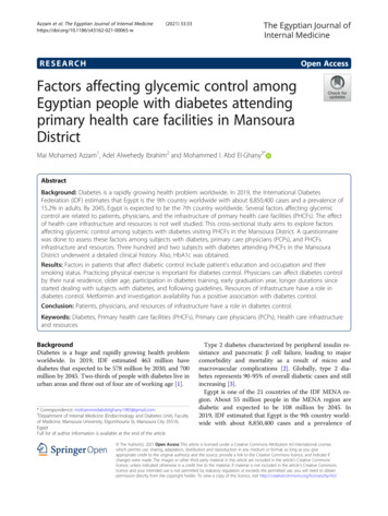
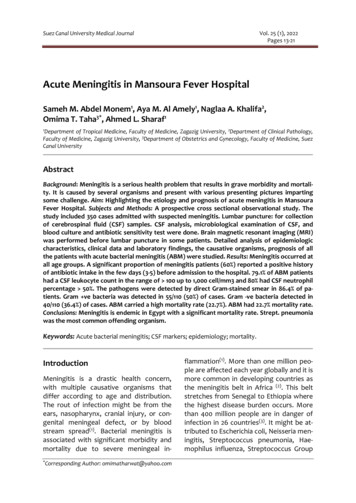
16Meningitis updates in Egyptage 50%. 85.5% of ABM patients had elevated CSF protein ( 80mg/dl). 81.8% ofABM patients had decreased CSF glucose( 50mg/dl). 90% of ABM patients had elevated CSF lactate ( 26mg/dl). Out of 110CSF samples, 95 cases (86.4%) pathogenswere detected by direct Gram-stained sm-ear. Gram ve bacteria were detected in55/110 (50%) of cases and Gram -ve bacteria in 40/110 (36.4%). Gram ve cocci werethe commonest (25/110) microorganismisolated in the present study. S. pneumonia was the most common isolated pathogen (24 isolates) (Table 3, and figure 2).Table 1: Demographic data of 110 ABMpatients admitted to MFHAge (Yrs.) N (%)5 (4.5%)6 -12 month10 (9.1%) 1 -612 (10.9%) 6 -18 18 -4025 (22.7%) 40 -6030 (27.3%) 6028 (25.5%)Sex N (%)68(61.8%)MaleFemale42 (38.2%)Residence N (%)77 (70.0%)UrbanRural33 (30.0%)Antecedent illness N (%)16 (14.5%)Otitis media21 (19.1%)Sinusitis37 (33.6%)PneumoniaTonsillitis8 (7.3%)None28 (25.5%)Spinal anesthesia N (%)1 (0.9%)Recent travel N (%)6 (5.5%)Antibiotic intake N (%)66 (60.0%)Culture and sensitivity testing revealedampicillin- sulbactam (53%), cefotaxime(51.2%), and ceftriaxone (47.3%) as the effective antibiotics against gram ve isolates. Vancomycin (70%), piperacillin/tazobactam (28%), and levofloxacin(27.6%) were revealed as sensitive antibiotics for those with gram -ve isolates.Blood cultures revealed the same organism as obtained by CSF culture in 73(66.4%) of cases. There was a different organism obtained in 14 (12.7%) of cases.While negative culture was obtained in 23(20.9%) of cases. MRI was indicated beforelumbar puncture if the case was suspected meningitis but with atypical presentation. MRI findings were as follows:- Obstructive hydrocephalic changes atthe level of aqueduct of Silvius withdilated both lateral ventricles and ballooned 3rd ventricle with normal sized4th ventricle.
Abdel Monem SM et al.--Ventriculitis which appeared as thickependymal enhancement in ependymal lining of lateral ventricles on postcontrast study as well as debris seenin both lateral ventricles forming fluid-fluid level. Theses debris displayedintermediate SI on both T1 and T2 WI,restriction on DWI and no enhancement on post-contrast studies.Infarction along the territory of themiddle cerebral artery (MCA) whichappeared as wedge shaped areas cortical and subcortical in location fol-17-lowing MCA territory displaying lowSI on T1 high SI on T2 and FLAIR images and restriction on DWI. It displayed mass effect in the form of effacement of the related cortical sulciand compression of ipsilateral lateralventricle.Subdural empyema which appearedas well- defined crescent extra axialcollection displaying low SI on T1 highSI on T2 and FLAIR, restriction onDWI, and thick marginal enhancement on post contrast study.Table 2: Clinical symptoms and signsof the study patientsClinical 8.2Vomiting3027.3%Photophobia1816.4%Neck rigidity10090.9%Irritability3531.8%Skin rash109.1%Altered consciences1816.4%Seizures1614.5%Kernig sign8072.7%Brudzinski sign7669.1%Local nerve signs87.3%AF bulge (N 5)360%Abnormal cry (N 10)440%Weak suckling (N 8)675%Data is expressed as percentage and frequency. AF (anterior fontanel)ABM carried a high mortality rate (22.7%).One infant 6m, one female patient 40 yrs.old, one girl 16 yrs. old, and all other caseswere above 55 years old. 21.8% of ABM patients developed permanent sequelae ashearing deficit, paralysis, cognitive slowness, and memory trouble (Table 4).DiscussionABM occurred at a rate of 31.43% while viral meningitis affected 37.14% of the studied population. The ratio of bacterial toaseptic meningitis cases vary between dif-ferent studies. In an earlier study conducted in Egypt, the majority of meningitiscases were possibly viral meningitis(42.86%) while bacterial meningitis withno positive culture growth occurred at arate of 17.13%, and finally, bacterial meningitis cases that showed positive culturegrowth affected 5.08%(15). However, another study reported a lower rate of bacterial meningitis (10.3%)(16). This differencein the percentage of bacterial versusaseptic meningitis can be attributed todifferences in the place and time of stud-
18ies done, so, in developing countries bacterial meningitis was the major constituent of meningitis cases, while in developed countries and especially after theimplementation of anti-capsular vaccines,bacterial meningitis became less event inrelation to viral meningitis. Cases of ABMwere distributed at all age group asagreed with previous results(15,17).Table 3: Frequency of pathogens causingABM detected by CSF direct stained filmDirect stained filmFrequency%Gram ve cocci2522.7%Gram ve diplococci2018.2%Gram ve bacilli54.5%Gram ve chain54.5%Gram –ve diplococci1715.5%Gram – ve coccobacilli1311.8%Gram –ve bacilli109.1%No detected strains1513.6%Table 4: Prognosis of the cases of .8Data is expressed as percentage and frequency. *(Oneinfant 6m, one female patient 40 years old, one girl16 years old, and all other cases were above 55 yearsold). ** Most of these sequelae hearing deficit, paralysis, cognitive slowness, and memory trouble.A previous study reported increased prevalence of meningitis among infants andchildren(18). This may be due to environmental factors and the availability of children vaccines against common pathogenscausing meningitis, as H. influenzae vaccine of children at the United States in1990(19). Also, the current study included afew numbers of infants and young children which affects the true reflection ofdisease distribution as most of them werereferred to Mansoura University ChildHospital. About three- quarters of ABMpatients had antecedent illnesses withMeningitis updates in Egyptpneumonia the most common antecedentcondition (33.6%). This agreed with a previous study where lung infections preceded pneumococcal meningitis in 40% of patients(20). Clinical features give the clue tothe early diagnosis of ABM by a physician;therefore, there are various clinical symptoms which can be quite helpful in the diagnosis process. The majority of patientspresented with the classic clinical featuresof bacterial meningitis includes fever,headache, and stiff neck. This agreed withpreviously reported results(21,22). Othersigns occurred at variable rates acrossstudies(20,23,24). Some patients with meningitis have atypical presentation e.g. infants younger than one year and elderlypatients(18). These neurologic manifestations are related to the severity of the disease and the time interval before arrivalto the hospital(16). Accordingly, none ofthe symptoms and signs could accuratelydiscriminate between patients with meningitis from those without it. Patients withsuspected meningitis on clinical groundsshould be referred for lumber punctureand CSF examination to settle the correctdiagnosis(25). Sixty- one cases of the recruited patients were referred for MRI before LP because of atypical presentation.Pathological MRI findings are frequentlyfound in patients with acute bacterialmeningitis such as leptomeningeal enhancement, hypo intensity of subcorticalwhite matter underlying intense meningeal inflammation(26). Additionally sometimes the edema of the brain or wideningof the extra-axial CSF spaces may be evident in meningitis which can be evident bynon-contrast MRI sequences(27). MRI isuseful for diagnosis of patients with atypical presentation as well as its complications(28). Bacterial meningitis is a lifethreatening disease that is associatedwith significant mortality and morbidity.This was supported by previous results(17).
Abdel Monem SM et al.19Figure 2: The frequency of bacteria causing ABM by culturesWithout any treatment, the case-fatalityrate can reach 70%, and one in five survivors of bacterial meningitis may be leftwith permanent disability including hearing loss, neurologic disability, or limbloss(29). The epidemiology of acute bacterial meningitis has changed markedly sincethe introduction of conjugate vaccines.However, the disease continues to cause aheavy burden even in developed countries, causing substantial morbidity andmortality(30).Strength and limitationsWe recruited many patients. The diagnosiswas done according to strict diagnosticcriteria. MRI was done for patients withatypical presentation. However, the correlation between MRI findings and the clinical presentation was not done.Conflict of interest: NoneReferences1-23-4-5-ConclusionAcute bacterial meningitis affects a widerange of patients' age. Physicians shouldnot neglect the atypical presentation ofthe disease to avoid drastic complications.MRI is a useful tool for the diagnosis ofatypical cases.6-7-WHO. Meningococcal meningitisfact sheet. World Health Organization : Geneva. 2017.1Pfister W, Roos KL, 2003: Bacterialmeningitis. In: Principles of Neurologic Infectious Diseases. Roos KL(ed.), McGraw-Hill.Patel JC, Soeters HM, Diallo AO, etal. A Network Supporting CaseBased Meningitis Surveillance andVaccine Evaluation in the Meningitis Belt of Africa, J Infect Dis, 220,Suppl 4, 1 Dec 2019, S148–S154.Thigpen MC, Whitney CG, Messonnier NE, et al. Bacterial Meningitis in the United States, 1998–2007. N Engl J Med. 2011; 364(21):2016-25.2Nath A. Meningitis: bacterial, viral,and other. In: Goldman L, editor.Goldman-Cecil Medicine, 25th ed.Philadelphia, PA: Saunders; 2016.p. 2480-95.3Jarvis JN, Meintjes G, Williams A,
20Meningitis updates in Egypt8-9-10-11-12-13-14-15-16-et al. Adult meningitis in a settingof high HIV and TB prevalence:findings from 4961 suspectedcases. BMC Infect Dis. 2010; 10(1):67.4Brouwer M, Beek Dv. Epidemiology of community-acquired bacterial meningitis. Curr Opin InfectDis 2018; 31 (1):78–84:Jeffery KJ, Read SJ, Peto TE, et al.Diagnosis of viral infections of thecentral nervous system: clinicalinterpretation of PCR results.Lancet 1997; 349, 313–317. doi:10.1016/S0140-6736(96)08107-X.Afifi S, Wasfy MO, Azab MA, et al.Laboratory-based surveillance ofpatients with bacterial meningitisin Egypt (1998-2004). Eur J ClinMicrobiol Infect Dis 2007; 26(5):331‐340.Van de Beek D, de Gans J, TunkelAR, et al. Community-acquiredbacterial meningitis in adults. NEngl J Med 2006; 354:44-53.Theodoridou MN, VasilopoulouVA, Atsali EE, et al. Meningitisregistry of hospitalized cases inchildren: epidemiological patternsof acute bacterial meningitisthroughout a 32-year period. BMCInfect. Dis. 2007; 7:101-6.Pelkonen T, Roine I, Monteiro L, etal, 2009: Risk factors for deathand severe neurological sequelaein childhood bacterial meningitisin sub-Saharan Africa. Clin. Infect.Dis. s-explained/index.php/Glossary:Antecedent causeBrivet FG, Ducuing S., Jacobs F. etal. Accuracy of clinical presentation for differentiating bacterialfrom viral meningitis in adults: amultivariate approach. IntensiveCare Med 31, 1654–1660 (2005).Ibrahim AA, Abdel Hakam SM,Ahmed AMH, et al. UpdatedEpidemiological Aspects and out-17-18-19-20-21-22-23-24-25-come of Meningitis Cases atAbbassia Fever Hospital during thePeriod 2006-2017. Afro-Egy J InfectEndemic Dis 2020;10(4):379-388Abdelkader MM, Aboshanab KM,El-Ashry MA, et al. Prevalence ofMDR pathogens of bacterialmeningitis in Egypt and newsynergistic antibiotic combinations. PLOS ONE. 2017; (2):1-17.Amarilyo G, Alper A, Ben-Tov A, etal. Diagnostic Accuracy of ClinicalSymptoms and Signs in ChildrenWith Meningitis. Pediatric Emergency Care. 2011; 27 (3):196-9.Troendle M, Pettigrew A. A systematic review of cases of meningitis in the absence of cerebrospinalfluid pleocytosis on lumbar puncture. BMC Infect Dis. 2019; 19(1):692-703.Aneja S. Acute Bacterial MeningitisEarly Diagnosis and Complications.Ind J Ped. 2015;82(4):303-5.Dery M, Hasbun R. Changingepidemiology of bacterial meningitis. Curr Infect Dis Reports. 2007;9 (4):301-7.Brouwer MC, Thwaites GE, TunkelAR, et al. Dilemmas in the diagnosis of acute community-acquiredbacterial meningitis. The Lancet.2012;380 (9854): 1684-92.Afifi S, Wasfy MO, Azab MA, et al.Laboratory-based surveillance ofpatients with bacterial meningitisin Egypt (1998–2004). Euro J ClinMicrobiol & Infect Dis. 2007; 26(5):331-40.Arda B, Sipahi OR, Atalay S, et al.Pooled Analysis of 2,408 Cases ofAcute Adult Purulent Meningitisfrom Turkey. Med Principles andPractice. 2008;17(1):76-9.Elmore JG, Horwitz RI, Quagliarello VJ. Acute meningitis with anegative gram's stain: Clinical andmanagement out-comes in 171episodes. Am J Med. 1996;100(1):78-84.
Abdel Monem SM et al.26- Heydarian F, Ashrafzadeh F, Rostazadeh A. Predicting factors andprevalence of meningitis in patients with first seizure and feveraged 6 to 18 months. Neurosciences. 2014; 19 (4): 297-300.27- Brouwer MC, van de Beek D.Earlier Treatment and ImprovedOutcome in Adult Bacterial Meningitis Following Guideline RevisionPromoting Prompt Lumbar Puncture. Clin Infect Dis. 2015; 61(4):664-5.28- Saberi A, Roudbary S-A, Ghayeghran A, et al. Diagnosis ofMeningitis Caused by PathogenicMicroorganisms Using MagneticResonance Imaging: A SystematicReview. Basic and Clin Neurosci J.2018;9(2):73-86.29- Lummel N, Koch M, Klein M, et al.Spectrum and Prevalence ofPathological Intracranial MagneticResonance Imaging Findings inAcute Bacterial Meningitis. ClinNeuroradiol. 2014;26(2):159-67.30- Tokimura R, Iguchi M, Ito E, et al.The duration of antibiotic therapyin bacterial meningitis with pyogenic ventriculitis. Rinsho Shinkeigaku. 2019;59(3):133-8.31- Gudina EK, Tesfaye M, Wieser A, etal. Outcome of patients with acutebacterial meningitis in a teachinghospital in Ethiopia: A prospectivestudy. PLOS ONE. 2018; 13(7):e0200067.21
in Mansoura fever hospital from April 2019 to March 2020. Informed consent was ob-tained from all patients and the study pro-tocol was approved by the ethical com-mittee of Faculty of Medicine, Zagazig University. The study included 350 pa-tients admitted to Mansoura fever hospi-tal with suspected meningitis. Bacterial