Transcription
Egyptian Journal of Ophthalmology (MOC) 2022;2:19-28Ranibizumab Response in Diabetic Macular Edema at Mansoura Ophthalmology CenterRoaa N. Elbeheiri, Sahar M. Eltarshouby, Hossameldin Y. Abouelkheir, Amr M. AbdelkaderOphthalmology Department, Faculty of Medicine, Mansoura University, Egypt.Correspondence to: Roaa N. Elbeheiri. Mansoura Ophthalmic Center, Faculty of Medicine, Mansoura University, ElgomhoriaStreet, Mansoura, Egypt. P.O: 35516. Tel. 00201025469011. E mail: mezoroka@gmail.comReceived: 7-11-2021, Accepted: 28-2-2022, Published online: 16-3-2022EJO(MOC) 2022;2:19-28.Short tile: Ranibizumab Response in DME at MOCAbstractPurpose: Diabetic macular edema (DME) is a complicated disease due to a multifactorial process comprising the breakdown ofthe blood retinal barrier with a subsequent fluid accumulation in the macula. Treatment of DME depends recently on moresuccessful therapies such as anti-vascular endothelial growth factor (VEGF) therapies that could stabilize or improve vision inseveral cases. Ranibizumab has been submitted for approval as a treatment of visual impairment owing to DME.Objective: To assess the response of intravitreal injection (IVI) of ranibizumab in DME at Mansoura ophthalmology center.Patients and methods: This were a prospective study carried out on a total of 50 diabetic cases with DME. Full history wastaken from included cases and full ophthalmological examination was conducted in addition to evaluation of the macular retinalmaps using optical coherence tomography (OCT) (Spectral domain OCT 2000). Three monthly consecutive intravitrealinjections of anti-VEGF of ranibizumab at a dosage of 0.5mg/0.05 ml were administered to all the cases.Results: There was highly statistically significant improvement in the visual acuity (VA) and highly statistically significantreduction in the macular thickness in the included cases after one month and after three months as compared with the pretreatmentvalue (P 0.001). The degree of diabetic control didn’t appear to affect the change of the macular thickness.Conclusion: The intravitreal ranibizumab injection effectively decreased macular thickness and improved VA. The structuraland functional effects of ranibizumab appeared as early as after 1 month of treatment and maintained at 3 months followingtreatment.Key words: Diabetes, Macular edema, Optical coherence tomography, Macular thickness.Of note, VEGF was reported to be responsible for theINTRODUCTION:Diabetic retinopathy (DR) is considered the main etiologyabnormal vascular permeability in DME5. The DME treatmentof blindness among working-age adults, and DME is the mainhas been shifted from the laser photocoagulation to anti-VEGF1reason for vision loss related to DR .therapy6.Retinal oedema is responsible for retinal micro-structuralThe advantages of anti-VEGF therapy in decreasing DMEalterations, retinal atrophy of photoreceptors and ganglion celland improving patient’s vision have been reported in many2disorders . In addition, it might be considered consensual thatthe best improvements in VA could be accomplished whenretinal oedema is managed. In the context of a chronic andprogressive disease, DME has to be faced as a state to controlas effectively and rapidly as possible3.studies7-10.Ranibizumab, in addition to aflibercept, have been reportedas the first line therapies among the other anti-VEGF11-13.There are several data demonstrating the efficiency ofranibizumab in treatment of patients with DME14-16. On theThe more increase in duration of diabetes mellitus (DM)other hand, there are studies that revealed poor response ofand the higher level of glycosylated hemoglobin (HbA1c) aresome patients to anti-VEGF therapies even after 3 or morethe main predisposing factors for DME development ininjections17, 18.diabetic cases4.Egyptian Journal of Ophthalmology, a publication of Mansoura Ophthalmic Center.Address: Mansoura Ophthalmic Center, Mansoura University, Mansoura, Egypt.Tel. 0020502202064.Fax. 0020502202060.E-mail: ejo@mans.edu.eg
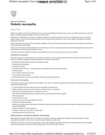
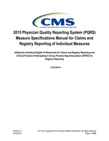
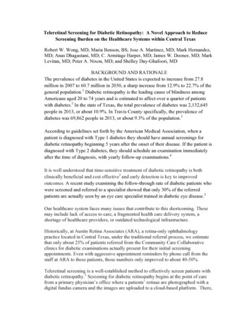
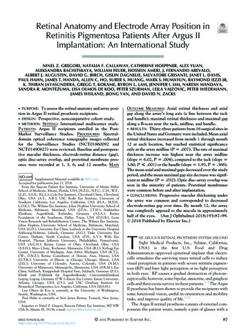
Ranibizumab Response in Diabetic Macular Edema at Mansoura Ophthalmology CenterEJO(MOC) 2022;2:19-28Optical coherence tomography Spectral domain (SD-PATIENTS AND METHODSThis is a prospective and interventional study carried out atMansoura ophthalmology center, Mansoura University,OCT) 2000 [Topcon, Inc., Paramus, NJ, USA] was used toassess the state of the retina and macula (Fig. 1).Mansoura, Egypt from January 2020 to December 2020.This study included 50 diabetic patients with DME (centralmacular thickness 300 μm) from both genders. The caseswith the following conditions were excluded; Patients withhistory of previous of IVI of anti-VEGF and steroids withduration less than one year, history of Grid or focal macularlaser and history of pars plana vitrectomy.The current study was submitted for approval from theinstitutional review board of Mansoura Faculty of Medicineand obtaining an informed written consent from theparticipants (code number MS.19.10.860), date (3\12\2019).All cases were subjected to complete history taking andFigure 1: Topcon3D OCTthrough full general examination.The following laboratory investigations were done forFull detailed ophthalmic examination was performed forall the cases comprising assessment of the visual acuity (VA)using Landolt’s VA chart and after that transformed forstatistical analysis to Log MAR. The objective refraction wasdone using a streak retinoscopy, followed by subjectiverefraction with trial frame and Snellen eye chart placed at 6-Mwhole the cases (HBA1C, lipid profile and serum creatinine).Entire blood samples were divided into two aliquots: the firstpart was collected in a vacationer tube containing Na2-EDTAfor the assay of HbA1c; the second was collected in a plainvacationer tube and centrifuged (3000 r.p.m) for serumpreparation.distance.Three consecutive monthly IVI of anti-VEGF ofSlit lamp biomicroscopy (Haag Streit BP 900) (HaagStreit, Koeniz, Switzerland) was utilized to evaluatecornealtransparency,anterior chamberfordepth andregularity, pupil shape, size, regularity and reactivity, state ofthe lens and complications of DM which include recurrentstye, xanthelasma, accelerated senile cataract, rubeosisiridis.Posterior segment examination was conducted byutilizing indirect ophthalmoscope and slit lamp biomicroscopyranibizumab (Lucentis; Novartis, Basel, Switzerland) at adosage of 0.5mg/0.05ml were administered in a sterile mannerusing a 30-G needle toward the center of the vitreous at 4mminphakicor 3.5mmin pseudophakiceyesandinferotemporally from the limbus.The cases were assessed at 1 month & 3 months regardingthe VA and macular thickness. Example for oct of a casebefore and after IVI injection of ranibizumab (Fig. 2).with auxiliary contact lens.Egyptian Journal of Ophthalmology (EJO), a publication of Mansoura Ophthalmic Center (MOC)20
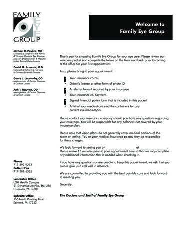
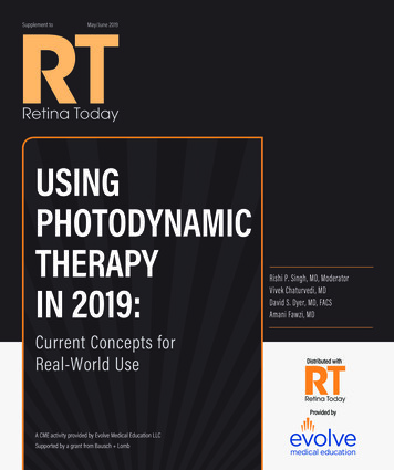
Ranibizumab Response in Diabetic Macular Edema at Mansoura Ophthalmology CenterA)EJO(MOC) 2022;2:19-28OCT scan and colour thickness map of first IVI injection of ranibizumabB) OCT scan and colour thickness map of second IVI injection of ranibizumabFig. 2: Example for oct of a case before and after IVI injection of ranibizumab.Pearson’sStatistical analysiscorrelationcoefficientandspearman’sData analysis was performed by Statistical Package for thecorrelation coefficient were utilized to test the associationSocial Sciences (SPSS 26.0, IBM/SPSS Inc., Chicago, IL)between two quantitative parametric and non-parametricsoftware. Categorical data were expressed as frequencies andvariables correspondingly. The positive coefficient indicatespercentages (%) while in the quantitative data, we used meandirect correlation while negative coefficient indicates inverseand standard deviations (SD) as well as median (range).correlation with increasing the strength of correlation withFor quantitative data, independent-Samples t-test andapproximating to 1 and -1. Probability (p value) 0.05 wasMann-Whitney U test were utilized to compare 2 groups ofconsidered to be statistically titativedatacorrespondingly.As demonstrated in table (1), The mean age in the casesFor comparing the quantitative data at two time points, wewas 56.92 7.27 years. There is 28 males (56%) and 22used paired samples t-test and Wilcox on signed rank test forfemales (44%). The mean duration of DM was 14.15 4.41.parametric and non-parametric data correspondingly. ForThere were 48 cases (96%) with IDDM and 2 cases (4%) withcomparing the quantitative data at three time points, we usedNIDDM. The mean HBA1C was 9.12 2.04.repeated measures ANOVA test and Friedman’s test forparametric and non-parametric data correspondingly.There were 21 cases (42%) with no Hypertension and 29cases (58%) represented with Hypertension. There were 48Egyptian Journal of Ophthalmology (EJO), a publication of Mansoura Ophthalmic Center (MOC)21
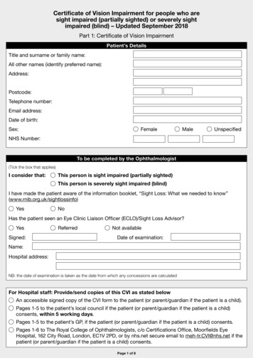
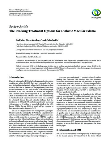
Ranibizumab Response in Diabetic Macular Edema at Mansoura Ophthalmology CenterEJO(MOC) 2022;2:19-28cases (96%) without renal affection and only 2 affected (4%).surgery. The mean Triglycerides level was 175.19 86.01, theThere were 45 cases (90%) with no Hepatitis C virus and 5mean cholesterol was 221 53.58, the mean Low densitycases with HCV (10%). There were 43 cases (86%) withoutlipoprotein level LDL was 145.39 27.27 and High-densityprevious surgery and only 7 cases (14%) with previouslipoprotein level HDL was 49 15.46.Table (1): Demographic, medical history, surgical history and laboratory data in study casesTotal number 30Age/yearsSexMedianRange56.92 7.2757(44-74)Males28 (56%)Females22 (44%)14.15 4.41Duration of DM (Years)Type of DMmean SDIDDM14(7-20)48 (96%)NIDDM2 (4%)Medical and surgical historyHTN29 (58%)Renal affection2 (4%)HCV5 (10%)Previous surgery7 (14%)Laboratory dataHbA1C (%)9.12 2.048.73(5.72-13.2)TGs (mg/dl)175.19 86.01151(80-415)221 53.58219(135-323)LDL (mg/dl)145.39 27.27149(100-184)HDL (mg/dl)49 15.4648(25-75)Cholesterol (mg/dl)As demonstrated in table (2), The mean VA beforeThe mean Macular thickness before treatment was 467.39treatment was 1.22 0.36, at 1 month was 1.03 0.31 and at 120.04, at 1 month was 362.89 130.33 and at 3 months was3 months was 0.86 0.25. There was a highly statistically292.83 80.88. There was a highly statistically significantsignificant improvement in the VA in the included cases afterdecrease in the macular thickness in the included cases afterone month and after three months as compared with beforeone month and after three months as compared with beforetreatment value (p 0.001). Also, there was a highlytreatment value (p 0.001). also, there was a highly statisticallystatistically significant improvement in the VA after threesignificant decrease in the macular thickness after threemonths of treatment as compared with at 1 month valuemonths of treatment as compared with at 1 month value(p 0.001).(p 0.001).Egyptian Journal of Ophthalmology (EJO), a publication of Mansoura Ophthalmic Center (MOC)22
Ranibizumab Response in Diabetic Macular Edema at Mansoura Ophthalmology CenterEJO(MOC) 2022;2:19-28Table (2): Analysis of Visual acuity and Macular thickness (µm) in the cases of the study along the duration of follow upFollow upVariablesTest of significanceBefore treatmentAt 1 monthAt 3 Months(N 50)(N 50)(N 50)1.22 0.361.03 0.310.86 0.25P 0.001*(-51.9:0)(-62.58:0)P1 0.001*Visual acuity (VA)Mean SDP2 0.001*Percent of changeP3 0.001*Macular thickness (µm)467.39 120.04Mean SD362.89 130.33292.83 80.88P 0.001*(-54.42:0)(-56.51: -17.14)P1 0.001*P2 0.001*Percent of changeP3 0.001* statistically significant if P 0.05P1: Significance between before treatment value and value at 1 monthP2: Significance between before treatment value and value at ٣ monthsP3: Significance between value at 1 month and value at ٣ monthsAccording to the degree of diabetes control, there was 8Table (4) shows that, there was a statisticallycases (16%) with controlled diabetes and there were 42 casessignificant positive correlation between change of VA with(84%) with uncontrolled diabetes. As demonstrated in tablecholesterol and change of macular thickness.(3), there was no statistically significant difference in theTable (4): Correlation between change of VA and otherpercent of change of macular thickness at 1 moth and 3 monthsfactors in the studybetween the cases with controlled versus uncontrolleddiabetes.change of VA at 3 months(Log mar)VariablesTable (3): Relation between diabetic control and change ofrPAge0.0320.825Duration of DM- 0.2560.111HBA1C- 0.1450.330TGs0.0600.726Cholesterol0.4190.011*LDL- 0.2680.168HDL0.1050.649Change of Macular0.4500.001*macular thicknessItemsControlledUncontrolledn 8n 42P valuePercent of change of macular thickness at 1 monthMean SDRange-16.26 9.44(-23.74:-4.88)-23.74 17.440.427(-54.42:0)Percent of change of macular thickness at 3 monthsMean SD-42.87 3.75-36.26 essEgyptian Journal of Ophthalmology (EJO), a publication of Mansoura Ophthalmic Center (MOC)23
Ranibizumab Response in Diabetic Macular Edema at Mansoura Ophthalmology CenterEJO(MOC) 2022;2:19-28injections, and this significance was maintained up to threeDISCUSSION:This study was conducted to evaluate response of IVI ofmonths following the last injection28.ranibizumab in DME at Mansoura ophthalmology center. TheThe results of our study concerning the relationshipstudy comprised 50 diabetic cases with DME who werebetween VA or macular thickness and intravitreal ranibizumabrecruited along a period of one year.was also confirmed in the study11The mean age in the cases was 56.92 7.27 years. There is28 males (56%) and 22 females (44%).Comparable outcomes were defined by Ashraf and hiscolleagues who recorded that in spite of a significant reductionPrevalence of either males or females showed greatdifferences between the different studies. Females constitutednearly about two thirds (64%) of diabetic population, inprevious epidemiological researches conducted in Egypt[19, 20]in CFT following the switch to ranibizumab in eyes with DMErefractory to bevacizumab29.Within the same context, Lai et al. (2020) have.demonstrated that IVI of ranibizumab was demonstrated to beHowever, gender wasn't demonstrated to be asignificantassociated with a significant improvement in BCVA andpredisposing factor for developing DR in the majority of on of the CFT over one year5.researchesIn a recent meta-analysis, there was a significant elevationdemonstrated that; males were more liable for DRin values in comparison with the pretreatment value. Asdevelopment in comparison with females, as in UAE24 andregards whole study types, the largest total increase from basal25Oman .value was noticed at 36 months, with a change in BVCA of 10In the current study, the average duration of DM was 14.15letters. The authors also reported that ranibizumab had a 4.41. There were 48 cases (96%) with IDDM and 2 casessignificant impact on CFT at all-time points, demonstrating a(4%) with NIDDM. The mean HBA1C was 9.12 2.04.significant reduction in comparison with the basal value. In theIn the same line, comparable duration was recorded bycontext of all study types, the average CFT decrease wasTang and his colleagues who have demonstrated that the159µm at 12 months, 135µm at 24 months, and 223µm at 36duration of DM among their patients was 13.5 (SD 9.37)months30.years26.The REFINE study demonstrated anaveragedrop ofThis was shorter than the duration recorded in a research146.5μm for CFT after one year of therapy under “3 PRN”carried out in Saudi Arabia where the average age of onset andregimen[31]. The RISE and RIDE study recorded an averageaverage duration of DM were 43.91 and 13.4 years,reduction of 249.3μm for CFT following monthly IVI of27correspondingly .The discrepancies among researches could beexplained by difference in inclusion criteria of the includedpatients.In the current study, there was a highly statisticallysignificant drop in the VA and macular thickness in theranibizumab for one year32 where as Nepomuceno and hiscolleagues have recorded an average reduction of 126μm forCFT under monthly IVI of ranibizumab33.The current study demonstrated that, there was astatistically significant positive association between change ofVA with cholesterol and change of macular thickness.included cases after one month and after three months asPrior researches recorded that VA improvement from basalcompared with before treatment value (p 0.001). also, therelevel was demonstrated to be accompanied by reductions in thewas a highly statistically significant reduction in the VA andCMT from basal value34.macular thickness after three months of treatment as comparedwith at 1 month value (p 0.001)Our results were also in accordance with Sarhan et al.(2019) who demonstrated that there was a significant (P 0.01)The current results came in accordance with Sarhan et al.correlation between the average change in CMT of all study(2019) who observed that ranibizumab significantly improvedstages and the average change in BCVA. One month after eachBCVA and CMT after three consecutive monthly intravitrealinjection, BCVA improved significantly, as a result of reducedmacular edema and vascular leakage.Egyptian Journal of Ophthalmology (EJO), a publication of Mansoura Ophthalmic Center (MOC)24
Ranibizumab Response in Diabetic Macular Edema at Mansoura Ophthalmology CenterEJO(MOC) 2022;2:19-28Our results also came in accordance with Minami et al.correlation is only moderate throughout the initial year of(2017) who demonstrated that there was a significanttherapy40, 41, implying that loss of VA is likely multifactorial(p 0.05) correlation between the Δ CMT-1w and ΔVA-1w inand might depend mainly on the disturbance of the retinalthe present study. Further researches with a large sample sizearchitecture or direct photoreceptor damage owing toare required to verify the association between thelongstanding oedema or other factors42.improvements in BCVA and improvements in CMT35.Certain researches have demonstrated that patientsThese outcomes are in accordance with prior studies'accomplish brilliant anatomical outcomes with anti-VEGFoutcomes in terms of the correlation between CMT and VAtreatment but don't accomplish corresponding functionalafter ranibizumab36.improvement. In addition, disorganization of the inner retinalBrowning and his colleagues have demonstrated that,although there was a significant correlation between VA andlayers, determined by SD-OCT, is accompanied by a worsebaseline BCVA and less promising outcomes43.CMT, there was a great change in VA at any given retinalOn the other hand, in certain cases with persistent DME,thickness, and OCT measurement solely mightn't be a niceBCVA might be kept or improved after the therapy. Actually,replacement for VA as the primary result in researches ofin cases with DME, marked diurnal alterations we rerecordedDME. OCT could only record the degree of oedema; thein retinal thickness measurements, that might be, partially,duration of oedema and the damage to cells cannot be assessed.owing to factors which include alterations in blood pressure asFurther study with more functional macular tests is warrantedwell as retinal metabolism44, 45.to study the relations between VA, macular state, and CMT37.The present study had certain limitations. First, theIn the current study, the most significant correlation amongrelatively small sample size to carry out a subgroup analysis.the three injections was observed between the baseline CMTThus, further major clinical research is required. Second, theand at 1 month after the first injection. These results were induration of follow up was short, thus additional research withaccordance with Sarhan et al. (2019).a long follow up period is essential to determine whether orIt was recorded that the basal value of CMT may predictthe structural outcomes following IVI of ranibizumab[38]not the short-term effects of an IVR injection on the BCVA. Theand CMT are associated with the long-term effects of the IVRmost significant correlation among the three injections, also,injection on the BCVA. Third, the present study had no controlwas observed between the basal value of BCVA and at 1group.month. As formerly recorded, the basal value of BCVA mightThe effects of the natural disease course or previouspredict the functional outcomes following IVI ranibizumabtreatments on the current results can’t be excluded. In thetherapy39.present study, we couldn’t assess the effect of circadianTaken together, the study reported that measuring thefluctuation, the reproducibility and alterations in the retinalefficiency as early as one month after an IVI of ranibizumabthickness measurements in healthy individuals and patients.in cases with DME may be predictive of the structural andFurther clinical study that comprises a control group andfunctional effects of the IVI of ranibizumab in addition to therepeated measurements is required.prediction from the basal value of CMT and BCVA.The current study had some limitations. First, the numberIn addition, a less CMT and BCVA effect was observed atof patients in this case series was too small to perform athree months than at one month. This recommends that othersubgroup analysis. Another larger clinical study is needed.inflammatory or angiogenic cytokines down the cascade at aSecond, the current follow-up period was short and anotherlater phase might participate in the pharmacodynamics ofclinical study with a long follow-up period is necessary to11ranibizumab .determine whether or not the short-term effects of an IVRAlthough several studied have demonstrated that; thereinjection on the BCVA and CMT are correlated with the long-was a significant association between BCVA letter scores andterm effects of the IVR injection on the BCVA. Third, theCFT in DME cases managed with anti-VEGF therapy, suchcurrent study had no control group.Egyptian Journal of Ophthalmology (EJO), a publication of Mansoura Ophthalmic Center (MOC)25
Ranibizumab Response in Diabetic Macular Edema at Mansoura Ophthalmology CenterEJO(MOC) 2022;2:19-28It could not exclude the influences of the natural diseaseEthics declarationscourse or previous treatments on the current results. In theConflict of interestcurrent study, we could not evaluate the effect of circadianRoaa N. Elbeheiri, Sahar M. Eltarshouby, Hossameldin Y.fluctuation and the reproducibility and variations in the retinalAbouelkheir, Amr M. Abdelkader all authors have no conflictsthickness measurements in both healthy subjects and patients.of interest that are directly relevant to the content of thisAnother clinical study that includes a control group andreview.repeated measurements is needed.Funding: No sources of funding were used to conduct atintravitrealReviewer disclosures: No relevant financial or otherranibizumab effectively decreased macular thickness andrelationships to disclose.improved VA. The results were maintained up to 3 monthsDeclaration of interest: No financial affiliations or financialfollowing the last dosage of ranibizumab.Structural andinvolvement with any organization or entity with a financialfunctional effects of intravitreal ranibizumab injection mightcompeting with the subject matter or materials discussed in thebe detectable as early as one month following the therapy.Thereviewdegree of diabetic control didn’t affect the degree of change ofReferencesvisual acuity and macular thickness.1. Abràmoff MD, Lavin PT, Birch M, Shah N, Folk JC.Recommendations: Strict and regular control and follow upPivotal trial of an autonomous AI-based diagnostic systemof cases with diabetes to prevent the occurrence of associatedfor detection of diabetic retinopathy in primary carecomplications especially diabetic retinopathy.offices. NPJ digital medicine. 2018;1(1):1-8.Further studies should be performed including larger number2. Bressler S, Ayala A, Bressler N, Melia M, Qin H, Ferrisof patients from more than a single center.3rd F, et al. Diabetic Retinopathy Clinical ResearchControl of modifiable risk factors could have a positiveNetwork Persistent macular thickening after ranibizumabimpact on progression of DR and on DME onset. Kawasakitreatment for diabetic macular edema with visionR et al., observed an association between lipid-loweringimpairment. JAMA Ophthalmol. 2016;134(3):278-85.medication and a decrease of DR and its complications.3. Ashraf M, Souka A, Adelman R. Predicting outcomes toFurthermore, an association between statins medication andanti-vascular endothelial growth factor (VEGF) therapy invitamin C (as an antioxidant agent) supplementation coulddiabetic macular oedema: a review of the literature. Britishhave a synergistic role in lowering DME onset and DRJournal of Ophthalmology. 2016;100(12):1596-604.progression.4. Chen Y-P, Wu A-L, Chuang C-C, Chen S-N. Factorsall persons with diabetes receive at least yearly dilated eyeinfluencing clinical outcomes in patients with diabeticexaminations and are offered appropriate treatment whenmacular edema treated with intravitreal ranibizumab:indicated.comparison between responder and non-responder cases.DATA AVAILABILITYScientific reports. 2019;9(1):1-8.All data are included in this article.5. Lai K, Huang C, Li L, Gong Y, Xu F, Zhong X, et al.ACKNOWLEDGEMENTAnatomical and functional responses in eyes with diabeticNonemacular edema treated with “1 PRN” ranibizumab: one-Corresponding authoryear outcomes in population of mainland China. BMCCorrespondence to: Roaa N. Elbeheiriophthalmology. 2020;20(1):1-7.Email: mezoroka@gmail.com6. Elman MJ, Aiello LP, Beck RW, Bressler NM, BresslerAffiliationsSB, Edwards AR, et al. Randomized trial evaluatingRoaa N. Elbeheiri, Mansoura University, Egypt.ranibizumab plus prompt or deferred laser or triamcinoloneEgyptian Journal of Ophthalmology (EJO), a publication of Mansoura Ophthalmic Center (MOC)26
Ranibizumab Response in Diabetic Macular Edema at Mansoura Ophthalmology almology. 2010;117(6):1064-77. e35.EJO(MOC) 2022;2:19-28Resistance to Anti-VEGF Therapy. Ophthalmology inRussia. 2018;15(4):382-7.7. Bressler NM, Varma R, Mitchell P, Suñer IJ, Dolan C,17. Koytak A, Altinisik M, Sari ES, Artunay O, Akkan JU,Ward J, et al. Effect of ranibizumab on the decision to driveTunçer K. Effect of a single intravitreal bevacizumaband vision function relevant to driving in patients withinjection on different optical coherence tomographicdiabetic macular edema: report from RESTORE, RIDE,patterns of diabetic macular oedema. Eye. 2013;27:716-21.and RISE trials. JAMA ophthalmology. 2016;134:160-6.8. Mitchell P, Bandello F, Schmidt-Erfurth U, Lang GE,Massin P, Schlingemann RO, et al. The RESTORE study:ranibizumab monotherapy or combined with laser halmology. 2011;118(4):615-25.9. Boyer DS, Nguyen QD, Brown DM, Basu K, Ehrlich JS,18. Jampol LM, Bressler NM, Glassman AR. Revolution to anew standard treatment of diabetic macular edema. Jama.2014;311(22):2269-70.19. Macky TA, Khater N, Al-Zamil MA, El Fishawy H,Soliman MM. Epidemiology of diabetic retinopathy inEgypt: a hospital-based study. Ophthalmic research.2011;45(2):73-8.Ride, et al. Outcomes with as-needed ranibizumab after20. Herman W, Aubert R, Engelgau M, Thompson T, Ali M,initial monthly therapy: long-term outcomes of the phaseSous E, et al. Diabetes mellitus in Egypt: glycaemic controlIII RIDE and RISE trials. Ophthalmol. 2015;122:2504-13.and10. Li X, Dai H, Li X, Han M, Li J, Suhner A, et al. iabetic Medicine. 1998;15(12):1045-51.and safety of ranibizumab 0.5 mg in Chinese patients with21. El-Asrar AMA, Al-Rubeaan KA, Al-Amro SA, Kangavevisual impairment due to diabetic macular edema: resultsD, Moharram OA. Risk factors for diabetic retinopathyfrom the 12-month REFINE study. Graefe's archive foramong Saudi diabetics. International ophthalmology.clinical and experimental ophthalmol.2019;257:529-41.1998;22:155-61.11. Nguyen QD, Brown DM, Marcus DM, Boyer DS, Patel S,22. Bamashmus MA, Gunaid AA, Khandekar R. Regular visitsFeiner L, et al. Ranibizumab for diabetic macular edema:to a diabetes clinic were associated with lower magnituderesults from 2 phase III randomized trials: RISE and RIDE.of visual disability and diabetic retinopathy—a hospital-Ophthalmology. 2012;119(4):789-801.based historical cohort study in yemen. Diabetes12. Korobelnik J-F, Do DV, Schmidt-Erfurth U, Boyer DS,Holz FG, Heier JS, et al. Intravitreal aflibercept for diabeticmacular edema. Ophthalmology. 2014;121(11):2247-54.13. Avery RL, Gordon GM. Systemic safety of prolongedtechnology & therapeutics. 2009;11(1):45-50.23. Waked N, Nacouzi R, Haddad N, Zaini R. Epidemiologyof diabetic retinopathy in Lebanon. Journal francaisd'ophtalmologie. 2006;29(3):289-95.monthly anti–vascular endothelial growth factor therapy24. Al-Maskari F, El-Sadig M. Prevalence of diabeticfor diabetic macular edema: a systematic review and meta-retinopathy in the United Arab Emirates: a cross-sectionalanalysis. JAMA ophthalmology. 2016;134(1):21-9.survey. BMC ophthalmology. 2007;7(1):1-8.14. Katz G, Moisseiev E, Goldenberg D, Moisseiev J,25. El Haddad OA, Saad MK. Prevalence and risk factors forLomnicky Y, Abend Y, et al. Ranibizumab for persistentdiabetic retinopathy among Omani diabetics. Britishdiabetic macular edema after bevacizumab treatment.journal of ophthalmology. 1998;82(8):901-6.European journal of ophthalmology. 2017;27(2):210-4.15. Rahimy E, Shahlaee A, Khan MA, Ying G-s, Maguire JI,26. Tang FY, Ng DS, Lam A, Luk F, Wong R, Chan C, et al.DeterminantsofquantitativeopticalcoherenceHo AC, et al. Conversion to aflibercept afte
Egyptian Journal of Ophthalmology (EJO), a publication of Mansoura Ophthalmic Center (MOC) 20 PATIENTS AND METHODS This is a prospective and interventional study carried out at Mansoura ophthalmology center, Mansoura University, Mansoura, Egypt from January 2020 to December 2020. This study included 50 diabetic patients with DME (central