Transcription
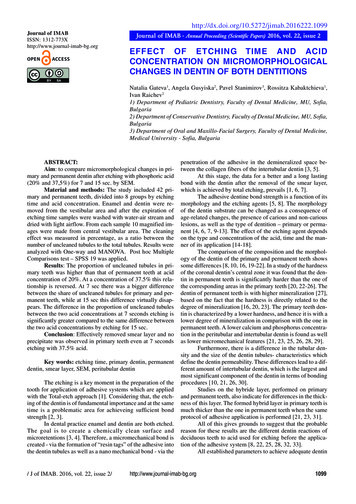
l of IMABISSN: 1312-773Xhttp://www.journal-imab-bg.orgJournal of IMAB - Annual Proceeding (Scientific Papers) 2016, vol. 22, issue 2EFFECT OF ETCHING TIME AND ACIDCONCENTRATION ON MICROMORPHOLOGICALCHANGES IN DENTIN OF BOTH DENTITIONSNatalia Gateva1, Angela Gusyiska2, Pavel Stanimirov3, Rossitza Kabaktchieva1,Ivan Raichev21) Department of Pediatric Dentistry, Faculty of Dental Medicine, MU, Sofia,Bulgaria2) Department of Conservative Dentistry, Faculty of Dental Medicine, MU, Sofia,Bulgaria3) Department of Oral and Maxillo-Facial Surgery, Faculty of Dental Medicine,Medical University - Sofia, BulgariaABSTRACT:Aim: to compare micromorphological changes in primary and permanent dentin after etching with phosphoric acid(20% and 37,5%) for 7 and 15 sec. by SEM.Material and methods: The study included 42 primary and permanent teeth, divided into 8 groups by etchingtime and acid concentration. Enamel and dentin were removed from the vestibular area and after the expiration ofetching time samples were washed with water-air stream anddried with light airflow. From each sample 10 magnified images were made from central vestibular area. The cleaningeffect was measured in percentage, as a ratio between thenumber of uncleaned tubules to the total tubules. Results wereanalyzed with One-way and MANOVA. Post hoc MultipleComparisons test – SPSS 19 was applied.Results: The proportion of uncleaned tubules in primary teeth was higher than that of permanent teeth at acidconcentration of 20%. At a concentration of 37.5% this relationship is reversed. At 7 sec there was a bigger differencebetween the share of uncleaned tubules for primary and permanent teeth, while at 15 sec this difference virtually disappears. The difference in the proportion of uncleaned tubulesbetween the two acid concentrations at 7 seconds etching issignificantly greater compared to the same difference betweenthe two acid concentrations by etching for 15 sec.Conclusion: Effectively removed smear layer and noprecipitate was observed in primary teeth even at 7 secondsetching with 37.5% acid.Key words: etching time, primary dentin, permanentdentin, smear layer, SEM, peritubular dentinThe etching is a key moment in the preparation of thetooth for application of adhesive systems which are appliedwith the Total-etch approach [1]. Considering that, the etching of the dentin is of fundamental importance and at the sametime is a problematic area for achieveing sufficient bondstrength [2, 3].In dental practice enamel and dentin are both etched.The goal is to create a chemically clean surface andmicroretentions [3, 4]. Therefore, a micromechanical bond iscreated - via the formation of “resin tags” of the adhesive intothe dentin tubules as well as a nano mechanical bond - via the/ J of IMAB. 2016, vol. 22, issue 2/penetration of the adhesive in the demineralized space between the collagen fibers of the intertubular dentin [3, 5].At this stage, the data for a better and a long lastingbond with the dentin after the removal of the smear layer,which is achieved by total etching, prevails [1, 6, 7].The adhesive dentine bond strength is a function of itsmorphology and the etching agents [5, 8]. The morphologyof the dentin substrate can be changed as a consequence ofage-related changes, the presence of carious and non-cariouslesions, as well as the type of dentition – primary or permanent [4, 6, 7, 9-13]. The effect of the etching agent dependson the type and concentration of the acid, time and the manner of its application [14-18].The comparison of the composition and the morphology of the dentin of the primary and permanent teeth showssome differences [8, 10, 16, 19-22]. In a study of the hardnessof the coronal dentin’s central zone it was found that the dentin in permanent teeth is significantly harder than the one ofthe corresponding areas in the primary teeth [20, 22-26]. Thedentin of permanent teeth is with higher mineralization [27],based on the fact that the hardness is directly related to thedegree of mineralization [16, 20, 23]. The primary teeth dentin is characterized by a lower hardness, and hence it is with alower degree of mineralization in comparison with the one inpermanent teeth. A lower calcium and phosphorus concentration in the peritubular and intertubular dentin is found as wellas lower micromechanical features [21, 23, 25, 26, 28, 29].Furthermore, there is a difference in the tubular density and the size of the dentin tubules- characteristics whichdefine the dentin permeability. These differences lead to a different amount of intertubular dentin, which is the largest andmost significant component of the dentin in terms of bondingprocedures [10, 21, 26, 30].Studies on the hybride layer, performed on primaryand permanent teeth, also indicate for differences in the thickness of this layer. The formed hybrid layer in primary teeth ismuch thicker than the one in permanent teeth when the sameprotocol of adhesive application is performed [21, 23, 31].All of this gives grounds to suggest that the probablereason for these results are the different dentin reactions ofdeciduous teeth to acid used for etching before the application of the adhesive system [8, 22, 25, 28, 32, 33].All established parameters to achieve adequate dentinhttp://www.journal-imab-bg.org1099
surface for bonding by application of adhesive systems havebeen tested in permanent teeth [4-6, 19, 34]. The same clinical protocol is transmitted directly in primary teeth, withouttaking into consideration the differences in the compositionand morphology of dentin that exists between the teeth fromboth dentitions [21, 23, 26, 35, 36].The aim of this study is to compare the micromorphological changes in dentin in primary and permanent teeth after etching with 20% and 37,5% phosphoric acid for 7 and 15seconds by using Scanning Electron Microscope (SEM).In order to achieve the goal the following task was set:- to determine the degree of cleaning the smear layerwith different concentrations of phosphoric acid and for different etching time.The working hypothesis is that there are no differencesbetween the dentin in permanent and primary teeth, as wellsas that etching time and acid concentration have no influenceon the degree of smear layer removal.MATERIAL AND METHODSSelection and preparation of experimental samples:Thestudy used intact extracted teeth from both dentitions. Theprimary teeth were collected from healthy children betweenthe age of 7 and 9 after their parents signed informed consentfor the use of the teeth in the experiment. The permanent teethwere also collected from healthy patients aged 55-65 yearswho also signed informed consent. The deciduous teeth wereextracted due to physiological exfoliation or because of orthodontic treatment and the permanent - due to periodontalproblems. After extraction, the teeth were placed in 10% formalin solution for 10 minutes, then until the time of execution of the task were stored in saline.Grouping of the experimental samples. The study included 42 intact teeth (primary and permanent incisors andcanines). The teeth were divided randomly into 8 groups of 5teeth in each group (only primary and only permanent), depending on the etching duration and phosphoric acid concentration (table 1).Table 1. Grouping of the experimental samples.EtchingGroup/tooth typeGroup/primary teethGroup/permanent teeth20% phosphoric acid37,5% phosphoric acid7 sec15 sec7 sec15 secGroup 1n 5Group 2n 5Group 3n 5Group 4n 5Group 5n 5Group 6n 5Group 7n 5Group 8n 5n number of samplesPreparation the tooth surface. With a turbine round bur(ISO 806 314 001 534 012 for primary teeth and ISO 806314 001 534 014 for permanent teeth) and water-air coolinga cut in medio-distal direction along the vestibular surface ismade. The purpose was pre-marking the depth of removal ofenamel and dentine. With a turbine fissure diamond bur (ISO806204108524835010) and the water-air cooling the enameland dentin parallel to the long axis of the tooth, at the depthof the round bur marking, are removed. Diamond burs werechanged after every three teeth. The surface after drilling waspolished with an abrasive disk (ISO 625900372523) eachused only for one tooth.All prepared samples were observed with an opticalmicroscope OLYMPUS VANOX-T under magnification of25x to 100x, to establish whether the enamel was completelyremoved from the vestibular surface.Dentin etching. The etching agents 20% (Pekaetch 20,Heraeus Kulzer GmbH) and 37,5% (Esticid - Gel, HeraeusKulzer GmbH) phosphoric acid were applied for 7 or 15 sec.After the etching time expired each sample waswashed with water-air stream for 15 sec and dried with gentle scattered airflow at a distance of 20-25 cm for 5 sec. Theprepared samples were left at room temperature for 24 hoursin separate sterile petri dishes for each group to avoid contamination before the SEM observation.Control samples. One tooth from each dentition wasnot etched after the removal of enamel and dentin from thevestibular surface in order to be used as a control sample.1100Development of SEM images. Ten SEM images weretaken of each sample at magnifications of 1,500 from a zonewith dimensions of 114µm \ 35.2µm in the central part of thevestibular surface of the researched object. The analysis ofall 420 SEM images served for evaluation of the etching effect for each sample.The criteria for assessing the cleaning effect of the acids are:- Degree of removal of the smear layer by comparingthe number of dentin tubules’ orifices without plugs and thosewhich are partially or completely obstructed from smear layeron each image;- Presence of a smear layer within the intertubular dentin - presence of precipitates and deposits on the surface ofthe intertubular dentin.The cleaning effect is measured in percentages – as aratio between the number of uncleaned (fully or partially obstructed by a smear layer) tubules and the whole amount oftubules in each image.For a statistical measuring of the results single-variate(One-way ANOVA) and multivariate (MANOVA) dispersionanalysis for comparison of quantifiable indicators in morethan two groups and assessment of the combined impact ofseveral factors were used. PostHocMultipleComparison testafter establishing a statistically significant difference betweenthe groups for analyzing the differences in pairs - packetSPSS 19 was also used.http://www.journal-imab-bg.org/ J of IMAB. 2016, vol. 22, issue 2/
RESULTSControl samplesDentin control samples without etching indicated asmear layer in both types of teeth – primary and permanent(figure 1).Fig. 1. Representative SEM images of a smear layer on the dentin surface of a primary (A) and a permanent (B)tooth.The non-etched dentin surface was covered with asmear layer with identical characteristics in both types ofsamples. The smear layer could be seen as a veil that covers the treated dentin surface. The inherent microcanal structure of the dentin cannot be seen. In some places the smearlayer was cracked. Visible cracks, which probably correspond to the entrances of the dentin tubules, could be observed. The surface of the smear layer was scattered withparticles with irregular shapes and different sizes whichwere visibly not well attached to it.When 20% phosphoric acid was applied the following results were observed:Groups 1 and 2 - 20% phosphoric acid for 7 secondsThe etching of the dentin surface in the two groups- primary and permanent teeth, breaks, but does not completely remove the smear layer (figure 2).Fig. 2. Representative SEM images of dentin surface of a primary (A) and permanent (B) tooth etched with 20%phosphoric acid for 7 seconds. A preserved smear layer on the intertubular dentin can be observed.The smear layer was removed primarily over the dentin tubules and preserved over the intertubular dentin (pins,fig. 2A and B). In some places some of the dentin tubulesorifices were not exposed. That’s why the number of openeddentin tubules was smaller than their real quantity. Manyof them remain obscured from the smear layer (pointer fig./ J of IMAB. 2016, vol. 22, issue 2/2A and B).Groups 3 and 4 - 20% phosphoric acid for 15 secondsThe results in group 3 primary and group 4 permanent teeth with increased etching time of 15 sec are similarto the previous ones. Some of dentin tubules remain by par-http://www.journal-imab-bg.org1101
tially or completely obscured by smear plugs (pointer fig.3). In both groups residues from the smear layer and pre-cipitates within the intertubular dentin are observed (pins fig. 3A and B).Fig. 3. Representative SEM images of dentin surface of a primary (A) and permanent (B) tooth which was etchedwith 20% phosphoric acid for 15 sec. There were residues of a smear layer (pins) and single, dentin tubules that areobscured with smear layer plugs (pointer).Dentin tubules in Group 3 which were obscured withsmear plugs were rarely observed when comparing the results between Group 1 and Group 3a (figure 2 and figure3), but remnants of the smear layer and precipitates on theintertubular dentin were preserved. The results are similarin the groups of permanent teeth - 2 and 4. Uncleaned dentin tubules in group 4 were more rarely observed, but stillremnants of smear layer on the intertubular dentin in bothgroups (fig. 2B and fig. 3B) could be seen.The following results when applying 37,5% phos-phoric acid were observed:Groups 5 and 6 – 37,5% phosphoric acid for 7 secondsA fully removed smear layer of the intertubular dentin and of the dentin tubules’ orifices can be observed inthe tested primary teeth from group 5 (fig. 4A). In the permanent teeth from Group 6 precipitates and residues of thesmear layer on the intertubular dentin surface can still beobserved (pins, fig. 4B). There are also remnants of plugsin the orifices of the dentin tubules (fig. 4B pointer).Fig. 4. Representative SEM images of dentin surface which was etched with 37,5% phosphoric ac
Considering that, the etch-ing of the dentin is of fundamental importance and at the same time is a problematic area for achieveing sufficient bond strength [2, 3]. In dental practice enamel and dentin are both etched. The goal is to create a chemically clean surface and microretentions [3, 4]. Therefore, a micromechanical bond is created - via the formation of “resin tags” of the adhesive .