Transcription
World J. Surg. 26, 1222–1227, 2002DOI: 10.1007/s00268-002-6486-2WORLDJournal ofSURGERY 2002 by the SociétéInternationale de ChirurgieCollis-Nissen Procedure in Patients with Esophageal Atresia: Long-termEvaluationHisayoshi Kawahara, M.D.,1 Kenji Imura, M.D.,2 Makoto Yagi, M.D.,2 Akio Kubota, M.D.,2 Akira Okada, M.D.112Department of Pediatric Surgery, Osaka University Graduate School of Medicine, 2-2 Yamadaoka Suita, Osaka 565-0871, JapanDivision of Pediatric Surgery, Osaka Medical Center for Maternal and Child Health, 840 Murodo-cho, Izumi-City, Osaka 594-1101, JapanPublished Online: September 4, 2002Abstract. A subset of patients with esophageal atresia (EA) have anassociated short esophagus and require an antireflux procedure. TheCollis-Nissen procedure, which consists of a combination of Collis gastroplasty and Nissen fundoplication, is considered an option in suchconditions. The long-term results of EA patients undergoing this procedure have rarely been reported. The results of the Collis-Nissen procedure were examined in four EA patients with follow-up for an average of9 years. The Collis-Nissen procedure was conducted concurrently withsegmental esophageal resection for an associated congenital esophagealstenosis close to the esophagogastric junction in two postoperative Grosstype C patients (7 months, 2 years), as an antireflux operation in apostoperative Gross type A patient with an unreducible sliding hiatalhernia (10 years), and with primary repair of EA in a Gross type A patientwith a long gap (4 months). There were no significant complicationsexcept minor anastomotic leakage and an anastomotic stenosis that required postoperative dilation. Significant reflux was negative, as shown byan upper gastrointestinal study and scintigraphy. Limited esophagitisjust above the esophagogastric junction was observed in a Gross type Apatient with a long mediastinal gastric segment. Swallow-related pressurereduction at the wrapping cuff was detected by manometric examination.The Collis-Nissen procedure is a useful option for treating EA patientswho require esophageal lengthening and control of gastroesophagealreflux.A subset of patients with esophageal atresia (EA) require esophageal lengthening in various situations because of a short esophagus. It is often difficult to ensure the safety of the intraabdominalesophagus by mobilizing the distal esophagus during esophagealsurgery following primary repair of EA because of the upwardtension due to tight adhesion of the esophagus to the mediastinum[1].It has been a therapeutic challenge for pediatric surgeons torepair Gross type A EA with a long gap between the proximal anddistal ends of the esophagus. Various procedures have been performed for surgical treatment of EA with a long gap, but thelong-term results of those procedures remain controversial [2].This International Society of Surgery/Société Internationale de Chirurgie (ISS/SIC) article was presented at the 39th World Congress of Surgery International Surgical Week (ISW01), Brussels, Belgium, August26 –30, 2001.Correspondence to: H. Kawahara, M.D., e-mail: kawahara@pedsurg.med.osaka-u.ac.jpCollis gastroplasty, which creates a gastric tubular segmentfrom the lesser curve of the stomach, is an option for lengtheningthe esophagus [3]. This procedure has been reported to be usefulto control gastroesophageal reflux (GER) in patients with a shortesophagus when conducted concurrently with Nissen fundoplication [4, 5]. The results of EA patients undergoing this procedurehave rarely been reported [6]. We report here the long-termresults of the Collis-Nissen procedure applied in EA patients.Materials and MethodsSubjectsDuring 1989 –1999 the Collis-Nissen procedure was performed infour EA patients by one pediatric surgeon (H.K.) in Osaka University Hospital and Osaka Medical Center for Maternal andChild Health. The clinical details of each patient were as follows.Case 1. A female baby was born at 39 weeks’ gestation weighing2821 g. She was diagnosed with Gross type C EA and underwentprimary repair at 4 days of age (Fig. 1). The association ofcongenital esophageal stenosis (CES) was suspected during theoperation because of difficulty inserting an 8F catheter into thestomach, which was confirmed by postoperative esophagram at 17days of age. Postprandial emesis and stridor became significant at2 months of age. A 2 cm segmental resection of the esophagealnarrowing and primary end-to-end anastomosis were performedat 7 months of age, when she weighed 7 kg. A Collis-Nissenprocedure was conducted concurrently with the repair of the CES.The tracheobronchial remnants, including cartilage, were documented histologically in the resected specimens.Case 2. A male baby was born at 37 weeks’ gestation weighing3046 g. He was diagnosed with Gross type C EA and underwentprimary repair at 2 days of age (Fig. 2). CES was suspected duringthe operation because an 8F catheter could not pass through thedistal esophagus into the stomach; it was confirmed when anesophagram showed a tapered narrowing close to the esophagogastric junction at 14 days of age. The frequency of food impaction
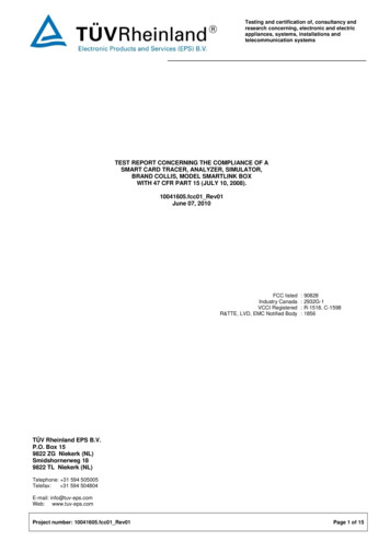
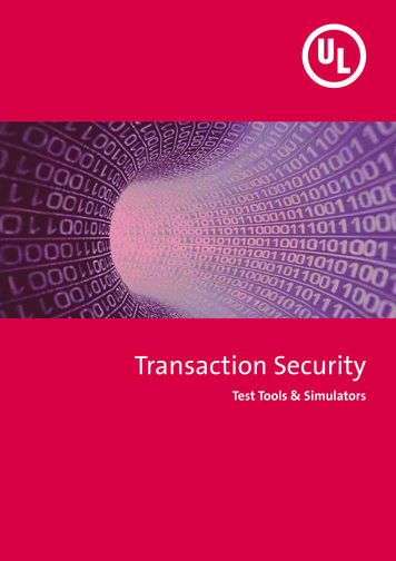
Kawahara et al.: Collis-Nissen for Esophageal AtresiaFig. 1. Case 1. A. Free gastroesophageal reflux (GER) and stasis ofcontrast medium in the esophageal body proximal to the narrowing (whitearrow) were observed at 2 months of age. B. A 2 cm segmental resectionof the narrowing and primary end-to-end anastomosis with Collis-Nissenprocedure was performed at 7 months of age. C. After repeated balloondilatation for 1 year, swallowing difficulties and postprandial stridor weredecreased at 19 months of age. D. Esophagram at 9 years of age shows anintact wrapping cuff and the appropriate passage of contrast medium fromthe esophagus into the intraabdominal stomach.Fig. 2. Case 2. A. Initial esophagram at 14 days of age showed a taperednarrowing (white arrow) close to the esophagogastric junction. B. Persistent narrowing (white arrow) and free GER of contrast medium wereobserved at 20 months of age, when he had frequent respiratory infectionsand emesis. C. Same procedure (3 cm segmental resection) as in case 1 wasperformed at 2 years of age, and his symptoms were relieved by theoperation. D. Esophagram at 10 years of age shows an intact wrapping cuffand no esophageal dilatation.at the site of the narrowing increased after the age of 16 months.The same procedure (3 cm segmental resection) as in case 1 wasperformed at 2 years of age, which relieved his preoperativesymptoms. The tracheobronchial remnants, including cartilage,were documented histologically in the resected specimens.Case 3. A female patient with a Gross type A EA and a fivevertebral body length gap underwent primary repair at 5 monthsof age by right thoracotomy and reoperation for anastomoticstricture at 2 years of age by left thoracotomy (Fig. 3). She hadcomplained of frequent emesis, especially during sleeping, andhad been unable to sleep in the flat, supine position. An esophagram at 9 years of age demonstrated GER into the upper esophagus and an unreducible sliding hernia, indicating that the esophagus was too short to conduct Nissen fundoplication withoutesophageal lengthening. The acid exposure value was 12% in thedistal esophagus and 55% in the stomach herniated into themediastinum by 24-hour pH monitoring. Endoscopic examinationshowed advanced esophagitis in the distal esophagus. The Collis-1223Fig. 3. Case 3. A. Esophagram at 9 years of age demonstrated GER in theupper esophagus and an unreducible sliding hernia. B. Collis-Nissen procedure performed at 10 years of age constructed a 7 cm gastric neoesophagus. As a result, an approximately 8 cm tubular stomach was located in themediastinum. C. Appropriate esophagogastric passage and no reflux fromthe intraabdominal stomach into the neoesophagus were observed at 17years of age. Dotted white arrows show the location of the esophagogastricjunction.Fig. 4. Case 4. A. Four-vertebral-body length gap between the proximaland distal ends of the esophagus remained after 3-month bougienage tolengthen the esophagus. B. Primary repair with Collis-Nissen procedurewas performed at 4 months of age. Mild anastomotic leakage was noted onan esophagram 2 weeks after the operation. C. Swallowing difficulties dueto anastomotic stricture developed at 9 months of age. D. Esophagealballoon dilatation performed intermittently over 18 months eliminated thestricture. E. Esophagram at 3 years of age showed appropriate esophagogastric passage of contrast medium given orally. Dotted arrow shows theesophagogastric junction; white arrow shows an anastomotic stricture.Nissen procedure was conducted at 10 years of age. A 7 cm gastricneoesophagus was constructed; and as a result, an approximately8 cm tubular stomach was located in the mediastinum.Case 4. A male infant had a four-vertebral body length gapbetween the proximal and distal ends of the esophagus after3-month esophageal elongation with bougienage (Fig. 4). Heunderwent primary repair combined with the Collis-Nissen procedure at 4 months of age, when he weighed 6 kg. Livaditismyotomy in the proximal pouch of the esophagus and pyloromyotomy were performed concurrently. Esophageal lengthening withCollis gastroplasty and Livaditis myotomy made it possible toperform primary esophageal end-to-end anastomosis.Comments. The characteristics of each subject are summarized inTable 1. The age when the procedure was conducted ranged from4 months to 10 years. Cases 1, 2, and 3 were diagnosed preoperatively with GER, as the acid exposure value in the distal esophagus was higher than 5% during 24-hour esophageal pH monitor-
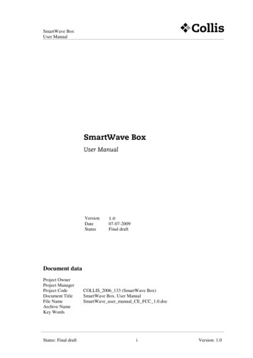
1224World J. Surg. Vol. 26, No. 10, October 2002Table 1. Characteristics of subjects.CaseGrosstypeAge atoperationaFollow-up(years)Indications for Collis-Nissenprocedure1234CCAA7 months2 years10 years4 months111293Associated CES at distal esophagusAssociated CES at distal esophagusGER with unreducible sliding herniaLong gap at primary repair for EAConcurrent operative proceduresbCurrent conditionSegmental resection of CESSegmental resection of CES—Esophageal anastomosis withLivaditis myotomyAsymptomaticAsymptomaticChest pain (infrequent)Emesis (infrequent)CES: congenital esophageal stenosis; GER: gastroesophageal reflux; EA: esophageal atresia.Age when subjects underwent Collis-Nissen procedure.Operative procedures performed concurrently with Collis-Nissen procedure.ab360 degrees around the gastric neoesophagus with five nonabsorbable sutures. The total length of the wrap depends on the age ofthe patient, but it is usually 3 to 5 cm. Fixation of the wrap to thediaphragm and approximation of the crus are routinely performed. Heineke-Mikulicz’s pyloroplasty is added when damageto the vagal nerve along the esophagus is considered. The pyloroplasty was conducted because of the possible vagal damageduring segmental esophageal resection in cases 1 and 2 andextensive dissection of the distal esophagus in case 4. Tube gastrostomy is carried out routinely to decompress the stomachduring the early postoperative stage and for supplemental feedingafter enteral nutrition is commenced.Follow-up StudyFig. 5. Standard Collis-Nissen procedure. A. Operation is performedthrough a left subcostal incision. B. Vertical gastroplasty is created withstaplers parallel to the lesser curve with the esophageal bougie in place inthe esophagus. C. Fundoplication is wrapped loosely 360 degrees aroundthe gastric neoesophagus with five nonabsorbable sutures. D. HeinekeMikulicz’s pyloroplasty is added when damage of the vagal nerve along theesophagus is considered. Tube gastrostomy is carried out routinely.ing. The Collis-Nissen procedure was done to: (1) reconstruct thedistal esophagus and fundus after segmental resection of the CESlesion located close to the esophagogastric junction (cases 1 and2); (2) construct an antireflux barrier below the diaphragmatichiatus to control advanced GER associated with a short esophagus (case 3); and (3) create an end-to-end anastomosis betweenthe ends of the esophagus with a long gap (case 4). During thestudy period, Nissen fundoplication alone was used in other EApatients who required antireflux surgery, and primary esophagealend-to-end anastomosis with Livaditis myotomy was performedfollowing esophageal elongation with bougienage in other Grosstype A EA patients. The interposition of the intestine or the colonwas not chosen as a definitive operation for Gross type A EA.Collis-Nissen ProcedureThe Collis-Nissen operation is performed through a left subcostalincision (Fig. 5). The esophagus is mobilized at the hiatus afterdivision of all the short gastric vessels. Vertical gastroplasty iscreated with staplers (Autosuture TA 55; US Surgical, Norwalk,CT, USA) parallel to the lesser curve with the esophageal bougiein place in the esophagus. The fundoplication is wrapped looselyThe subjects were followed up for 3 to 12 years. Clinical symptomsand radiographic findings were analyzed in all subjects. Endoscopy, esophageal pH monitoring, esophageal manometry, andscintigraphy were performed on clinical demand. Esophageal manometry was performed with the infusion method using a multichannel assembly associated with a sleeve sensor, the details ofwhich were reported previously [7]. Manometric recording wasperformed for 30 minutes in each subject after a 4-hour fast.ResultsThere were no deaths in this series. Early postoperative complications were minor leakage in the anastomosis in one case (case 4)and anastomotic stenosis in three cases (cases 1, 2, 4). The anastomotic leakage was treated conservatively with total parenteralnutrition for 2 weeks in case 4.Esophageal balloon dilatation was performed repeatedly foranastomotic stenosis located just above the wrapping cuff over 1year and over 3 months in cases 1 and 2, respectively (Figs. 1, 2).Swallowing difficulty and postprandial stridor continued until 19months of age in case 1. Cases 1 and 2 were asymptomatic at theend of the follow-up period.Case 3 had an uneventful course during the early postoperativeperiod, and the regurgitation of intragastric contents experiencedpreoperatively was completely eliminated (Fig. 3). However, shedeveloped a postoperative complaint of occasional nocturnalchest pain a couple of months after the operation. Twenty-fourhour pH monitoring revealed that the pH at the esophagealanastomotic site remained higher than 4.0, whereas the acid exposure value was 81% in the gastric neoesophagus and 75% justabove the esophagogastric junction. Scintigraphic examinationwith 99mTc-labeled Sn-colloid in the intraabdominal stomach
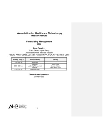
Kawahara et al.: Collis-Nissen for Esophageal AtresiaFig. 6. Scintigraphic examination of case 3. A. Intravenous injection of99mTc-pertechnetate showed the location of the gastric mucosa in theneoesophagus; it was located close to the level of the tracheal carina(white arrow) and in the intraabdominal stomach. B. Administration of99mTc-labeled Sn-colloid into the intraabdominal stomach showed noreflux into the neoesophagus during the 60-minute measurement.showed no reflux from the intraabdominal stomach into the gastric neoesophagus during a 60-minute measurement (Fig. 6). Intravenous injection of 99mTc pertechnetate showed the location ofgastric mucosa in the neoesophagus, which reached the level ofthe tracheal carina and the intraabdominal stomach (Fig. 6).Ulcerative esophagitis of less than 1 cm was continuously observed just above the esophagogastric junction by repeated endoscopic examinations during the follow-up period, whereas an endoscopic retrograde view from the intraabdominal stomachconfirmed intact cuff wrapping over the gastric neoesophagus.Microscopic findings in esophageal specimens obtained 5 yearsafter the operation showed a low-degree dysplastic change in thearea of the esophageal ulcer despite continuous administration ofhistamine-2 antagonist agents.Esophageal balloon dilatation was performed intermittently forstricture in the anastomosis of the proximal and distal ends ofesophageal atresia over 18 months in case 4 (Fig. 4). Case 4required additional feeding via gastrostomy until the age of 3years, when he could ingest the proper volume of food orally. Herarely exhibits emesis, except when he develops respiratory tractinfections. His physical development remained approximately 1SD below the average value for age compared with the Japanesestandard.Manometric examinations were performed in cases 1, 2, and 4.Basal pressure of the neo-lower esophageal sphincter (LES) at thewrapping cuff was usually 10 mmHg or less. Swallow-associatedtransient pressure reductions in the neo-LES were observed (Fig.7). The amplitude of contractions was usually less than 30 mmHgin the esophagus and neoesophagus.DiscussionWe previously reported that an association of CES is a moreimportant factor that influences the patients’ subsequent clinical1225Fig. 7. Representative manometric tracing after Collis-Nissen procedurein case 1. The neo-lower esophageal sphincter (LES) at a wrapping cuffhad 10 mmHg or less basal tone, as shown in the LES channel. Transientneo-LES pressure reductions occurred concurrently with swallow signalsshown in the pharyngeal channel (pharynx). The amplitude of contractionswas usually less than 30 mmHg, as shown in the esophageal and neoesophageal channels.course after the repair of EA than has generally been appreciated[1]. In patients with a narrow segment in the distal esophagus,possible ways to reach the lesion are a transthoracic or transabdominal approach. A transabdominal approach was chosen fortreating the CES in cases 1 and 2, as a right thoracotomy is usuallyperformed to repair the EA before treating the CES, and postoperative EA patients might develop a chest deformity or respiratory malfunction by repeated (ipsilateral or bilateral) thoracotomy. Considering the poor acid-clearing capacity frequentlyassociated with EA [8] and the impaired anti-reflux LES functioncaused by abdominal dissection of the distal esophagus, fundoplication is a useful adjunct for preventing postoperative GER whena distal esophageal lesion is treated by the transabdominal approach [9, 10]. In addition to cases 1 and 2, we have treated apostoperative Gross type A EA patient with CES, who underwentNissen fundoplication following resection of a narrowing segmentand primary end-to-end anastomosis [1]. This patient subsequently exhibited wrap herniation and swallowing difficulty. Theupward tension on the esophagogastric junction caused by esophageal shortening after segmental resection of the distal esophagusmay induce wrap failure. Esophageal lengthening with a Collisgastroplasty should be considered with Nissen fundoplication during surgical treatment of CES located in the distal esophagus inpostoperative EA patients.It is well known that GER is a major cause of postoperativecomplications seen in EA patients. The usefulness of Nissenfundoplication in the treatment of EA patients with GER hasgained wide acceptance [11–13]. Evans reported a Gross type AEA patient with a 6 cm gap, who underwent primary repair afterlengthening the distal esophagus with Collis gastroplasty at 3months of age and who subsequently required Nissen fundoplication at 6 months of age [14]. Orringer et al. reported a 16-year-oldEA patient with recurrent anastomotic stricture whose symptomswere eliminated by intraoperative esophageal dilatation and acombined Collis-Besley hiatal hernia repair [15]. Cameron et al.reported application of the Collis-Nissen procedure in EA patients but not in those with a congenital short esophagus, such ascase 3 [6]. From a technical point of view, the standard Nissen
1226procedure was not applicable in case 3 because of the shortesophagus and the tight adhesion of the esophagus with themediastinal tissue, which was caused by the primary repair and thesecond operation for anastomotic stricture. The possible antireflux procedures for patients with such a short esophagus may bethe Collis-Nissen procedure or the intrathoracic fundoplicationprocedure. Collis-Nissen procedure was considered preferable forcase 3, as fundoplication procedures that leave the stomach in thechest exposes the patient to the well documented serious complications associated with paraesophageal hiatal hernia [16].The cause of the low-degree dysplasia seen just above theesophagogastric junction in case 3 remains unclear. Advancedendoscopic esophagitis was observed before the Collis-Nissenprocedure, but histologic examination was not performed at thattime. These mucosal changes are possibly attributable to theesophageal acid exposure induced by GER prior to the CollisNissen procedure, which had continued for 10 years until theprocedure was performed. However, the influence of the gastricjuice secreted from the tubular gastric segment on the esophagealmucosa cannot be ignored. The scintigraphic and pH data in thiscase indicated a continuous acid environment in the tubular gastric segment consisting of the gastric neoesophagus and the original tubular stomach located in the mediastinum. The esophagealmucosa close to the esophagogastric junction may be frequentlyexposed to gastric acid, even when reflux from the intraabdominalstomach across the wrapping cuff to the distal esophagus wasprevented. Collis stated that the area of the cardia and lessercurve has the definite ability to produce acid, but that it is deficient in its ability to produce pepsin [3, 17]. However, Lindahl etal. reported that gastric metaplasia in the cervical esophagus isfrequent in gastric tube patients and is probably caused by the acidsecreted by the parietal cells of the tubular stomach [18]. It may bespeculated that pepsin and gastric acid secreted in the gastrictubular segment proximal to the wrapping cuff may damage themucosa of the esophagus close to the gastric tubular segmentwhen this segment is too long, such as in case 3. Parietal cellvagotomy combined with the Collis-Nissen procedure was reported to reduce acid secretion in the mediastinal tubular gastricsegment [19]. However, acid secretion is not completely abolishedeven after parietal cell vagotomy because gastrin stimulates acidsecretion in the vagotomized gastric neoesophagus [18]. Longterm endoscopic follow-up was necessary to detect the malignantchanges that developed in the distal esophagus in case 3.There are many options for the surgical management of EAwith a long gap: circular or spiral esophageal myotomies, interposition with colon or jejunum, gastric transposition, gastric tubeesophagoplasty, extrathoracic esophageal elongation [2, 20 –24].Despite various surgical procedures, no consensus has beenreached with respect to the treatment of choice for EA patientswith a long gap. We propose a primary esophageal anastomosisconcurrent with Collis-Nissen procedure as a reasonable option.Collis gastroplasty provides sufficient length of the neoesophagusand prevents excessive GER when conducted with Nissen fundoplication. This procedure is not difficult in EA patients weighingmore than 6 kg when the stomach has been used for enteralnutrition preoperatively. In case 4, there have been no significantpostoperative symptoms except infrequent emesis. Further experience with the Collis-Nissen procedure concurrent with primaryesophageal repair of EA may be necessary to assess the usefulnessWorld J. Surg. Vol. 26, No. 10, October 2002of this procedure as the definitive operation for Gross type A EAwith a long gap.Manometric examinations to evaluate esophageal motor function have rarely been performed after the Collis-Nissen procedure. Cooper et al. demonstrated a transient pressure reduction inthe neo-LES coincident with peristaltic esophageal contractionsusing a three-transducer built-in catheter [25]. We have shownpressure changes in the esophagus, including the neoesophagusand neo-LES, using a manometric assembly with a sleeve sensor,which has been widely accepted as the most appropriate methodfor measuring sphincter pressure changes [26]. The postoperativebasal pressure in the neo-LES reported by Cooper et al. wasapproximately 20 mmHg [25], whereas it was mostly 10 mmHg orless in our patients. Considering esophageal dysmotility in EApatients, a loose fundoplication may be better for avoiding postoperative swallowing difficulties.ConclusionsThe Collis-Nissen procedure is a safe, useful option for surgicaltreatment of EA patients who require esophageal lengtheningwith reflux control, so long as the length of the mediastinal gastricsegment is limited.Résumé. Parmi les patients présentant une atrésie de l’œsophage (AO), ilexiste un sous groupe avec œsophage court et qui nécessite un procédéanti-reflux. Le procédé de Collis-Nissen, qui consiste en la combinaisond’une gastroplastie de type Collis combinée à une fundoplicature de typeNissen est considérée comme une option valable dans ces conditions. Lesrésultats à long terme chez les patients porteurs d’AO n’ont été querarement rapportés. On a analysé les résultats du procédé de CollisNissen chez quatre patients ayant une AO ayant un suivi d’un moyen de9 ans. Le procédé de Collis-Nissen a été réalisé en même temps qu’unerésection segmentaire de l’œsophage pour sténose oesophagiennecongénitale associée située près de la jonction oesogastrique chez deuxpatients de type C de Gross postopératoires (7 mois, 2 ans), commeprocédé anti-reflux chez un patient de type A de Gross postopératoireayant une hernie hiatale par glissement irréductible (10 ans), et commeméthode de cure primitive d’AO chez un patient de type A de Gross avecun intervalle long (4 mois). Il n’y avait aucune complication importante,sauf un cas de fistule anastomotique et une sténose anastomotiquenécessitant une dilatation postopératoire. On n’a pas mis en évidence dereflux sur le transit eosphagogastrique ou la scintigraphie. Uneoesophagite limitée juste au-dessus de la jonction a été observée chez unpatient Gross-A porteur d’un long segment gastrique dans le médiastin.Une réduction des pressions au niveau de la valve a été détectée par lamanométrie en rapport avec la déglutition. Le procédé de Collis-Nissenest une option utile dans le traitement d’AO nécessitant un rallongementde l’œsophage et le contrôle d’un reflux gastro-oesophagien.Resumen. Un subgrupo de pacientes con atresia esofágica (AE) presentaesófago corto y requiere elongación esofágica más un procedimientoanti-reflujo. El procedimiento de Collis-Nissen, que consiste en lacombinación de la gastroplastia de Collis (que crea un tubo gástrico apartir de la curvatura menor del estómago para elongamiento esofágico)con la fundoplicación de Nissen, es considerado como una opción para seraplicada en tal condición. Se dispone de escasos informes sobreresultados a largo plazo en pacientes con AE sometidos a esteprocedimiento. Los resultados a largo plazo con el procedimiento deCollis-Nissen fueron analizados en 4 pacientes con AE con unseguimiento promedio de 9 años. El procedimiento de Collis-Nissen fuepracticado concomitantemente con una resección segmentaria del esófagopor estenosis congénita asociada del esófago en la región vecina a la unióngastroesofágica en 2 pacientes postoperatoirs con AE del tipo Gross C (7meses, 2 años), como una operación anti-reflujo en un pacientepostoperatorio con AE del tipo Gross A con una hernia por deslizamientoirreductible (10 años) y con una reparación primaria de la AE en un
Kawahara et al.: Collis-Nissen for Esophageal Atresiapaciente del tipo Gross A con un defecto largo (4 meses). No sepresentaron complicaciones importantes excepto escape anastomóticomenor y estenosis anastomótica que requirió dilatación postoperatoria.No se detectó reflujo significante ni en el estudio radiográfico del tractoGI superior ni en la escintigrafía. Se observó esofagitis limitada en elpaciente del tipo Gross A con un largo segmento gástrico mediastinal. Seregistró reducción de la presión de deglución a nivel del manguito antireflujo mediante examen manométrico. El procedimiento de Collis-Nissenes una opción útil en el tratamiento de pacientes con AE que requierenalargamiento esofágico y el control de reflujo gastroesofágico.References1. Kawahara H, Imura K, Yagi M, et al. Clinical characteristics ofcongenital esophageal stenosis distal to associated esophageal atresia.Surgery 2001;129:29 –382. Rescorla FJ, West KW, Scherer LR, et al. The complex nature of typeA (long gap) esophageal atresia. Surgery 1994;116:658 – 6643. Collis JL. An operation for hiatus hernia with short oesophagus.Thorax 1957;12:181–1884. Orringer MB, Sloan H. Combined Collis-Nissen reconstruction of theesophagogastric junction. Ann. Thorac. Surg. 1978;25:16 –215. Richardson JD, Richardson RL. Collis-Nissen gastroplasty for shortened esophagus. Ann. Surg. 1998;227:735–7426. Cameron BH, Cochran WJ, McGill CW. The uncut Collis-Nissenfundoplication: results for 79 consecutively treated high-risk children.J. Pediatr. Surg. 1997;32:887– 8917. Kawahara H, Imura K, Yagi M, et al. Mechanisms underlying theantireflux effect of Nissen fundoplication in children. J. Pediatr. Surg.1998;33:1618 –16228. Tovar JA, Diez Pardo JA, Murcia J, et al. Ambulatory 24-hourmanometric and pH metric evidence of permanent impairment ofclearance capacity in patients with esophageal atresia. J. Pediatr. Surg.1995;30:1224 –12319. Nielson IR, Croitoru DP, Guttman FM, et al. Distal congenital esophageal stenosis associated with esophageal atresia. J. Pediatr. Surg.1991;26:478 – 48210. Yeung CK, Spitz L, Brereton RJ, et al. Congenital esophageal stenosisdue to tracheobronchial remnants: a rare but important associationwith esophageal atresia. J. Pediatr. Surg. 1992;27:852– 855122711. Fonkalsrud EW. Gastroesophageal fundoplication for reflux followingrepair of esophageal atresia. Arch. Surg. 1979;114:48 –5112. Wheatley MJ, Coran AG, Wesley JR. Efficacy of the Nissen fundoplication in the management of gastroesophageal
results of the Collis-Nissen procedure applied in EA patients. Materials and Methods Subjects During 1989-1999 the Collis-Nissen procedure was performed in four EA patients by one pediatric surgeon (H.K.) in Osaka Uni-versity Hospital and Osaka Medical Center for Maternal and Child Health. The clinical details of each patient were as follows .