Transcription
Healing of Retinal Photocoagulation LesionsYannis M. Paulus, ATul Jain, Ray F. Gariano, Boris V. Stanzel, Michael Marmor,Mark S. Blumenkranz, and Daniel PalankerPURPOSE. To systematically assess the changes in retinal morphology during the healing of retinal photocoagulation lesionsof various clinical grades.METHODS. Rabbits were irradiated with a 532-nm Nd:YAG laserwith a beam diameter of 330 m at the retinal surface, a powerof 175 mW, and pulse durations between 5 and 100 ms. Retinallesions were clinically graded 1 minute after placement asinvisible, barely visible, light, moderate, intense, very intense,and rupture and were assessed histologically at six time pointsfrom 1 hour to 4 months.RESULTS. At all pulse durations, the width of the retinal lesionsdecreased over time. At clinical grades of light and more severe(pulse durations, 10 –100 ms), retinal scarring stabilized at 1month at approximately 35% of the initial lesion diameter.Lesions clinically categorized as barely visible and invisible(pulse durations of 7 and 5 ms) exhibited coagulation of thephotoreceptor layer but did not result in permanent scarring.In these lesions, photoreceptors completely filled in the damaged areas by 4 months.CONCLUSIONS. The decreasing width of the retinal damage zonesuggests that photoreceptors migrating from unaffected areasfill in the gap in the photoreceptor layer. Laser photocoagulation parameters can be specified to avoid not only the innerretinal damage, but also permanent disorganization and scarring in the photoreceptor layer. These data may facilitate studies to determine those aspects of laser treatment necessary forbeneficial clinical response and those that result in extraneousretinal damage. (Invest Ophthalmol Vis Sci. 2008;49:5540 –5545) DOI:10.1167/iovs.08-1928From the Department of Ophthalmology, Stanford UniversitySchool of Medicine, Stanford, California.Supported by an Alcon Research Institute grant, the Horngren andMiller Family Foundations, OptiMedica Corp., the Angelos andPenelope Dellaporta Research Fund, and the L. Boltzmann Institute forRetinology and Biomicroscopic Laser Surgery, Vienna, Austria.Submitted for publication February 24, 2008; revised June 29,2008; accepted September 26, 2008.Disclosure: Y.M. Paulus, None; A. Jain, None; R.F. Gariano,None; B.V. Stanzel, None; M. Marmor, None; M.S. Blumenkranz,OptiMedica Corp. (I, C, P); D. Palanker, OptiMedica Corp. (I, C, P)The publication costs of this article were defrayed in part by pagecharge payment. This article must therefore be marked “advertisement” in accordance with 18 U.S.C. §1734 solely to indicate this fact.Corresponding author: Daniel Palanker, Stanford University Department of Ophthalmology, 300 Pasteur Drive, Room A157, Stanford,CA 94305-5308; palanker@stanford.edu.retinal scarring, resulting in scotomas and decreased peripheral, color, and night vision.7 It remains largely unknown howthese putative benefits of PRP or its many deleterious sideeffects7 relate to parameters of laser treatment and subsequentretinal healing. Several investigators have presented evidencethat light PRP (minimum-intensity photocoagulation)8,9 oreven subvisible treatment (micropulse photocoagulation)10,11has an efficacy equivalent to that of conventional PRP in causing regression of high-risk proliferative diabetic retinopathyand is associated with fewer complications and fewer treatment sessions. In a recent report, at 12 months, light PRP wasequivalent to classic PRP in reduction or elimination of diabeticmacular edema, visual improvement, change in contrast sensitivity, and decreased foveal retinal thickness on OCT.8 Both ofthese approaches claim to offer the therapeutic benefit ofconventional therapy without many of its side effects.12 However, many physicians remain skeptical about these claims, anda prospective randomized clinical trial is needed to provideconvincing evidence one way or the other.Recent studies have begun to explore the evolution oflaser-induced retinal lesions over time.13 Retinal lesions in ratswere shown to stabilize by 60 days.14 Immunohistochemicalstudies of the laser lesions in mice 4 weeks after treatment haveshown that the healing processes are driven by cells andvessels that are retinal in origin.14,15 It has also been found thatthe lesion size in rodents decreases over time due to migrationof photoreceptors from the untreated surrounding areas, fillingin the damaged outer retina.16 Healthy adjacent photoreceptors have been shown to repopulate regions of photoreceptordamage in a process associated with actin.17 The lesions of 200 m showed resolution of damage in the photoreceptor layer,whereas 800- m lesions did not.16 This observation suggeststhat photoreceptor migration may have a finite limit.16 Postinjury proliferation and migration of neurons have also beenrecently described in several other neuronal systems.18 –21 Although rodent studies have provided important insight into thedynamics of retinal healing and remodeling, the lack of retinalscarring in rodents makes it a suboptimal model for retinalphotocoagulation lesions in humans.In this study we provide a systematic histologic analysis ofthe effects of retinal photocoagulation and describe the dynamics of healing and reorganization of retinal lesions of variousclinical grades in rabbits. This study of laser-induced retinallesions was facilitated by the use of a recently developedsemiautomated retinal photocoagulation system (PASCAL; OptiMedica Inc., Santa Clara, CA).22 This delivery system allowsfor creating well-aligned arrays of retinal lesions with variabledurations of laser exposure and power. The effects of pulseduration and laser power on the initial size and character ofretinal photocoagulation lesions in rabbits have been described.22,23 The purpose of the present study was to determine the treatment parameters for retinal photocoagulationthat allow avoiding not only inner retinal damage, but alsopermanent disorganization and scarring in the photoreceptorlayer. This study is a step toward determination of those aspects of laser treatment necessary for clinical response andthose that result in extraneous retinal damage.5540Investigative Ophthalmology & Visual Science, December 2008, Vol. 49, No. 12Copyright Association for Research in Vision and OphthalmologyRetinal laser photocoagulation was first described 45 yearsago1 and remains the standard of care for many retinaldiseases. Panretinal photocoagulation (PRP) for proliferativediabetic retinopathy involves the purposeful destruction of asignificant fraction of the photoreceptors, as well as othermore superficial retinal layers.2 Several mechanisms have beensuggested to underlie the efficacy of PRP and other laser treatments, including improved retinal oxygenation, reduction inmetabolic activity, inhibition of angiogenic stimulators, increased production of angioinhibitory factors, and oxidativestress.3– 6 Side effects of PRP treatment include permanent
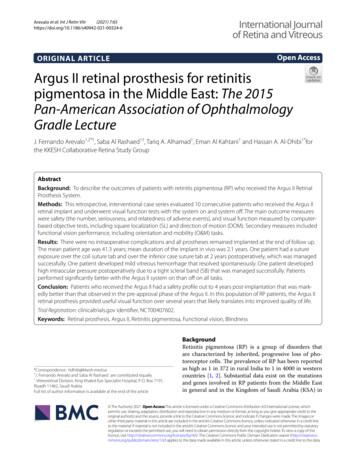
IOVS, December 2008, Vol. 49, No. 12Healing of Retinal Photocoagulation Lesions5541METHODSPattern Scanning PhotocoagulationThis laser system (PASCAL; OptiMedica Inc.) provides 532 nm opticalradiation from a diode-pumped, continuous wave, frequency-doubledNd:YAG laser coupled to a multimode step index optical fiber. The exitsurface of the fiber is telecentrically imaged through the scanningsystem onto the retina with variable magnification, thus providing avariety of spot sizes with nominally top-hat intensity profiles. At theaerial image plane of the slit lamp microscope the laser spots measure500 m in diameter, with the laser intensity transition from 10% to 90%occurring over 6 m. Variation of the beam intensity within the top-hatarea does not exceed 7%. A touch-screen graphic user interface isused to control laser parameters including the spot size, laser power,pulse duration, and pattern geometry. Once the treatment parametersare appropriately selected, a foot pedal is used to activate the laser.Retinal Laser ApplicationEighteen Dutch Belted rabbits (weight, 2–3 kg) were used in accordance with the ARVO Statement for the Use of Animals in Ophthalmicand Vision Research, after approval from the Stanford University Animal Institutional Review Board. The rabbits were anesthetized withketamine hydrochloride (35 mg/kg, IM), xylazine (5 mg/kg, IM), andglycopyrrolate (0.01 mg/kg, IM), administered 15 minutes before theprocedure. Pupillary dilation was achieved by 1 drop each of 1%tropicamide and 2.5% phenylephrine hydrochloride. Topical tetracaine0.5% was instilled in the treated eye before treatment. Only one eye ofeach rabbit was treated, unless the rabbit was to be killed immediately.A standard retinal laser contact lens (Mainster model OMRA-S;Ocular Instruments, Bellevue, WA) was used to focus the laser on therabbit fundus. Taking into account the combined magnifications of thecontact lens and rabbit eye of 0.66,24 the aerial image of 500 mcorresponded to a retinal spot size of 330 m. Using modified scanningsoftware, columns consisting of six exposures with pulse durations of5, 7, 10, 15, 20, 50, and 100 ms were applied. Five identical columnswere applied in each eye to broaden the statistical base of the study.Laser power was maintained at 175 mW for all lesions. Placement oflarger marker burns adjacent to the patterned lesions facilitated laterhistologic localization of the barely visible and invisible lesions.The clinical appearance of the laser lesions were graded by a singlemasked observer within 1 minute after the treatment according to thefollowing scale: invisible, barely visible, light, moderate, intense, veryintense, and rupture. The barely visible lesions were defined as a faintlightening of the fundus pigmentation. Light burns exhibited furtherblanching but without frank whitening. Moderate lesions exhibiteddefinite retinal whitening without adjacent edema, and intense burnsdisplayed central whitening and a halo of translucent edema. Veryintense lesions had a more marked and complete central retinal whitening and a larger ring of edema. A rupture was assumed to haveoccurred if small bubbles with or without hemorrhage appeared at thelesion site.FIGURE 1. Figures 1 through 5 show progression of the lesions produced with a retinal beam size of 330 m in diameter, with laser powerof 175 mW. Sections were stained with toluidine blue. Photographswere taken via 20 microscope objective, except for a 1-hour lesion,which was photographed using 10 magnification due to its large size(and has its own scale bar). Yellow bar: the extent of damage at theRPE-photoreceptor junction. GCL, ganglion cell layer; INL, inner nuclear layer; ONL, outer nuclear layer; RPE, retinal pigment epithelium;CHO, choroid. Shown in this figure is a representative intense lesionproduced by 100-ms pulse. The lesion encompassed all retinal layers.Pigmented cells invaded the full thickness of the retina from 1 week to2 months. At 4 months, a scar occupied the 36% of the initial lesion.sured using a microscope measuring scale. The lesion size was measured as the furthest extent of damage at the RPE-photoreceptorjunction. These three measurements for each of the four burns wereaveraged, and representative lesions for each time point were photographed. The mean and SD of these 12 numbers were calculated(Excel; Microsoft, Redmond, WA). The total size of the measurementsdatabase was 504 numbers (168 3).Fluorescein AngiographyFor fluorescein angiography (FA), 0.3 mL of contrast (Fluorescite 10%;(Alcon Laboratories, Hünenberg, Switzerland) was slowly (10 seconds)injected into the marginal ear vein as described elsewhere.26 Fundusphotographs were taken with a fundus camera (TRC 50 [Topcon,Tokyo, Japan], using a Kodak Megaplus Camera, Model 1.4 [EastmanKodak, Rochester, NY], operated by Winstation imaging software[Ophthalmic Imaging Systems, Inc, Sacramento, CA]), starting a fewseconds after the injection, and every 20 seconds for up to 5 minutes.Fluorescein angiography was performed 1 hour and 1, 3, and 7 daysafter the placement of lesions with all clinical grades: very intense,intense, moderate, light, barely visible, and invisible.Retinal HistologyRESULTSRabbits (three in each group) were killed 1 hour, 1 day, 1 week, 1month, 2 months, or 4 months after treatment with a lethal dose of anIV barbiturate (Beuthanasia; Schering-Plough Animal Health Corp.,Kenilworth, NJ) in an ear vein. The eyes were enucleated and fixed in1.25% glutaraldehyde/1% paraformaldehyde in cacodylate buffer at pH7.2 overnight at room temperature. The eyes were then postfixed inosmium tetroxide, dehydrated with a graded series of ethanol, processed with propylene oxide, embedded in an epoxy resin, and sectioned into 1 m-thick sections. Samples were stained with toluidineblue and examined by light microscopy.25Serial sections of the retina were examined, and four burns fromtwo different animals were analyzed for each of the six time points andseven pulse durations (168 in total). By visually scanning the serialsections, three widest lesions corresponding to each burn were mea-HistologyIntense Lesions. As shown in Figure 1, at 1 hour aftertreatment, intense lesions were sharply demarcated and encompassed all retinal layers, as well as the retinal pigmentepithelium (RPE) and choroid. Retinal edema was present, andthe normal laminar architecture of the retina was disorganizedby displacement of the outer retina.At 1 day, edema was nearly resolved in the inner and outerretina. The inner and outer nuclear layers (INL and ONL) weresparse and contained pyknotic nuclei. Photoreceptor innersegments appeared shortened, and the outer segments appeared in disarray. Even though RPE cells were clearly affected,the RPE pigmentation appeared largely normal. Hyaline (pale
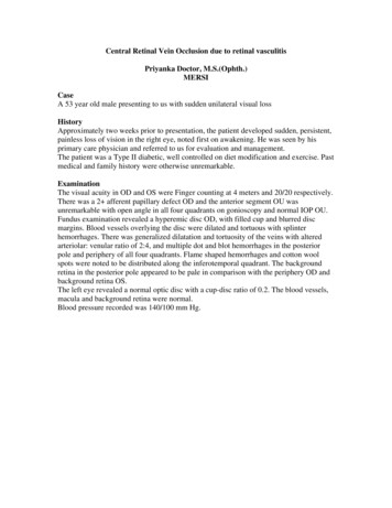
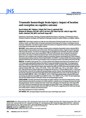
5542Paulus et al.IOVS, December 2008, Vol. 49, No. 12TABLE 1. Relative Width of the Retinal Lesions of Various Grades over Time, as Measured at theRPE-Photoreceptor JunctionDays after TreatmentClinical GradePulse Duration ghtBarely 850608782676934Data are expressed as percentage of the initial lesion size. For each pulse duration, the width for alltime points was normalized to the size of the retinal lesion at 1 hour. Intense to light lesions stabilized atapproximately 40% of the initial lesion size at 2 months. Barely visible and invisible lesions continued toheal, so that there was no visible defect at the RPE–photoreceptor junction at 4 months.blue) vacuoles were present between the basal RPE andBruch’s membrane. There appeared to be a loss of choroidalstructures accompanied in part by vascular engorgement andexudation. The lateral extent of the damage zone became lessclear because of scattered, enlarged INL and ONL cells at thelesion borders.At 1 week, intense lesions had contracted to 54% of theirinitial diameter (Table 1). INL cells remain enlarged, but extracellular edema was now absent. Pigmented cells invaded thefull thickness of the retina. Retinal architecture was now difficult to discern because of partial loss of cells and their replacement with gliotic tissue in all retinal layers. The RPE wasdiscontinuous, with large pigmented clumps interspersed withdepigmented areas. Multilayered hypopigmented cells werepresent on an intact Bruch’s membrane. Vascular caliber hadnormalized in larger choroidal vessels, but remained decreasedwithin the choriocapillaris, which was less dense. Choroidalthickness had returned to normal.At 1 month, edema was absent and the lateral/horizontalwidth of the lesion was 40% of its initial size. Gliosis andpigmented cells are present in all retinal layers, and there wassome central thinning. Hypertrophied and hyperpigmentedRPE was present.FIGURE 2. Light-moderate lesions (20-ms pulse duration) at initialstage (1 hour) involved strong edema in the inner retina above thedestroyed layer of photoreceptors. Edema largely subsided by 1 day,and damaged photoreceptors were replaced with hypertrophied gliaby 1 week. The scar was largely stabilized after 1 month and covered40% of the initial lesion size at 4 months.At 2 months, the lesions further contracted to 37% of theoriginal lesion diameter, and otherwise appeared similar tolesions at 1 month.At four months, the lesion diameter was still approximately36% of the original lesion and similar in appearance as at 2months, with the exception that intraretinal pigmented cellshad disappeared and the lesion largely consisted of hypocellular matrix.Light and Moderate Lesions. One hour after treatmentthe light-to-moderate lesion exhibits strong edema in the innerretina, damaged inner and outer nuclear layers, and large vacuoles in the inner segment layer (Figs. 2, 3). Choroidal changesare less pronounced than in the intense lesion.At day 1, retinal edema had decreased considerably. Innerand outer segments of photoreceptors were strongly reduced,and nuclei in the ONL were small and pyknotic within theboundaries of the lesion. The INL nuclei appeared more roundand enlarged, compared with the surrounding untreated retina.The central defect in ONL and INL of intense lesions wasabsent.At 1 week, there was no significant choroidal involvement.ONL and most of the INL were replaced by gliotic tissue andwere invaded by pigmented cells. Continuous hypopigmentedRPE layer can be clearly seen in Figure 3.FIGURE 3. In the light lesions (15-ms pulse duration), initial damagewas localized to the photoreceptors. The RPE layer was restored by 1week, although the cells appeared hypopigmented. The width of thegliotic scar in the outer retina stabilized by 1 month at approximately40% of its original size.
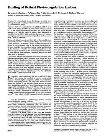
IOVS, December 2008, Vol. 49, No. 12FIGURE 4. Barely visible lesions produced at 7-ms pulse duration exhibited perfect localization of the retinal damage to photoreceptors.Gliotic replacement of the damaged photoreceptors continued to contract over time, showing an outer retina with almost continuous normal morphology at 4 months.At 1 month, the lesion had contracted asymmetrically andappeared larger toward the inner retina and smaller at the RPE.The INL was thickened, and its cells were enlarged in thecenter of the lesion. Photoreceptors filled the peripheral partof the lesion, leaving a remaining damage zone of 40% of theoriginal width. The RPE was hyperpigmented in the lesionperiphery with zones of hypopigmentation and pigmentedcellular aggregates more centrally.At 2 months, the cellular accumulation in the center of thelesion remained displaced toward the RPE, patchy gliosis filledin the cellular gaps in the photoreceptor layer, and a decreasednumber of hyperpigmented cells remained in the retina. RPEpigmentation appeared more continuous, with some smallclumps of pigmented cells remaining. The width of the lesioncontinued to be 40% of its original size.At 4 months, the lesion maintained its size at 40%, althoughthe INL regained its normal structure at the periphery. Thecentral portion of the INL remained displaced toward the RPE,and the gap in the photoreceptor layer remained filled in withgliosis and displaced INL cells.Barely Visible and Invisible Lesions. As shown in Figure4, the barely visible lesions differed both quantitatively andqualitatively from the more intense lesions. Initial lesion diameters were smaller than those of lesions created with longerpulse durations (see Fig. 6).At 1 hour, the inner retina appeared largely unaffected,whereas the ONL was hyperchromatic. RPE seemed collapsed,but its pigmentation remained unchanged. Outer segments ofphotoreceptors were disorganized, and the nuclei were pyknotic.At 1 day, there was slight edema in the inner retina, butboth INL and GCL seemed to be only minimally affected. Innersegments of photoreceptors were shortened, the outer segments depigmented, and the ONL displaced toward the RPE.The RPE had a hypochromatic cytoplasm.At 1 week, the INL was slightly expanded, and missingphotoreceptors were probably replaced by glial elements. Retinal edema was mild and localized primarily to the outer retina.The RPE layer was restored, but seemed hypopigmented in thecenter of the lesion. The choroid appeared normal. The lesioncontracted to 69% of its original size.At 1 month, the lesion had contracted to 10% of its initialdiameter. Histologic changes were localized to the outer retinaand consisted of a thinned ONL and disorganized photorecep-Healing of Retinal Photocoagulation Lesions5543FIGURE 5. Invisible lesion produced with 5-ms pulse duration exhibited laser damage localized to photoreceptors and RPE. Damagedphotoreceptors were replaced within a few weeks, with normal retinalmorphology completely restored by 4 months.tors in the center of the lesion. Gliosis and downward displacement of the INL were notably absent. Intraretinal pigmentedcells, present in more severe lesion grades, were absent aswell.At 2 months, the lesion was similar to that at 1 month. A fewhyperpigmented RPE cells were evident only in the center ofthe lesion.At 4 months, the lesion was distinguished from adjacentnormal retina only by a few-cells-wide area of decreased density within the ONL with occasional adjacent vacuoles in theinner segments, and slight elevation. Mild and variable hyperpigmentation at the RPE level persisted in some lesions. Photoreceptor morphology within lesions was otherwise indistinguishable from that in untreated retina.Histology of ophthalmoscopically invisible lesions (5-msexposures) revealed even more localized damage to photoreceptors and RPE, with excellent preservation of the otherretinal layers (Fig. 5). Healing of the lesion followed stepssimilar to those of the barely visible lesion, with even moreFIGURE 6. The width of the retinal lesions of various grades over time,as measured at the RPE–photoreceptor junction. All lesions decreasedin size, and more intense lesions are wider initially. Moderate andintense burns stabilized at 150 m and light to light-moderate burnsstabilized at 100 m by 2 months, with minimal change in size at 4months. Barely visible and invisible lesions continued to heal to complete closure by 4 months. Error bars show the SD for each time pointof each of four burns measured per time point.
5544Paulus et al.complete resolution by the end of 4 months. In additionalexperiments, pulse durations of 3 ms and shorter failed toproduce ophthalmoscopically visible burns. Retinal histologyappeared entirely normal within the treated region (data notshown).Changes in the lateral extent of the retinal lesions over timewere summarized in the plot shown in Figure 6. Table 1 showsthe relative lesion sizes over time, normalized on initial lesionsize at 1 hour.Fluorescein AngiographyAt 1 hour and 1 day after laser application, all clinical grades oflaser lesions (from invisible to very intense) exhibited hyperfluorescence with fluorescein angiography. At 3 days, light andmore intense lesions remained hyperfluorescent, whereas clinically invisible and barely visible lesions were not evident onangiography. By 7 days after laser application, none of thelesions remained hyperfluorescent.DISCUSSIONThe present study provides a systematic assessment of histologic changes over time in retinal photocoagulation lesions ofvarious clinical grades in rabbits. Use of the pattern deliverysystem (PASCAL; OptiMedica Inc.) helped, with rapid andwell-aligned application of multiple lesions with variable pulsedurations. We achieved consistent clinical levels of lesion intensities by varying pulse duration while maintaining constantspot size and power settings. Since laser power, spot size, andpulse duration affect the resulting lesion character in a different fashion,23 our results may not be entirely generalizable tolesions that differ in their clinical grade due to variations inspot size and/or power settings. Since lesion size decreaseswith decreasing pulse duration and laser power, more burns orlarger laser spot sizes are necessary to treat an equivalentretinal area that may be essential to reach a similar clinicalefficacy.For all clinical grades, the lateral extent of retinal damagedecreased over time, with the extent of reduction dependenton the initial width and the severity of the lesion. Within 2months the intense, moderate, and light lesions decreased inwidth by a factor of 3, whereas the barely visible and invisiblelesions disappeared nearly entirely. Retinal gliosis appearedonly temporarily between 1 and 4 weeks in barely visiblelesions, whereas in more intense burns, it stabilized by 1month, and persisted at 4 months. The end-stage histology ofthe light, moderate, and intense burns is similar. Similar healingrates have been observed during experimental RPE debridement in rabbits.27 Resolution of fluorescein angiography hyperfluorescence by 1 week corroborates the anatomic demonstration that RPE continuity was restored by 1 week (see forexample, Fig. 3). For invisible and barely visible lesions, thiscontinuity occurred even faster— by 3 days.Lateral photoreceptor migration to replace initial cell losshas been suggested by previous studies in the rat retina.16However, restoration of the photoreceptor layer in rats wasmore rapid, reaching its maximum extent by 3 weeks, whereaswe found this process continuing in rabbits throughout the4-month period of observation, particularly in the first 2months. In addition, retinal lesions in rats exhibit less gliosisthan in rabbits. Observation of the photoreceptors migration inboth the merangiotic rabbit model and the holangiotic ratmodel of retinal photocoagulation, indicate the likelihood ofsimilar phenomenon in humans. High-resolution OCT imagingof the evolution of retinal lesions of various grades in patientscould provide such evidence.IOVS, December 2008, Vol. 49, No. 12Photoreceptor outer segments damaged during selectiveRPE photocoagulation in rabbits have been shown to recoverby 4 weeks after laser irradiation after initial disc disorganization and vacuolization.28 However, it is unlikely that, in ourcase, photoreceptor restoration after retinal photocoagulationwas due to the recovery of damaged outer segments sinceouter nuclear layer pyknosis at 1 day proceeded to large visiblelesions devoid of nuclei at 1 week.Remaining unknown is the role of neuronal regeneration inthe restoration of the photoreceptor layer observed in ourstudy, either through differentiation of resident neuronal stemcells at the ciliary margin29 –36 or the transdifferentiation ofMüller glia.37Regardless of the mechanism, restoration of retinal continuity has clinically important implications. If the therapeuticbenefit of PRP involves reduction in the number of photoreceptors, it can be achieved more selectively using barely visibleor invisible lesions without the permanent scarring of moreintense lesions. Restoration of retinal continuity and absence ofscarring might reduce microscotomas typical of conventionalPRP and may also allow for retreatment.In addition, such minimally traumatic therapy reduces thelevel of pain and discomfort associated with PRP. Applicationof the barely visible or invisible lesions is facilitated usingarrays that can incorporate higher intensity visible markerburns at the corners (Pascal; OptiMedica Inc.). Such lesionsenable proper placement of successive applications of arrays.Our results may also form the basis for a useful model ofretinal fibrosis. Fibrosis remains a major clinical problem inophthalmology and is the key factor causing irreversible blindness in several diseases, including subretinal scarring in endstage macular degeneration. We have shown that several features of retinal healing (e.g., cell death, edema, RPE invasion,gliosis, and matrix deposition) can be predictably isolated orcombined within lesions by varying the parameters of lasertreatment. Our descriptions of these graded responses maythus facilitate scientific investigation into discrete steps inretinal wound healing and allow for the assessment of potentialtherapies to prevent or resolve retinal fibrosis.In summary, we have described the histopathology of retinal photocoagulation lesions across a wide spectrum of clinicalgrades and over an extended time course. These data may beuseful in further evaluations of mechanisms of retinal healingand may form the basis for a rational approach to optimizingparameters for clinical retinal laser therapy.AcknowledgmentsThe authors thank Roopa Dalal for histologic preparations and retinallesion measurements; Philip Huie for help with animal housing andadministration; and Dan Andersen, Georg Schuele, and Hiroyuki Nomoto for stimulating discussions.References1. Kapany NS, Peppers NA, Zweng HC, Flocks M. Retinal photocoagulation by lasers. Nature. 1963;199:146 –149.2. Little HL, Zweng HC, Peabody RR. Argon laser slit-lamp retinalphotocoagulation. Trans Am Acad Ophthalmol Otolaryngol.1970;74(1):85–97.3. Jennings PE, MacEwen CJ, Fallon TJ, et al. Oxidative effects of laserphotocoagulation. Free Radic Biol Med. 1991;11(3):327–330.4. Sanchez MC, Luna JD, Barcelona PF, et al. Effect of retinal laserphotocoagulation on the activity of metalloproteinases and the alpha(2)-macroglobulin proteolytic state in the vitreous of eyes withproliferative diabetic retinopathy. Exp Eye Res. 2007;85(5):644 – 650.5. Spranger J, Hammes HP, Preissner KT, et al. Release of the angiogenesis inhibitor angiostatin in patients with proliferative diabeticretinopathy: association with retinal photocoagulation. Diabetologia. 2000;43(11):1404 –1407.
IOVS, December 2008, Vol. 49, No. 126. Matsumoto M, Yoshimura N, Honda Y. Increased production oftransforming growth factor-beta 2 from cultured human retinalpigment epithelial cells by photocoagulation. Invest OphthalmolVis Sci. 1994;35(13):4245– 4252.7. Fong DS, Girach A, Boney A. Visual side effects of successfulscatter laser
laser-induced retinal lesions over time.13 Retinal lesions in rats were shown to stabilize by 60 days.14 Immunohistochemical studies of the laser lesions in mice 4 weeks after treatment have shown that the healing processes are driven by cells and vessels that are retinal in origin.14,15 It has also been found that