Transcription
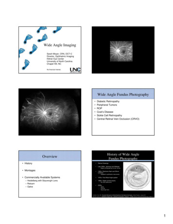
Wide Angle ImagingSarah Moyer, CRA, OCT-CDirector, Ophthalmic ImagingKittner Eye CenterUniversity of North CarolinaChapel Hill, NCNo financial interest.Wide Angle Fundus Photography Diabetic RetinopathyPeripheral TumorsROPCoat’s DiseaseSickle Cell RetinopathyCentral Retinal Vein Occlusion (CRVO)History of Wide AngleFundus PhotographyOverview History Retinal Drawings Late 1800s- Jackman and Webster Montages 1950s- Electronic flash and 35mmcameras Commercially Available Systems 1970s- First Wide Angle system– Heidelberg with Staurenghi Lens– Retcam– Optos 1990s- Digital camera backs 2000s––––––Began photographing human retinasAdapted to ophthalmic instrumentsNo more darkrooms!OptosPanoretStaurenghi lensTextSchefler A, Flynn HW. Pneumatic Retinopexy for Retinal Detachment After Macular Hole Surgery. Retinal Physician. January 2011.Bennett TJ, Barry CJ. Ophthalmic imaging today: ophthalmic photographer’s viewpoint – a review. Clinical & Experimental Ophthalmology. 2009;37:2-13.1
Equator-Plus Traditional Fundus cameraoptically Transscleral Fiber Opticillumination Spring-loaded Contact Lens Calculated to exert a maxpressure of 20mm Hg. Buzzer indicates if pressureis exceeded. 148 degree field of viewTextPomerantzeff, Oleg. Equator-plus camera. Investigative Ophthalmology. May 1975: Vol 14. Num 5. pp 401-406.Department of Retina Research, Eye Research Institute of Retina Foundation, Boston, MA.Pomerantzeff, Oleg. Equator-plus camera. Investigative Ophthalmology. May 1975: Vol 14. Num 5. pp 401-406.Department of Retina Research, Eye Research Institute of Retina Foundation, Boston, MA.Panoret Medibell Medical VisionTechnologies Ltd Early 2000sCiliochoroidal effusion with serous nonrhegmatogeneousretinal detachment in a 55-year-old man Named for capturing“panoramic” retinal images Handheld Fundus camera Transscleral Fiber Opticillumination 130 degree field of viewMacular hole and peripheral retinoschisis with an outerlayer hole in a 36-year-old woman.TextChoroidal metastases from breast cancer, withshifting subretinal fluid, in a 54-year-old woman.Shields, C, et al. Panoramic Imaging of the Ocular Fundus. Arch Ophthalmol. 2003;121(11):1603-1607.PanoretCombined hamartoma of the retina and retinalpigment epithelium in a 12-year-old boyMushroom-shaped choroidalmelanoma in a 46-year-old woman.AdvantagesDisadvantages Great with people of all ages Portable Steep learning curve No longer commerciallyavaialbleOcular histoplasmosis syndrome in a 47-year-old womanShields, C, et al. Panoramic Imaging of the Ocular Fundus. Arch Ophthalmol. 2003;121(11):1603-1607.2
MontagesThink twice before trusting a montage .TextThink twice before trusting a montage .TextThink twice before trusting a montage .Think twice before trusting a montage .TextText3
MontagesThink twice before trusting a montage .AdvantagesDisadvantages Software modification of currentsystem Easy to change eye Easy to learn technique Time consuming Artifacts Hard to perfect techniqueTextHeidelberg with Staurenghi LensImaging Technique Additional lens used withHeidelberg HRA / Spectralis 2005 Contact lens 150 degree field of viewTextTextPhoto Courtesy of Ocular InstrumentsPhoto Courtesy of Amanda ByeLens PlacementAfter Acquisition TextImages are inverted and reversed just like an indirect ophthalmoscope.Heidelberg software has a button to invert the image back to “normal”.TextPhoto Courtesy of Amanda ByePhoto Courtesy of Amanda Bye4
Coat’s DiseaseCRVOChoroidal MelanomaROPImages courtesy Eric KegleyImages courtesy Eric KegleyWith Lens150 30 100 50 20 10 Without LensStaurenghi, G. et al. Scanning Laser Ophthalmoscopy and Angiography with a Wide-FieldContact Lens System. Archives of Ophthalmology. February 2005: Vol 123 pp 224-252.ICGFABrain Tumor1 year later30 PaintingStaurenghi, G. et al. Scanning Laser Ophthalmoscopy and Angiography with a Wide-FieldContact Lens System. Archives of Ophthalmology. February 2005: Vol 123 pp 224-252.Staurenghi, G. et al. Scanning Laser Ophthalmoscopy and Angiography with a Wide-FieldContact Lens System. Archives of Ophthalmology. February 2005: Vol 123 pp 224-252.5
ICGFAHeidelberg with Staurenghi LensMacular exudates from capillary hemangiomaAdvantagesDisadvantages Modification of current system Can manipulate to get very farinto periphery Video of FA / ICG Difficult to switch between eyesPossibility of corneal abrasionSteep learning curveDifficult for some patients totolerateComplete closure after laserStaurenghi, G. et al. Scanning Laser Ophthalmoscopy and Angiography with a Wide-FieldContact Lens System. Archives of Ophthalmology. February 2005: Vol 123 pp 224-252.RetcamAdvantagesDisadvantages Designed for children Portable Can be used in operating room/ incubator Can get into far periphery Video capability Steep learning curve Low resolution Possibility of corneal abrasionClarity RetcamImages courtesy of www.claritymsi.com/Handheld FundusClarity Retcam Captures images with video. Transpupillary Fiber Optic illumination First system sold in 1997 Portable 110 degree field of viewLid SpeculumContact with gelImages courtesy of www.claritymsi.com/6
Handheld FundusPediatric ImagingPositionsCaptures ImagesFocusExposureCoat’s Disease09-2008 Coat’s DiseaseFamilial Exudative Vitreoretinopathy (FEVR)Cystoid Macular Edema (CME)Retinopathy of Prematurity (ROP)RetinoblastomaNon accidental traumaGonio imagingCoat’s Disease06-200909-200902-201111-2009Coat’s DiseaseFamilial Exudative Vitreoretinopathy (FEVR)11-20097
Familial Exudative Vitreoretinopathy (FEVR)Retinopathy of Prematurity (ROP) Avascular TractionRetinopathy of Prematurity (ROP)Retinopathy of Prematurity (ROP)Retinopathy of Prematurity (ROP)Cystoid Macular Edema (CME)Secondary to Acute Myeloid Leukemia (AML)6 year old8
Non accidental Trauma4mAxenfeld RiegerAniridia9mTrauma18m12mUnknown EtiologyPhoto credit: Ditte Hess, CRA, FOPSOptos Scanning Laser Ophthalmoscope Non-mydriatic 200 degree field of view / 80% of retina Founded in 1992. Commerciallyavailable 2000.TextMagnification9
MagnificationMagnificationMagnificationBecause I’m a perfectionist .Because I’m a perfectionist .10
Let’s talk about color What’s missing?Blue Green filterPhotos Courtesy of Eric KegleyGreen filterBlue filterLet’s talk about color Blue Green filterGreen filterBlue Green filterBlue filterGreen filterSimulated WhiteBlue filter11
Tip: Use Chinrest to move up and downArtifactsTip: Use Chinrest to move up and down12
Artifact?13
ExposureExposureArtifact?Solution?14
15
Scanning artifactScanning artifactScanning artifactArtifact?16
Artifact?OptosAdvantagesDisadvantages Easy to learn Fast Easy for children Artifacts (lids, exposure, lens) Bulk export Personal spacePediatric ImagingIf I’m on time How Wide Angle imaging benefited genetictesting .17
Thank you!Debra Cantrell, COARona Lyn Esquejo-Leon, CRAEric Kegley, CRA18
Kittner Eye Center University of North Carolina Chapel Hill, NC Wide Angle Imaging No financial interest. Wide Angle Fundus Photography Diabetic Retinopathy Peripheral Tumors ROP Coat’s Disease Sickle Cell Retinopathy Central Retinal Vein Occlusion (CRVO) Overview History Montages Commercially Available Systems