Transcription
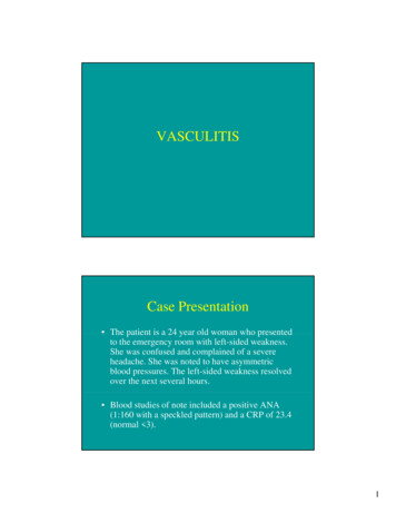
VASCULITISCase Presentation The patient is a 24 year old woman who presentedto the emergency room with left-sided weakness.She was confused and complained of a severeheadache. She was noted to have asymmetricblood pressures. The left-sided weakness resolvedover the next several hours. Blood studies of note included a positive ANA(1:160 with a speckled pattern) and a CRP of 23.4(normal 3).1
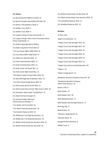
Case PresentationEvaluationva uat o includedc uded a transesophagealt a sesop agea echoec o whichw c showeds oweddiffuse thickening of the aorta and a CT and MRI whichshowed multiple abnormalities including: 1. thickened regions of the wall of the thoracic aorta whichshowed significant enhancement, suggestive of activeinflammation 2. encasement and narrowing of the right pulmonary arterywhichhi h alsol enhancedhd 3. marked narrowing of the proximal left carotid artery andthe left subclavian artery and diffuse narrowing of the rightsubclavian and proximal portion of the right carotid.Ultrasound B-mode and color-duplex flow imaging of the left common carotid artery(longitudinal section): homogeneous, midechoic, circumferential wall thickening("macaroni sign") with luminal stenosisMeini, S. et al. Circulation 2006;114:e544Copyright 2006 American Heart Association2
Case Presentation Findings were felt to be consistent withTakayasu’s arteritis, an inflammatorygranulomatous disease of the medium andlarge vessels that is prevalent in youngwomen.Vasculitis Vacsulitis is an inflammation of the vesselwall. Inflammation results from immune complexdeposition or from cell-mediated immunereactions directed against the vessel wall. It can involved small, medium and largeblood vessels.3
Vasculitis: A classification by size and type of involved vessel.Adapted from Jennette and Falk: Small-vessel vasculitis, NEJM 337: 1512, 1997.IntimaHuman AortaMediaAdventitia4
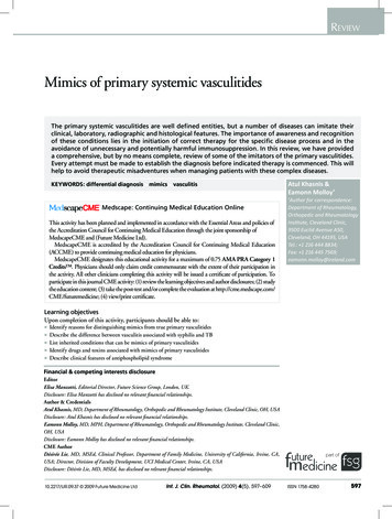
mediaintimaMultinucleate giant cell5
Multinucleategiant cells6
Inflammation in adventitiaTakayasu Arteritis Women 20-5020 50 years of age Symptoms: Weakened pulses in arms.Cold, numb fingers. Ocular disturbances.Hypertension. Neurologic deficits. Mayy have aneurysmy early;y; heal withfibrosis with narrowing of lumen.7
Aortitis: Takayasu v. Giant Cell Both show female predilectionTakayasu patients 20 – 50 years oldGiant cell patients age 70Giant cell has mild intimal scarringTakayasu has more adventitialinflammation, scarring and endarteritisobliterans.Giant Cell Arteritis Most common of the vasculitidesLarge, medium and small arteries involvedPrimarily temporal, vertebral, ophthalmicMost in persons 50 years of ageFever, fatigue, weight loss, facial pain, headache.Diagnose with temporal artery biopsy: Focalthickening with granulomatous inflammationfocused on the internal elastic lamina. Responds to anti-inflammatory therapy8
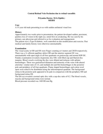
75 yr old woman: temporal artery biopsyMediaInternal elastic lamia89 year old woman: temporal artery biopsy9
75 year old woman withgiant cell arteritis intemporal arteryVasculitis: A classification by size and type of involved vessel.Adapted from Jennette and Falk: Small-vessel vasculitis, NEJM 337: 1512, 1997.10
Polyarteritis Nodosa (PAN) Medium to small muscular arteries in any organSSegmentall necrotizingi i inflammationi fliAneurysms and thrombosisMay heal with fibrosisAll stages of activity may be presentgy Immune complexp depositionp((30% areEtiology?Hepatitis B antigen positive in serum).No association with ANCA (antineutrophilcytoplasmic antibodies).Polyarteritis NodosaSymptoms: malaise,malaise fever,fever weight loss,losshypertension, abdominal pain and blood instool, muscular pain, peripheral neuritis,renal failure.Treatment: High dose immunosuppressionwith corticosteroids, cyclophosphamide.11
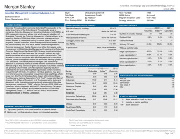
32 year old womanwith peripheralneuropathy:muscle biopsyActive vasculitiswith “fibrinoid”necrosis“Fibrinoid” necrosis of a small artery12
Active vasculitis in a small arteryyHealed lesion with narrowing oflumenVasculitis: A classification by size and type of involved vessel.Adapted from Jennette and Falk: Small-vessel vasculitis, NEJM 337: 1512, 1997.13
Kawasaki disease: Coronaryartery aneurysm with markedintimal proliferation.Kawasaki Disease Most under age 4 years Fever, skin erosions, enlarged lymph nodes,20% have coronary artery vasculitis. Death rate now 0.8% in Japan – due to giantaneurysmsyof coronaryy arteries. Aneurysm formation in 25% of untreatedcases; less than 1% with IV Ig14
Vasculitis: A classification by size and type of involved vessel.Adapted from Jennette and Falk: Small-vessel vasculitis, NEJM 337: 1512, 1997.Microscopic Polyangiitis Necrotizing vasculitis of small vessels(smaller than involved in PAN) Symptoms: skin nodules, hemoptysis,abdominal pain, hematuria, proteinuria. Glomerulonephritis in 90%. Often an immunologic reaction to drug(penicillin), microorganisms, administeredproteins, or tumor antigens.15
Wegener Granulomatosis1 Acute necrotizing granulomas of ear,1.earnose, throat, or lung.2. Necrotizing vasculitis of small to mediumsized vessels3. Renal disease - focal necrotizinggglomerulonephritisAntineutrophil CytoplasmicAntibodies (ANCA) Autoantibodies directed against enzymes ingranules in neutrophils, lysosomes ofmonocytes, and in endothelial cells. Cytoplasmic (cANCA): proteinase-3 Perinuclear (p(pANCA):C ) myeloperoxidasey p Wegener’s granulomatosis – cANCA Microscopic polyangiitis - pANCA16
Lung biopsy:granulomatousinflammationLung: small vessel vasculitis17
Kidney: GlomerulonephritisKidney: granulomatousinflammation18
Wegener’s Granulomatosis: Etiology: ? Hypersensitivity toundetermined antigens.g Prognosis: Untreated – 90% mortality in 2years 85-90% of patients respond tocyclophosphamide and prednisonebut 50% have relapses19
Vasculitis: A classification by si ze and type of involved vessel. Adapted from Jennette and Falk: Sma ll-vessel vasculitis, NEJM 337: 1512, 1997. Microscopic Polyangiitis Necrotizing vasculitis of small vesselsNecrotizing vasculitis of small vessels (smaller than involved in PAN) Symptoms: skin nodules, hemoptysis,