Transcription
184 Case reportVlaams Diergeneeskundig Tijdschrift, 2014, 83Immunological deep dermal vasculitis in a catImmunologische, diepe dermale vasculitis bij een katS. Gaisbauer, S. Vandenabeele, S. Daminet, D. PaepeDepartment of Medicine and Clinical Biology of Small Animals, Faculty of Veterinary Medicine, GhentUniversity, Salisburylaan 133, B-9820 Merelbekedominique.paepe@ugent.beABSTRACTIn this case report, a 13.5-year-old, neutered, female domestic shorthaired cat withimmunological deep dermal vasculitis is described. The patient was presented with lethargy,fever, polydipsia, anorexia and swollen distal limbs. Dermatological examination revealed partialalopecia, pitting edema and painfulness in all distal limbs. Several diagnostic examinations wereconducted to confirm the suspected diagnosis and to look for possible triggers of cutaneousvasculitis. Morphological changes that were indicative for deep dermal vasculitis were seen duringthe histological examination of the skin. The other examinations did not reveal an underlyingtrigger or cause of the dermal vasculitis. The cat was diagnosed with immunological deepdermal vasculitis. The cat was treated with antibiotics, infusion, tube feeding and prednisolone.Improvement and healing of the dermal symptoms were only noticed after the start ofprednisolone therapy.SAMENVATTINGIn deze casuïstiek wordt een 13,5 jaar oude, vrouwelijke, gesteriliseerde Europese korthaarbeschreven met immunologische, diepe dermale vasculitis aan de distale ledematen. De patiëntwerd aangeboden met lethargie, koorts, polydipsie, anorexie en opzetting van de distale ledematen.Op het dermatologisch onderzoek werd partiële alopecia, oedeem en pijn aan alle distale ledematenvastgesteld. Verschillende diagnostische onderzoeken werden uitgevoerd om het vermoeden vandermale vasculitis te bevestigen en een onderliggende oorzaak op te sporen. Op het histologischonderzoek van de huid werden veranderingen aangetoond die duidden op diepe dermale vasculitis.De andere onderzoeksresultaten wezen geen onderliggende oorzaak voor de dermale vasculitis aan.De diagnose van immunologische, diepe dermale vasculitis werd gesteld. De kat werd behandeld metantibiotica, infuus, nasoesofagale sondevoeding en prednisolone. Verbetering en genezing van devasculitisletsels werden slechts gezien na het opstarten van de prednisolonetherapie.INTRODUCTIONVasculitis is a process of inflammation and destruction of blood vessels mediated by neutrophils, lymphocytes or macrophages (Gross et al., 1992; Griffinet al., 1993). An abnormal immune response, which isnot a disease itself but a symptom, leads to cutaneousvasculitis (Innerå, 2013). Common dermal symptomsof vasculitis are crateriform ulcers, necrosis, pustulesor papules, hemorrhagic bullae, plaques and palpablepurpurae at distal extremities, such as the paws, tail,pinnae, scrotum and oral mucous membranes (Milleret al., 2013). The location of the lesions is typicallyassociated with the pathway of the vessels (Miller etal., 2013). To confirm the diagnosis of cutaneous vas-culitis, histology of deep dermal biopsies is needed(Innerå, 2013).In veterinary medicine, the classification of cutaneous vasculitis is based on histological inflammatory patterns (Gross et al., 2005). Vasculitides arecategorized in leukocytoclastic (nuclear cell dust inand around vessel walls) or non-leukocytoclastic,and are then subdivided according to the degree ofneutrophilic, eosinophilic or lymphocytic cell invasion of vessel walls (Gross et al., 2005). It must benoted that histological lesions evolve over time andthat infiltrates of mixed inflammatory cells are common in older lesions (Nichols et al., 2001).If vasculitis is diagnosed, a proper diagnostic searchfor an underlying cause is recommended (Innerå,
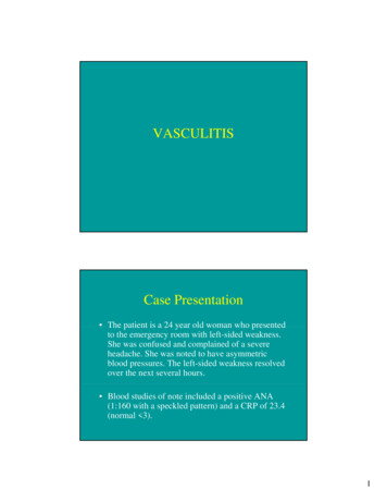
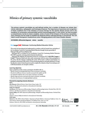
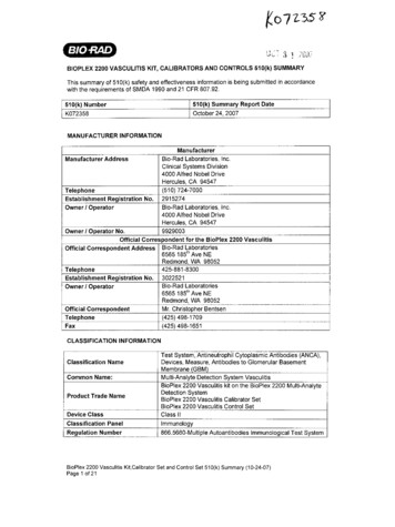
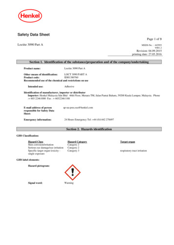
Vlaams Diergeneeskundig Tijdschrift, 2014, 832013). In human medicine, small vessel leukocytoclastic vasculitis has been attributed to a long list ofpossible causes (infections: bacterial, viral, parasitic;drugs; chemical substances; chronic persistent disorders: autoimmune connective tissue diseases, inflammatory bowel diseases; neoplasia; certain foods oradditives) (Carlson et al., 2005). However, in human aswell as in veterinary medicine, approximately 50 % ofthe cases are categorized as idiopathic vasculitis (Carlson, et al., 2005; Tai et al., 2006; Jasani et al., 2008).Cutaneous vasculitis has rarely been diagnosed in cats(Miller et al., 2013), and only few cases have beendescribed. Reported underlying causes are felineinfectious peritonitis (FIP), the application of fenbendazole, cimetidine and rabies vaccine. In severalcases, an underlying cause is not found (McEwan etal., 1987; Gross, 1999; Nichols et al., 2001; Cannonet al., 2005; Declerq et al., 2008; Jasani et al., 2008).In this article, a cat with immunological deep dermal vasculitis is described.CASE REPORTA 13.5-year-old, neutered, female domestic shorthair cat was brought to the referring veterinarianbecause of polydipsia, which had already been present for some months, acute lameness of the righthind limb and lethargy. The detailed history revealedthat the cat had been vaccinated six months before,the owner had not changed the diet before the symptoms started and that the cat had never been treatedwith medication except for tolfenamic acid (Tolfedine , Vétoquinol SA, Belgium) some years before.At physical examination (day 0), the cat was febrile(T 40.5 C) and the right hind limb was painful.Therapy with amoxicillin-clavulanic acid (Synulox ,Zoetis Belgium SA, Belgium; 8.75 mg/kg s.c., sid)and tolfenamic acid (Tolfedine , Vétoquinol SA, Belgium; 4 mg/kg s.c., sid) was instituted. At day 2, thecat was less lethargic, but a soft swelling of the distalright hind limb (metatarsus, digits) was noticed. Temperature was 39.2 C and the cat became anorectic.Therapy was changed to amoxicillin-clavulanic acid(Synulox , Zoetis Belgium SA, Belgium; 8.75 mg/kg s.c., sid), clindamycin hydrochloride (Antirobe ,Zoetis Belgium SA, Belgium; 25 mg bid, p.o.) andmeloxicam (Meloxidyl , Céva Santé Animale, Belgium; 0.2 mg sid, p.o.). At day 6, the cat was presented with lethargy and persistent anorexia. The referringveterinarian noticed a temperature of 37.4 C and softpainful swelling of the distal part of both hind limbs(metatarsus and digits). The owners were unable togive the clindamycin hydrochloride. The cat washospitalized and intravenous infusion with lactatedRinger’s solution (Hartmann , B. Braun Melsungen AG, Germany) was started. The treatment withamoxicillin-clavulanic acid was continued and thetreatment with meloxicam (Meloxidyl , Céva Santé185Animale, Belgium) was stopped. At day 7, the distal part of the left front limb (metacarpus, digits) wasswollen too, body temperature was within the normalrange and the other symptoms remained stable. Thenext day, force feeding was started (day 8). At day 9,serous fluid was oozing from the right hind limb andclindamycin hydrochloride (Antirobe , Zoetis Belgium SA, Belgium; 25 mg bid, p.o.) was started. Atday 10, all paws showed leakage of serous fluid. Tolfenamic acid (Tolfedine ,Vétoquinol SA, Belgium:4mg/kg s.c., sid) was given once and buprenorphine(Vetergesic , Alstoe Limited, United Kingdom; 10μg/kg i.v., tid) was started. During hospitalization,there was persistent anorexia, intermittent fever andpainful and swollen distal limbs. Several blood examinations were performed at day 5, 7, and 9, of whichthe last one at day 9 was the most comprehensivewith a complete blood count (CBC) and biochemistryincluding protein electrophoresis (Table 1). Bloodexamination revealed moderate leukocytosis (matureneutrophilia, monocytosis), mild anemia (decreasedhemoglobin and hematocrit), thrombocytopenia (notchecked on cytology), hypoalbuminemia, increased α1 and α 2 globulines, moderate hyperbilirubinemia,mild hyperchloremia, moderately increased lactatedehydrogenase, azotemia and hyperphosphatemia.Azotemia and hyperphosphatemia were normalized atday 9, hypoalbuminemia worsened over time.On day 12, the cat was referred to the internalmedicine service of the Department of Medicine andClinical Biology of Small Animals, Faculty of Veterinary Medicine, Ghent University, Belgium. Physical and dermatological examinations revealed dentalplaque, mild tachycardia and intense pitting edemaat all distal limbs, including warm and painful skin.The hair was easy to epilate from the abdomen andtarsi. Partial alopecia was present at the proximal ventral part of the tail and at the extremities (tarsi, metatarsi, metacarpi, digits) (Figure 1). On the tarsi anddigits, there were erosions and brown crusts. Therewas severe swelling and partial alopecia around thecuticles. On the dorsal metacarpal area of the left frontlimb, where the first catheter had been placed, the skinwas erythematous and crusty (Figure 2). Under digitalpressure, serous fluid passed through the skin of thedorsal side of the right distal front limb. The combination of fever with warm, painful edematous swellingof all distal extremities was suspicious for cutaneousvasculitis.Several diagnostic examinations were conductedto confirm the vasculitis, and look for possible triggers(e.g. infection, neoplasia, inflammation) of cutaneousvasculitis. An impression smear of the erythematousand crusty parts of the skin of the distal limbs wasmade and stained with Diff Quick solution. It showeddegenerated neutrophils and some cocci that werenot phagocytized. Anesthesia was required for theother diagnostic tests. The cat was premedicated withmethadone (Comfortan , Eurovet Animal Health BV,
186Vlaams Diergeneeskundig Tijdschrift, 2014, 83Figure 1. Brown crusts, erosions and partial alopecia ofthe tarsi and swollen paws.the Netherlands; 0.02mg/kg, i.v.). For induction andmaintenance, alfaxalon (Alfaxan , Jurox Limited,United Kingdom; induction: 5 mg/kg i.v., maintenance: on effect, in total including the induction 20mg/kg was used) were given as boli. Mouth inspection under anesthesia was done to assess if mucosallesions or infections were present. It presented noabnormalities besides moderate dental plaque. Thecomplete blood cell count and some biochemicalblood parameters were repeated. A mild hypoalbuminemia, leukocytosis, consisting of neutrophilia andmonocytosis, mild basophilia, and non-regenerativeanemia were present (day 12) (Table 2). A SNAP Combo test (IDEXX Laboratories Europe BV, theNetherlands) for feline leukemia virus antigen andfeline immunodeficiency virus antibody was negative.Serum was sent to Algemeen Medisch LaboratoriumDiergeneeskunde Antwerpen (MEDVET) for serology of toxoplasmosis. The titre of IgM was negativeand the titre of IgG was highly positive, which indicated that the cat had immunity against Toxoplasma,but had no active, clinical disease. An abdominalultrasound and thoracic radiographs were conductedto look for underlying neoplastic or inflammatorydiseases. Thoracic radiographs (ventrodorsal andright lateral) revealed a mild interstitial lung patternof the left caudal lung lobe which could have beencaused by the decubitus of the cat prior to the radiographs were taken. There was also a suspicion of mildpleural effusion, but there was too little fluid to con-Figure 2. Erythematous and crusty skin of the left frontlimb.duct a thoracocentesis. Reduced echogenicity of theliver parenchyma with prominent portal veins, mildheterogeneity of the pancreas, mildly reduced sizeand rounded appearance of the left kidney associatedwith a reduced corticomedullary definition and hyperechogenic cortex of the right kidney were noted onultrasonography. Cystocentesis and fine-needle aspi-
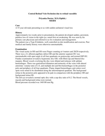
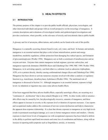
Vlaams Diergeneeskundig Tijdschrift, 2014, 83187Figure 3. Skin biopsy. Serocellular crusts on the epidermis, edema of the superficial and deep dermis,perivascular to interstitial mixed infiltration of inflammatory cells (mast cells, lymphocytes, neutrophils, feweosinophils), reactive fibroblasts (magnification 5x).Figure 4. Skin biopsy of the deep dermis. Small arteries and venules with perivascular infiltration of inflammatory cells (mast cells, lymphocytes, neutrophils, feweosinophils), edema of the surrounding dermis, reactivefibroblasts (magnification 40x).ration of the liver were performed to evaluate for thepresence of lymphoma and hepatic lipidosis. Cytology of the liver showed mild hepatocellular degeneration, the presence of non-degenerated neutrophils inand around clusters of hepatocytes. It was difficult todistinguish if the presence of neutrophils could indicate mild lymphocytic cholangitis or if these were dueto mild blood contamination. Urinalysis consisted ofurine specific gravity, microscopic sediment analysis,urine dipstick (protein, glucose, ketones, hemoglobin,bilirubin, urobilinogen, acetone, nitrite, leukocytes,pH), urinary protein/creatinine ratio and bacterialculture. This was done to exclude urinary tract infection and inflammation and to assess renal functionconsidering a possible glomerulonephritis or renalvasculitis as a representation of a more generalizedvasculitis. Urinalysis only revealed isosthenuric urine(USG 1.016) and very mild proteinuria (0.4). A dorsopalmar view of the distal right hind limb was takento evaluate bony structures, which showed no bonyabnormalities. Five punch biopsies of the skin (two ofthe left front limb in the area of the skin lesions, twodorsally of right front limb, one of left lateral tarsus)were taken. After taking the biopsies, all biopsy siteswere bandaged.Based on the clinical and dermatological findings, an immunological dermal vasculitis was considered to be the most likely differential diagnosis. Awaiting the results of the histopathology andtoxoplasmosis serology, therapy with prednisolone(Codipred , Codifar NV, Belgium; 1mg/kg i.m.,sid) was started. Amoxicillin and clavulanic acid(Synulox , Zoetis Belgium SA, Belgium; 8.75mg/kg s.c., sid), clindamycin hydrochloride (Antirobe , Zoetis Belgium SA, Belgium; 12.5 mg/kgp.o., bid), buprenorphine (Vetergesic , Alstoe Limited, United Kindom; 10 μg/kg i.v., qid) and infusion therapy with lactated Ringer’s solution (Hartmann , B. Braun Melsungen AG, Germany; 80 ml/kg/ 24 hours i.v.) were continued. A nasoesophagealfeeding tube was placed and tube feeding with a protein- and calorie-rich diet (Royal Canin Convalescence Support , Royal Canin, France) was started.On the second day in the Small Animal Clinic ofthe Faculty of Veterinary Medicine, (UGhent), thecat ate some food spontaneously, and the rest of itsdaily food requirement was tubefed. Body temperature was normal. The condition of the right front limbwas unaltered. Serous fluid passed through the skinof the left front limb under digital pressure. The otherlimbs were still bandaged. The level of anemia andhypoalbuminemia remained stable (day 13) (Table 2).The next day, the cat ate its complete nutritionalrequirement spontaneously. Infusion therapy andtube feeding were stopped. The paws were markedlyless swollen and less painful. There were still multiple crusts on the skin of the paws. The applicationFigure 5. At recheck one month later, the paws were notswollen and hair was regrowing.
188Vlaams Diergeneeskundig Tijdschrift, 2014, 83Table 1. Results of blood examinations performed by the referring veterinarian.ParameterDay 5Day 7Day 9Month 6Reference intervalHematologyErythrocytes7.865.797.295.5 - 10.0Leukocytes11.736.58.75.5 - 15.5Band neutrophils 1.830 - 0.42Segmented neutrophils10.526.25.63.0 - 11.5Eosinophils0.210.40.90.05 - 1.10Basophils0000 - 0.1Lymphocytes0.651.91.2 - 5.6Monocytes0.473.110.250 - 0.7Hemoglobin7.34.76.35.4 - 9.9Hematocrit269224272260 - 460MCV34393740 - 55MCH9898.00 - 10.6MCHC27212418.6 - 23.6Thrombocytes246126323190 - 430BiochemistryGlucose (sober)3.93.5-6.0Total protein655584.355.0 - 85.0Albumin27.522.618.435.631 - 40Alfa 1 globulin 20,5 - 1.5Alfa 2 globulin 15.89 - 15Beta globulin 7.97 - 11Gamma globulin 14.47 - 18Urea34.627.39.917.35.90 - 12.50Creatinine403.1287.3115.8225.4270 - 130LDH 42580 - 170AST344317 60ALT24274737 - 75Bilirubin total 11.622.50 - 3.50Bilirubin direct 11.281.71 - 2.50GGT 30-8ALP 393410 - 50Bile acid 317 20Lipase 11 250Sodium151150161146 - 158Potassium3.83.74.63.40 - 5.20Chloride105117123105 - 112Calcium2.22.12.631.80 - 3.00Phosphor2.61.51.951.10.- 1.60HormonesT4 6.413.612 - mmol/Lmmol/Lmmol/Lmmol/Lmmol/Lmmol/LBold written data are outside the reference interval. ALP alkaline phosphatase, ALT alanin aminotransferase, AST aspartateaminotransferase, GGT gamma-glutamyltransferase, LDH lactate dehydrogenase, MCH Mean corpuscular hemoglobin, MCHC mean corpuscular hemoglobin concentration, MCV mean corpuscular volume
Vlaams Diergeneeskundig Tijdschrift, 2014, 83of amoxicillin-clavulanic acid was changed to peros (Clavubactin , Le Vet BV, the Netherlands; 12.5mg/kg p.o., bid). On day 15, the clinical conditionimproved further. Only the toes remained painful andthe distal limbs were still mildly swollen. A completeblood cell count, albumin, creatinine and total bilirubin were repeated (day 15) (Table 2). The results confirmed the anemia and a mild hypoalbuminemia, butthe monocytosis and neutrophilia had improved. Onthe next day (day16), the cat was discharged. Therapywith prednisolone (Prednisolone , Kela Laboratoria,Belgium; 1.1 mg/kg p.o., sid), amoxicillin-clavulanicacid (Clavubactin , Le Vet BV, the Netherlands; 12.5mg/kg p.o., bid), clindamycin hydrochloride (Antirobe , Zoetis Belgium SA, Belgium; 12.5 mg/kg p.o.,bid) and buprenorphine (Vetergesic , Alstoe Limited,United Kindom; 10 μg/kg p.o.) in case of pain wascontinued awaiting the laboratory results. One weekafter discharge from the clinic, all laboratory resultswere available, and because there was no infection,the antibiotic therapy was discontinued. The histopathological results showed that all skin biopsies hadsimilar morphological changes (Figures 3 and 4).On the epidermis, serocellular crusts were seen. Thesuperficial and deep dermis were edematous. A perivascular to interstitial mixed infiltration of inflammatory cells (mast cells, lymphocytes, neutrophils,few eosinophils) and many reactive fibroblasts wereseen. Small foci of extravasation of red blood cells189and some thrombi in the blood vessels were present in the deep dermis and panniculus. Focally, therewere several blood vessel lumina in which numerousneutrophils were seen. Although mural infiltrationwas not seen, the fact that the alterations were worsein the panniculus suggests that there were mural infiltrations of the vessel walls in bigger, deeper vessels,which were not included in the biopsy. The observedchanges were suggestive for deep dermal vasculitis.One month later, the cat was presented for arecheck appointment. The cat owner had not givenbuprenorphine as the cat did not show signs of pain athome. Because the cat had suffered from polyuria andpolydipsia, the owner had decided himself to stepwisereduce prednisolone. At the time of the recheck, thecat received one quarter of a 5 mg tablet a day. Physical examination did not reveal significant abnormalities. The paws were not swollen anymore and hairwas regrowing (Figure 5). The plan was to recheckblood examination (CBC, biochemistry) and urinalysis to evaluate the former abnormalities. However, thecat was too aggressive to sample blood and urine, andthe owner refused sedation or anesthesia of the cat toperform these procedures. The advice was to continuetherapy for three weeks with one quarter of prednisolone (Prednisolone , Kela Laboratoria, Belgium) 5mg every other day before stopping the medical treatment. A control appointment 6 to 8 weeks later wasadvised.Table 2. Results of blood examinations performed at the Department of Small Animal Medicine, Faculty of VeterinaryMedicine, UGent, Belgium.ParameterDay 12Day 13Day 15Reference intervalHematologyErythrocytes5.47 5.086.54 - 12.2Leukocytes29.64 23.652.87 - 17.02Neutrophils23.47 20.211.48 - 10.29Eosinophils0.360.390.17 - 1.57Basophils0.40.260.01 - 0.26Lymphocytes4.392.030.92 - 6.88Monocytes1.02 0.760.05 - 0.67Hemoglobin6.6 6.29.8 - 16.2Hematocrit19.2262130.3 - 52.3Reticulocytes1222.43 - 50MCV35.13635.9 - 53.1MCH12,112.211.8 - 17.3MCHC34.433.928.1 - 35.8Thrombocytes202242151 - 600BiochemistryTotal protein6157 - 89Albumin20202123 - 39Creatinine14216771 - 212Bilirubin total12120 - 15Bold written data are outside the reference interval. MCH Mean corpuscular hemoglobin, MCHC meancorpuscular hemoglobin concentration, MCV mean corpuscular %109/LfLpgg/dL109/Lg/Lg/Lμmol/Lμmol/L
190After six months, the cat came for another controlto the referring veterinarian. According to the owner,all complaints had resolved and the cat was againvery active and able to hunt. Physical examinationdid not reveal abnormalities: the paws were not swollen anymore and hair was completely regrown. Bloodcould be taken, but it was not possible to take a urinesample. The blood examination (CBC, biochemistry and total thyroxine) showed that the number ofleukocytes (neutrophils and monocytes) had normalized and the anemia was dissolved. Moderate azotemiaand hyperphosphatemia were again present (Table 1).DISCUSSIONImmunological deep dermal vasculitis was diagnosed in a 13.5-year-old, neutered, female domesticshorthaired cat based on clinical findings, the histological examination of skin biopsies, exclusion ofpossible underlying causes and response to therapy.A detailed history was performed to detect possible triggers such as food, food additives, chemicalsubstances, vaccinations or drugs. Based on the history, these triggers could be excluded. In the literature, a case of a dog with deep dermal vasculitis afterthe administration of meloxicam has been reported(Niza et al., 2007). Meloxicam was also administeredin that case, but only after the onset of the symptoms.Physical examination, blood examination (CBC, biochemistry), a SNAP Combo Test (IDEXX Laboratories Europe BV, the Netherlands) for feline leukemia virus antigen and feline immunodeficiency virusantibody, serology of toxoplasmosis, mouth inspection, an impression smear of the altered skin, thoracicradiographs, abdominal ultrasound, fine-needle aspiration of the liver, cystocentesis and urinalysis wereconducted to look for infections (bacterial, viral, parasitic), inflammation or neoplasia as a possible causeof the deep dermal vasculitis.The age of reported cases range from seven monthsto seven years old (McEwan et al., 1987; Gross, 1999;Nichols et al., 2001; Cannon et al., 2005; Declerq etal., 2008; Jasani et al., 2008). In comparison to thesecases, the age of the cat of the present case is high.However, with the small number of cases that havebeen reported, it is difficult to say if there is a certainage predisposition for dermal vasculitis in cats. In thecurrent literature, there is no information about breedor sex predisposition.Dermal vasculitis is characterized by an immuneresponse concerning blood vessels (Innerå, 2013). Atype three hypersensitivity reaction is the most likelyand generally accepted mechanism of cutaneous vasculitis in animals (Innerå, 2013). Antigen and antibodies can form antigen-antibody complexes, which maylead to immune complex deposition on the endotheliumof blood vessels inducing vasculitis (Voie et al., 2012).Constitutional signs, such as anorexia, depression andVlaams Diergeneeskundig Tijdschrift, 2014, 83pyrexia, which also occurred in the present case, canprecede dermal symptoms (Miller et al., 2013). In thiscase, signs of dermal vasculitis involved all four distallimbs, which correspond to the distribution of dermalvasculitis in dogs (Miller et al., 2013). In humans,small vessel leukocytoclastic vasculitis also affectslower limbs and areas of the body, which are ventralof the heart (Pulido-Pérez et al., 2012). The clinicallyand histologically diagnosed edema, which was present in the four distal limbs of the patient of the presentcase, is a prevalent sign of dermal vasculitis (Miller etal., 2013; Innerå, 2013). Painful skin as was seen inthis case has also been reported in the literature as apossible symptom of dermal vasculitis (Innerå, 2013).Several abnormalities detected by blood examinations (mild anemia, moderate leukocytosis, increasedα 1 and α 2 globulins, moderately increased lactatedehydrogenase activity, decreased serum thyroxineconcentration) can be explained as consequences ofthe dermal vascular inflammation. In this case, hypoalbuminemia was probably caused by a combinationof dermal vascular inflammation (negative acutephase protein), persistent anorexia and potentiallyprotein loss by skin lesions.Only mild abnormalities were seen on the radiographs of the thorax. Despite these, the cat neverdeveloped respiratory signs during hospitalizationand follow-up. The suspicion of mild pleural effusioncould be an indicator for a more generalized vasculitis. Therefore, an examination of the pleural effusionwould have been ideal, but was impossible becauseof the little amount. However, there was no other evidence of involvement of internal organs or the presence of abdominal fluid, making generalized vasculitis unlikely. The ultrasonographic alterations of thekidneys and the initial azotemia may have indicatedrenal disease, such as glomerulonephritis or chronickidney disease, but were not typical of vasculitis. Alsothe isosthenuria could have confirmed the suspicionof renal disease, although it might have been influenced by the infusion therapy, which had been givenby the referring veterinarian until the day before urinalysis. Glomerulonephritis was ruled out by the lackof significant proteinuria. The initial azotemia couldalso have been caused by an acute component due toa prerenal cause (dehydration) and the therapy withnon-steroidal, anti-inflammatory drugs. The rapidnormalization of the azotemia after infusion therapymakes prerenal factors very likely. Although repeatedmeasurement of the urine specific gravity would beideal, the recurring azotemia at the last recheck by thereferring veterinarian indicated chronic kidney disease. Chronic kidney disease is a frequent disease inolder cats with a prevalence up to 30% in cats olderthan 15 years (Lulich et al., 1992). Chronic kidneydisease is mainly a degenerative disease with onlymild inflammation and thus less likely as a trigger ofdeep dermal vasculitis. Therefore, it is more likelythat chronic kidney disease is an incidental concur-
Vlaams Diergeneeskundig Tijdschrift, 2014, 83rent finding in this cat instead of a possible trigger ofdeep dermal vasculitis.The results of the abdominal ultrasound and fineneedle aspiration of the liver and hyperbilirubinemia could indicate a possible liver disease such aslymphocytic cholangitis. However, it is important torealize that fine-needle aspiration of the liver is not avalid diagnostic tool to diagnose inflammatory hepaticdiseases or the seriousness of these diseases. In a studyby Wang et al. (2004), cytology only agreed in 27.3%of feline cases of inflammatory hepatic disease withthe histopathologic diagnosis. Normal blood values ofalanin aminotransferase (ALT) and aspartate aminotransferase (AST), the quick decline of bilirubin andthe response to therapy make non-specific reactivehepatitis more likely. Non-specific reactive hepatitiscan occur secondary to extrahepatic diseases, causedby hepatic hypoxia and endotoxemia (Rothuizen,2010). When the primary disease is treated successfully, most of these reactive hepatic changes resolvewithout specific hepatic therapy (Rothuizen, 2010).Histological features of leukocytoclastic vasculitisinclude perivascular and mural infiltration by polymorphonuclear leukocytes, extravasation of red bloodcells, fibrinoid necrosis and occasionally thrombosis (Sams et al., 1976). The histopathological resultsshowed several of these changes, such as a perivascular mixed infiltration of inflammatory cells, somethrombi in the blood vessels and small foci of extravasation of red blood cells were present in the deep dermis and panniculus. Focally, there were several bloodvessel lumina, in which numerous neutrophils werepresent, but mural infiltration was not seen. The histological alterations, which were seen in the dermalpunch biopsies in this case suggested the presence ofneutrophilic mural infiltration of the vessel walls indeeper layers, which were not included in the punchbiopsy. The punch biopsies probably did not includethese deep layers, because of the intense dermal edema, making deeper biopsies than usually necessary.Using excision biopsies (e.g. amputation of a digit)could have solved this problem. In both veterinaryand in human medicine, the diagnostic value of a skinbiopsy increases with the depth of the biopsy, whendeep vasculitis is suspected (Carlson et al., 2005;Innerå, 2013).Although multiple diagnostic tests and a detailedhistory were performed, an underlying cause, whichcould have triggered deep dermal vasculitis, was notfound. This indicates an immunological cause of thedeep dermal vasculitis in this case. This was also supported by the complete response to the prednisolonetherapy, which would not have occurred if an underlying infectious or neoplastic cause was overlooked.The present case also shows that owner compliance isa problem in small animal medicine. The owner hadreduced the dosage of prednisolone without consulting the veterinarian, despite oral and written recommendations regarding treatment. Studies have shown191that about 10% to 25% of veterinary clients do notgive all the medication prescribed (Maddison, 2011).As pet owner compliance is essential for an effectivetherapy, a lack of compliance might be a reason fortreatment failure.In this case, the cat recovered completely
vasculitis. Several diagnostic examinations were conducted to confirm the vasculitis, and look for possible triggers (e.g. infection, neoplasia, inflammation) of cutaneous vasculitis. An impression smear of the erythematous and crusty parts of the skin of the distal limbs was made and stained with Diff Quick solution. It showed