Transcription
234Neuroscienee Letters, 109 (1990) 234 238Elsevier Scientific Publishers Ireland Ltd.NSL 06642Magnesium reduces N-methyl-D-aspartate(NMDA)-mediated brain injury in perinatal ratsJohn W. McDonald l, Faye S. Silverstein 2 and Michael V. Johnston 3 Neuroseience and Medical Scientist Training Program, :Departments f Pediatrics and Neurology,University )['Michigan Medical School, Ann Arbor, M I (U.S.A.) and- Departments c ['Neurology andPediatrics, The Johns Hopkins University t?f Medieine and the Kennedy Institute. Baltimore. MD r U.S.A. )(Received 25 July 1989; Revised version received 3 October 1989; Accepted 4 October 1989)Key words:Magnesium: N-Methyld)-aspartate: Neurotoxicity; Neuroprotection: Rat brain; PerinatalWe evaluated the neuroprotective effects of systemically administered magnesium against N-methyl-Daspartate (NMDA)-mediated brain injury in perinatal rats. Postnatal day (PND) 7 rats received unilateralintrastriatal injections of 25 nmol NMDA followed 15 min later by single or multiple doses of magnesiumintraperitoneally (i.p.). Animals were sacrificed five days later and the severity of brain injury was assessedby comparison of the weights of the injected and contralateral cerebral hemispheres. NMDA injectionreduced the weight of the injected cerebral hemisphere by 31 3%. Single doses of magnesium reducedthe severity of NMDA-induced brain injury in a dose-dependent fashion (2 mmol/kg 29 1I% protection:3 mmol/kg, 52 12% protection; 4 mmol/kg, 62 7% protection). Multiple doses of magnesium reducedbrain injury by 65 4%. These data demonstrate that systemically administered magnesium antagonizesthe neurotoxic effects of NMDA in vivo in perinatal rats.The N-methyl-D-aspartate (NMDA)-type excitatory amino acid (EAA) receptor iscoupled to a Ca 2 permeable cationic channel; the receptor/channel complex includes several regulatory sites [2, 24]. Physiological concentrations of magnesium reduce responses to N M D A by blocking the NMDA-associated ionophore in a voltage-dependent manner [2]. Intrastriatal injection of N M D A typically produces focalbrain injury in PND 7 rats [17, 19, 20]. Competitive and non-competitive N M D Areceptor antagonists attenuate this damage in PND 7 rats in a dose-dependent fashion [20, 21]. Furthermore, these antagonists also limit N M D A neurotoxicity inadult rats and in neocortical cultures [11, 12]. However, few of these antagonists areapproved for use in humans. Although its mechanism of action is unknown, magnesium has been used therapeutically for many years in obstetrics for the treatment ofeclampsia [4]. This study examines the neuroprotective effects of magnesium againstNMDA-mediated brain injury in PND 7 rats.Correspondence: M.V. Johnston, Kennedy Institute, Room 506, 707 N. Broadway, Baltimore, MD 21205,U.S.A.0304-3940/90/ 03.50 {'; 1990 Elsevier Scientific Publishers Ireland Ltd.
235PND 7 Sprague-Dawley albino rats, anesthetized with ether, received unilateralintrastriatal stereotaxic injections of 25 nmol NMDA (0.5 #1 injection vol., pH 7.4)using a Hamilton syringe (26 gauge) as detailed previously [18, 19, 22]. With bregmaas a reference, injection coordinates were AP 2.0 mm, ML 2.5 mm, at a depth of4 mm from dura. Magnesium (sulfate) was dissolved in isotonic saline (pH 7.4) andadministered by i.p. injection in single or multiple doses (injection vol. 0.05 ml) beginning 15 min after the NMDA injection. Two magnesium treatment protocols weretested; (1) a single dose (2 mmol/kg, n 4; 3 mmol/kg, n 4; 4 mmol/kg, n 4;) and(2) 6 doses (each 1.8 mmol/kg given at variable time points over a 6 h period, totaldose of 11 mmol/kg) as necessary to control behavioral signs of seizures ( n - 8). Isotonic saline-treated NMDA-injected controls were included in each protocol following the appropriate dosage schedule (protocol 1, n 4); protocol 2, n 8). A 3rdgroup of NMDA-injected rats (n 8) received equimolar NaCI injections in vehiclefollowing the dosage schedule of protocol 2 (six 1.8 mmol/kg injections). Additional,non-NMDA-injected rats were given i.p. injections (2 mmol/kg) of either magnesium(n 6) or NaC1 (n 6). Animals' body temperatures were maintained at normothermia (35.5 2 C surface temperature) for 2 h following treatment using warminglamps and electronic surface probes. All animals were decapitated on PND 12, fivedays after NMDA injection. The degree of resulting brain injury was quantitated bycomparison of the weights of the injected (/) and contralateral (C) cerebral hemisphere weights using the formula, %Damage IO0(C-I)/C, expressed as mean S.E.M. [18, 22]. This is a sensitive and reliable method for quantitating unilateralbrain injury in this experimental model; %Damage values are highly correlated(r2 0.99, P 0.001) with ipsilateral reductions in both choline acetyl transferase activity and regional cross-sectional areas [18, 20, 22]. To assess neuroprotective efficacy %Protection values were calculated as: %Protection 100"[1 -(%Damage magnesium-treated/%Damage saline-treated)]. Statistical comparisons were made usingone-way ANOVA except where otherwise noted. Saline-treated NMDA-injectedcontrols were pooled for statistical comparisons since no significant differences wereobserved between groups ( P N.S., independent t-test).Intrastriatal injection of NMDA (25 nmol) produced tonic-clonic seizure activityand in animals sacrificed five days later resulted in predominantly unilateral confluent brain necrosis involving the corpus striatum and extending into the thalamus,dorsal hippocampus, and adjacent neocortex [17, 19]. In saline-treated groups,NMDA (25 nmol) injections reduced the weight of the injected hemisphere by31.3 2.5% (mean S.E.M., n 10) relative to the contralateral cerebral hemisphereand resulted in 17% mortality rate. Administration (i.p.) of magnesium, but notNaC1, reduced the behavioral signs of seizures and decreased muscle tone. The highest dose of magnesium (4 mmol/kg) produced short periods (10-30 s) of apnea.Higher doses resulted in prolonged apnea and increased the mortality rate. Singledoses of magnesium significantly reduced the extent of NMDA-mediated braininjury in a dose-dependent manner (P 0.001, ANOVA, Fig. 1). Multiple doses ofmagnesium reduced the extent of brain injury by 65 4% relative to saline treated,NMDA-injected controls (P 0.001, ANOVA). In contrast, multiple doses of equi-
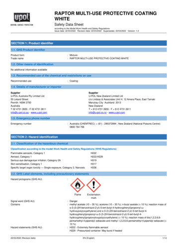
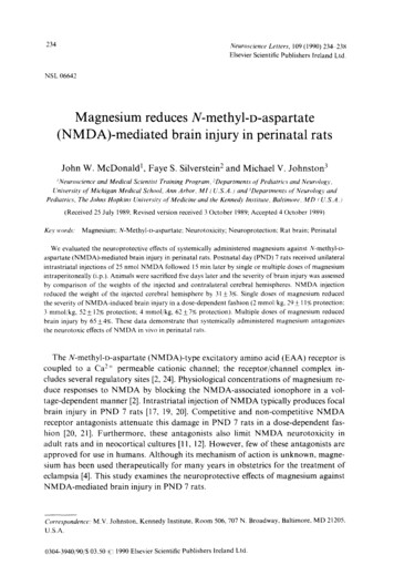
23630,6u20,OIE 1Q100NMDA23411Magnesium (mmol/kg) NMDAFig. I. Quantitative neuroprotective effects of single and multiple doses of magnesium against NMDAmediated brain injury. Unilateral intrastriatal stereotaxic injections of NMDA (25 nmol) were performedin ether anesthetized PND 7 rats. Fifteen min later, single (2, 3, 4 mmol/kg, n 4/dose) or multiple (6injections of 1.8 mmol/kg each over 6 h, n 8) doses of magnesium were administered i.p. in 0.05 ml saline.NMDA-injected littermate controls received equivalent volumes of saline (n 10). Animals were decapitated 5 days later on PND 12. The severity of resulting brain injury was assessed by comparison of theweights of the injected (I) and contralateral (C) cerebral hemisphere weights and expressed as %Damage(mean S.E.M.) using the formula on the vertical axis. "P 0.05, *'P 0.001, ANOVA, magnesiumtreated vs saline treated.molar NaC1 increased NMDA-induced brain injury by 25 - 7% (P 0.001, independent t-test; 39.2 2.2 %Damage, NaC1, n 8 vs 31.3 2.5 % Damage, isotonic salinetreated controls, n 10. The mortality rate was 0% in groups treated with magnesiumor NaC1.Both competitive and non-competitive NMDA receptor antagonists attenuateNMDA-mediated brain injury [11, 19-21]. These data demonstrate that magnesiumcan also limit NMDA-induced brain injury. In the same animal model, completeneuroprotection against NMDA toxicity is possible with MK-801 and TCP, twonon-competitive NMDA antagonists, and with CGS-19755, a competitive NMDAantagonist. The level of neuroprotection achieved by magnesium against NMDAtoxicity in this experimental model is equivalent to the maximal degrees of protectionproduced by the non-competitive NMDA antagonists, phencyclidine (PCP), ketamine and dextromethorphan,the competitive NMDA antagonist CPP, and the glycine modulatory site antagonist HA-966 in this experimental model [18, 20-22]. Itis possible that combined treatment with magnesium and other NMDA receptorantagonists may potentiate their neuroprotective effects.Several mechanisms could account for magnesium's neuroprotective properties [1,8]. High concentrations of magnesium may directly block the NMDA-associatedionophore [2]. Furthermore, magnesium acts as a Ca 2 antagonist and can inhibitneurotransmitter release and reduce seizure activity at high concentrations [3, 14].Our study does not directly address the mechanisms of magnesium's neuroprotective
237effects. However, several lines of evidence suggest that neither inhibition of neurotransmitter release nor anticonvulsant actions play a major role in magnesium's neuroprotective effects in this experimental model. The dosages of magnesium used in thisstudy did not block neuromuscular transmission. In addition, in this experimentalmodel adenosine, an inhibitor of synaptic neurotransmitter release, does not reduceN M D A toxicity [18]. Nor is it likely that magnesium's neuroprotective effects canbe attributed to anticonvulsant properties since anticonvulsants (e.g. pentobarbital,carbamazepine) which reduce the behavioral signs of seizures are not neuroprotective[18]. Further studies are necessary to determine if the protective effects of magnesiumare specific for NMDA-mediated damage or whether magnesium can also reduceother forms of excitotoxic brain injury. The mechanism by which multiple doses ofhyperosmolar NaC1 enhance NMDA-mediated brain injury are not clear but maybe related to intravascular hypernatremia and hypovolemia.Excessive activation of N M D A receptors has been implicated in the pathogenesisof neuronal injury in a number of acute neuroiogic disorders including hypoxiaischemia, physical brain trauma, and prolonged seizures [5, 9, 15, 16, 25]. The findingthat magnesium reduced NMDA-mediated brain injury supports in vitro and in vivostudies which indicate that magnesium can limit neuronal injury resulting fromhypoxia and ischemia and traumatic brain injury [5, 9, 13, 16, 23, 25, 26]. Magnesiumnormally does not readily penetrate the fully developed blood-brain barrier (BBB),but BBB permeability is greater in infants and during seizures, hypoxia-ischemia andother acute neurologic disorders in adults [4, 7, 10]. Thus, magnesium may provideeffective therapy in these conditions when administered systemically. The experimental evidence of magnesium's neuroprotective properties in the developing and adultbrain, together with its ability to penetrate the BBB under certain conditions, its proven clinical utility in obstetrics, and the absence of neurotoxicity suggest that thisagent could provide an important therapeutic approach to limit excitotoxic braininjury in humans [3, 4, 9, 10, 13, 23, 25, 26].Supported by USPHS Grants 1P01NS19613 (M.V.J.) and NS 26142 (F.S.S.).J.W.M. is a recipient of a MSTP fellowship (5 T32 6M07863-07).1 Aikawa, J.K., Magnesium: Its BiologicalSignificance,CRC Press, Boca Raton, F1, 1981,pp. 21-29.2 Ascher, P. and Nowak, L., Electrophysiologicalstudies of NMDA receptors, Trends Neurosci., 10(1987) 284-288.3 Borges,L.F. and Grucer, G., Effectof magnesium on epilepticfoci, Epilepsia, 19 (1978) 81-91.4 Chesley,L.C., Hypertensivedisorders in pregnancy,Appleton-Century-Crofts,New york, 1978.5 Clark, G.D. and Rothman, S.M., Blockadeof excitatoryamino acid receptorsprotects anoxic hippocampal slices, Neuroscience,21 (1987)6654571.6 Cortez, S.C., Mclntosh, T.K. and Noble, L.J., Experimentalfluid-percussion brain injury: vasculardisruption and neuronal and glial alterations, Brain Res., 482 (1989)271 82.7 Cutler, R.W.P., Lorenzo, A.V. and Barlow, C.F., Changes in blood-brain barrier permeabilityduringpharmacologicallyinduced convulsions,Brain Res., 29 (1968)367-384.8 Ebel, H. and Gunther, T., Magnesium metabolism: a review,J. Clin. Chem. Clin. Biochem., 18 (1980)257-2709 Fadden, A.I. and Simon, R.P., A potential role for excitotoxinsin the pathophysiologyof spinal cordinjury, Ann. Neurol., 23 (1988) 6234526.
23810 Fishman, R.A., Neurological aspects of magnesium metabolism, Arch. Neurol., 12 (1965) 562-569.11 Foster, A.C., Gill, R., Kemp, J.A. and Woodruff, G.N., Systemic administration of MK-801 protectsagainst ischemia-induced hippocampal neurodegeneration in the gerbil, J. Neurosci., 7 (1987)3343 3349.12 Goldberg, M.P., Viseskul, V. and Choi, D.W., Phencyclidine receptor ligands attenuate cortical neuronal injury after N-methyl-D-aspartate exposure or hypoxia, J. Pharmacol. Exp. Ther., 245 (1988)1081 1087.13 Kass, I.S., Cottrell, J.E., and Chambers, G., Magnesium and cobalt, not nimodipine, protect neuronsagainst anoxic damage in the rat hippocampal slice, Anesthesiology, 69 (1988) 710 715.14 Katz, B., Nerve, Muscle, and Synapse, McGraw-Hill, New York, 1989, pp. 129 141.15 Labuyere, J., Fuller, T.A., Olney, J.W., Price, M.T., Zormuski, C. and Clifford, D., Phencyclidine andketamine protect against kainic acid-induced seizure related damage, Soc. Neurosci. Abstr., 12 (1986)344.16 McDonald, J.W., Silverstein, F.S. and Johnston, M.V., MK-801 protects the neonatal brain fromhypoxic-ischemic damage, Eur. J. Pharmacol., 140 (1987) 359 36l.17 McDonald, J.W., Silverstein, F.S. and Johnston, M.V., Neurotoxicity of N-methyl- -aspartate is markedly enhanced in developing rat central nervous system, Brain Res., 459 (1988) 200-203.18 McDonald, J.W., Silverstein, F.S. and Johnston, M.V., Comparison of neuroprotective effects of competitive and non-comppetitive NMDA antagonists against NMDA mediated neurotoxicity in an invivo perinatal rat model. In E.A. Cavalheiro, J. Lehmann, L. Turski (Eds.), Recent Advances in Excitatory Amino Acid Research, Vol. 46, Liss, New York, 1988, pp. 601-604.19 McDonald, J.W., Silverstein, F.S., Cardona, D., Hudson, C., Chen, R. and Johnston, M.V., Systemicadministration of MK-801 protects against N-methyl-o-aspartate and quisqualate mediated neurotoxicity in perinatal rats, Neurosci., in press.20 McDonald, J.W., Sitverstein, F.S. and Johnston, M.V., Neuroprotective effects of MK-801, TCP,PCP, and CPP against N-methyl-D-aspartate induced neurotoxicity in an in vivo perinatal rat model,Brain Res., 490 (1989) 33 40.21 McDonald, J.W., Uckele, J., Silverstein, F.S. and Johnston, M.V., HA-966 (l-Hydroxy-3-aminopyrrolidone-2) selectively reduces N-methyl-D-aspartate (NMDA)-mediated brain damage, Neurosci. Lett.104(1989) 167 170.22 McDonald, J.W., Roeser, N.F., Silverstein, F.S. and Johnston, M.V., Quantitative assessment of neuroprotection against NMDA-mediated brain injury, Exp. Neurol., in press.23 Mclntosh, T.K., Vink, R., Yamakami, I. and Faden, A.I., Magnesium protects against neurologicaldeficit after brain injury, Brain Res., 482 (1989) 252 260.24 Reynolds, I.J. and Miller, R.J., [3H]MK-801 binding to the NMDA receptor/ionophore complex isregulated by divalent cations: evidence for multiple regulatory sites, Eur. J. Pharmacol., 151 (1988)103 I12.25 Rothman, S., Synaptic release of excitatory amino acid neurotransmitter mediates anoxic neuronaldeath, J. Neurosci., 4 (1984) 188 1891.26 Vacanti, F.X. and Ames, A., Mild hypothermia and Mg protect against irreversible damage duringCNS ischemia, Stroke, 15 (1984) 695 598.
Pediatrics, The Johns Hopkins University t?f Medieine and the Kennedy Institute. Baltimore. MD r U.S.A. ) (Received 25 July 1989; Revised version received 3 October 1989; Accepted 4 October 1989) . Appleton-Century-Crofts, New york, 1978. 5 Clark, G.D. and Rothman, S.M., Blockade of excitatory amino acid receptors protects anoxic hippo .