Transcription
Erzsébet Csernák 1, János Molnár 2, Gábor E. Tusnády 2, Erika Tóth 1Manuscript title: Application of targeted-Next-Generation Sequencing, TruSeq CustomAmplicon assay for molecular pathology diagnostics on formalin-fixed and paraffinembedded samples.Running title: Targeted- Next-Generation sequencing for diagnostic useAuthors: Erzsébet Csernák 1, János Molnár 2, Gábor E. Tusnády 2, Erika Tóth 11 National Institute of Oncology, Department of Surgical and Molecular Pathology, H-1122Budapest, Hungary2"Momentum" Membrane Protein Bioinformatics Research Group, Institute of Enzymology,Research Centre for Natural Sciences, Hungarian Academy of Sciences, H-1117 Budapest,HungaryCorrespondence: Erika Tóth, e-mail: erika66toth@gmail.com1
Erzsébet Csernák 1, János Molnár 2, Gábor E. Tusnády 2, Erika Tóth 1AbstractThe implementation of targeted therapies revolutionized oncology. As the number of newoncogenic driver mutations, which provide molecular targets for prediction of effective andselective therapies, is increasing, the implementation of fast and reliable methods bymolecular pathology labs is very important. Here we report our results with TruSeq CustomAmplicon (TSCA) assay performed on formalin-fixed and paraffin embedded (FFPE)material. The oligo capture probes targeted the hotspot regions of ten well known oncogeneslinked to clinical diagnosis and treatment of lung and colorectal adenocarcinomas, melanomasand gastrointestinal stromal tumors. Fifteen previously genotyped FFPE DNA samples fromdifferent tumor types were selected for massively parallel sequencing. A bioinformaticspipeline was developed to identify high quality variants and remove sequence artifacts. Withthe exception of one sample, which was of lower quality than the others, relevant mutationscorresponding to tumor types could be reliable detected by the developed bioinformaticalpipeline. This study indicates that the application of TSCA assay is a promising tool inmolecular pathology diagnostics, but it is important to standardize sample processing(including fixation, isolation procedure, sample selection based on quality assessment andrigorous variant calling) in order to achieve the highest success rate and avoid false results.2
Erzsébet Csernák 1, János Molnár 2, Gábor E. Tusnády 2, Erika Tóth 1IntroductionNGS has a fundamental importance in the genetic profiling of various diseases and in thetranslation of the obtained data back into medical applications. In the field of oncology,molecular characterization of tumors is growing rapidly and the number of new biomarkersimplicated in tumor progression and therapeutic sensitivity is expanding.As the number of new oncogenic driver lesions, which provide molecular targets forprediction of effective and selective therapies is increasing, the implementation of fast andreliable methods by molecular pathology departments is very important. The aim of molecularpathology labs is giving the most accurate and precise results within the shortest period oftime. „Turnaround time“(TAT) in molecular pathology is as much important as in surgicalpathology.This raises the need for deployment of high performance and rapid molecular tests that canscreen a large panel of genes for tumor actionable mutations both specifically and accurately.In this respect, standard PCR based approaches can only detect a limited number of genomicalterations owing to low-level multiplicity. The genetic analysis should also be appropriate forworking with DNA extracted from formalin fixed tissues, because this is the type of materialthat is generally available at routine pathological laboratories. Although numerous studieshave been successfully used FFPE material for NGS, several caveats and considerationsshould be taken into account when we use FFPE samples for diagnostic purposes.1–3 The mainissue with FFPE samples is DNA degradation caused by formalin fixation resultingincorporation of incorrect bases during PCR.4 Since artificial mutations are present at lowrate, there could be difficulties when we use sensitive techniques to detect low frequencyvariants or to improve analysis from tissue samples with very low tumor content on a normalbackground. As NGS method implements clonal sequencing and enables low detection limit,the probability of erroneously identifying mutations is higher in such cases, resulting inreduced specificity. Nevertheless, the use of efficient bioinformatics tools has been thought toovercome such problems.5In light of this, our aim was to investigate what conditions are necessary to achieve reliablemutation detection from FFPE material by targeted next generation sequencing. To do this,formalin-fixed tumor tissues with different tumor ratio, mutational status and varying degreesof quality were selected. The sequencing reaction was performed on Illumina MiSeqinstrument using TruSeq Custom Amplicon (TSCA) technology. For data evaluation wedeveloped our own bioinformatics pipeline to adapt to damaged samples with particular3
Erzsébet Csernák 1, János Molnár 2, Gábor E. Tusnády 2, Erika Tóth 1attention to removal of sequencing artifacts. Finally, we also assessed FFPE sample criteriaconcerning quality and tumor cell content required to obtain a successful sequencing result.Materials and methodsSample and testing selectionWe used TruSeq Custom Amplicon assay with the Illumina Miseq instrument. We expandedour currently used gene panels with new marker genes using Illumina DesignStudio whichcontains 3,493 Kb of cumulative sequence and 36 amplicons (Supplemental Table).The oligo probes targeted the hotspot regions of clinically relevant oncogenes includingKRAS, NRAS, BRAF, EGFR, KIT, PDGFRA, MAP2K1, AKT1, PIK3CA, and FGFR2(Supplemental Table). Fifteen FFPE tissue samples of colorectal cancer (CRC), melanoma,gastrointestinal stromal tumor (GIST), and non-small cell lung cancer (NSCLC) patients wereselected for targeted-NGS, harboring known mutations in oncogenes previously detected byin-house fluorescence probe based real-time PCR assay, Sanger sequencing and Cobas KRASand EGFR mutation tests (Roche Diagnostics).Sample preparation and DNA quality assessmentPrior to isolation, the tumor / normal cell ratio was estimated by our local pathologist onhematoxylin and eosin (H&E) stained slides and tumor tissues were macro dissected from asingle representative block. Depending on the diameter of the tumor area, three or five 5µmsections were deparaffinized and subjected to cell lysis with proteinase K treatment at 56 ºCfor 24 hours. After this, DNA extraction was carried out with the Cobas DNA SamplePreparation kit (Roche Diagnostics) according to the manufacturer’s instruction. The purifiedDNA concentration was determined by fluorescent method using the Quant-iT HighSensitivity DNA Assay Kit (Life Technologies). We used two PCR based quality controlmethods namely the Illumina FFPE QC assay (Illumina) and KAPA hgDNA Quantificationand QC Kits (Kapa Biosystems) in order to assess whether there is sufficient number ofamplifiable template in the DNA sample for library construction.Library preparation and sequencing analysisLibrary preparation was performed according to the TSCA protocol. The successfullyproduced samples were pooled and sequenced on the Illumina MiSeq instrument using theMiseq reagent nano kit v2 generating 2x150 paired-end reads at a level of 300Mb output. Thereads were aligned to the human (Homo sapiens) reference sequence version GRCh37.7,4
Erzsébet Csernák 1, János Molnár 2, Gábor E. Tusnády 2, Erika Tóth 1which was obtained from the Ensembl database.6 The alignments of paired-end reads weregenerated with the bwa-mem algorithm. Duplicated reads were removed from the alignmentby the Picard tool MarkDuplicates. The SAMtools7 pipeline was used in the variant-callingprocess.Filtering out sequencing artefactsGenerating the final variants from the deduplicated alignments, we used strict filteringoptions. In case of single nucleotide variants, the variant must be covered with high qualityreads from each sequencing strand at least one time and the Phred variant quality must beequal or higher than 45. Additionally, when the number of high quality reads is less than 5 thealternative variant containing high quality reads must be equal or greater than 2, else the ratioof alternative variant containing reads must be greater than 0.25. In case of short indels, thevariants must be covered with high quality reads containing the variant from each sequencingstrand at least one time and the Phred variant quality must be equal or higher than 45.Additionally, if the coverage of high quality reads is 4 than the alternative variant must becovered with high quality reads at least two times. Else the ratio of alternative variantcontaining reads must be greater than 0.25. These criteria were used to distinguish sequencingartefacts (e.g. formalin induced mutations) from real heterozygous variants. Finally thedetected variants were annotated by the Ensembl Variant Effect Predictor8.ResultsLibrary construction from FFPE samplesTable 1 shows the results of DNA quality assessment. We determined those parameters thatmainly affect the success of library preparation. The DNA concentration had minimalinfluence, because as little as 5 ng/µl worked properly (case 5, Table 1) which is favorablylower than 25 ng/µl specified in the protocol. This flexibility is likely because of the fact thatthe FFPE samples contain certain amount of single stranded DNA, which is also suitable foramplicon based assays unlike other types of library preparation protocols, where doublestranded DNA is required. However, it should be noted that if the concentration was too lowas in case 3 (0.4ng/µl), the sample didn’t function for TSCA even if it was of good quality(Table 1).We used two quantitative real-time PCR (qPCR) assays to assess the amplification efficiencyof the input DNA. The Illumina FFPE QC Kit calculates a Ct value, which is a subtractionof the quantification threshold cycle (Ct) of a control template included in the kit, from the Ct5
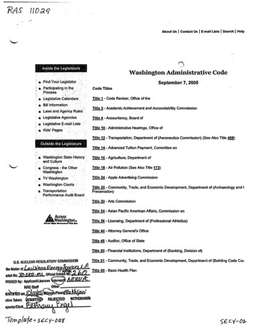
Erzsébet Csernák 1, János Molnár 2, Gábor E. Tusnády 2, Erika Tóth 1value of a given sample, both used in equal concentration. According to therecommendations, all samples with values below or equal to 2 can be selected for TSCA.Interestingly, case 1 with 1.7 Ct didn’t work; by contrast, case 14 with 2.4 Ct gave goodresult (Table 1). These minimal discrepancies can be attributed to the fact that the Ct valueonly characterizes the amplification capacity of a defined target length and does not give anyinformation if there is sufficient number of amplifiable template in wider size range, that isthe extent of fragmentation in damaged FFPE DNA. Using KAPA hgDNA Quantification andQC assay allows us to get this kind of quality information by measuring Q129/Q41 ratio. Forcalculating this value, standard curves were generated with amplifying targets of 41bp and129bp in the human genome. The relative quality can be derived by normalizing theconcentration obtained with the 129bp assay against the concentration from the 41bp assay.The value can vary between 0 and 1, depending on the sample quality. We found that aQ129/Q41 ratio below 0.2 was a negative predictor of library success. These values could alsoexplain the deviations mentioned above at cases 1 and 14, by modifying the expectedoutcome based on Ct. For sample 4, despite its poor quality (Q 0,104) the library workedwell probably due to its good amplification efficiency (Table 2). In spite of the low quality,we carried out the subsequent sequencing analysis to see its influence on final datainterpretation. Based on the 129bp assay, we also calculated a Ct value (by subtracting theexpected Ct value of a sample calculated from its concentration and standard curve, from themeasured Ct) that gave relatively small differences to Illumina QC Ct, which was likelycaused by the various lengths of targets used in qPCRs. The inconsistency of samples 1 and14 was also observed at the Q129 Ct value, which confirmed the necessity of using theQ129/Q41 ratio or a similar quality value for better selection of samples suitable forsequencing (Table 1).TSCA assay resultsSequencing metricsFull coverage was relatively high (6929 in average, range between 4764-8342) due to limitedsamples were running, but this could be corrected, and it did not affect the recovery of data.The TSCA sequencing run gave an excellent specificity, (defined as the percent of filteredreads mapping to target regions) and was very similar among formalin-fixed samples at anaverage value of 96.6%. We assessed the uniformity of target read distribution betweenamplicons and samples after duplicate removal (Figure 1). The coverage values deviated fromwhat we expected, with a range from 8.45 /- 3.77 to 21.34 /- 7.94 within the samples,6
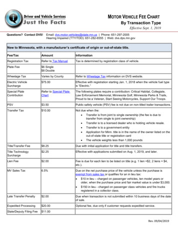
Erzsébet Csernák 1, János Molnár 2, Gábor E. Tusnády 2, Erika Tóth 1which may reflect the uneven quality of FFPE samples available for sequencing. Furthermore,this variation can be seen within the amplified regions as well (Figure 1). It should be notedthat due to the deduplication of redundant sequences (PCR duplicates) our relative coverageseems quite small (Figure 1), but this step was important to improve variant calling andeliminate bias influencing the real allelic representation of the sample. In the cases ofNRASex3, AKT1ex2, and EGFRex20 target regions, a high GC sequence content (above80%) was found near the TSCA oligo probes that likely caused this observation. However,these discrepancies did not affect the reference mapping and the subsequent variant calling.Detecting high quality variantsUsing our variant detection pipeline, we were able to identify high quality variants (Table 2).As our experience showed, it is crucial to remove the duplicated sequences, which caninterfere the variant calling by the biased read coverage (Figure 2). This was presented inother publication as well, i.e. the FFPE samples contain high number of PCR duplicateswithin the 60% - 85% range9. We made a test about the reliability of variant calling. Theselected loci (KIT ex11) from sample 11 was downsampled using the Picardtool DownsampleSam within the range of 100%-10% (the coverage was in the range from 16to 2), and we were able to identify the alternative allele when the covarege was as low as two.Using this knowledge, we achieved a high concordance in the identification of clinicallyrelevant mutations that were previously characterized by our currently used laboratory tests.In case of sample 4 we were not able to apply our strict automated variant detection pipelineto identify mutations, as it generated a proportionately high number of low-quality readsmaking it unsuitable for reliable analysis.DiscussionThe advents of next generation sequencing technologies have opened the possibility to getenormous amount of genetic information from a given sample and have a deeper knowledgeof the association between the genomic structure and function, and various biologicalprocesses or diseases. Applying this information in clinical practice has many benefitsincluding refining diagnosis, individualizing treatments, predicting drug effects or developingnew therapies.10 In light of these advantages, we considered the possibilities to replace ourcurrently used low scale diagnostic tests with a large scale NGS method. Using fifteen preselected FFPE tumor tissue samples as controls harboring clinically important mutations, weinvestigated the main criteria required to the successful adoption of NGS in clinical7
Erzsébet Csernák 1, János Molnár 2, Gábor E. Tusnády 2, Erika Tóth 1diagnostics. Basically, two target enrichment techniques can be applicable for this purpose:amplicon-based and sequence capture approaches. We chose an amplicon based method,because we focused on a small set of clinically important genes with defined hotspots. Wepreferred TruSeq custom amplicon (TSCA) assay as it has simple workflow, the entireprocedure takes only two days, thus allowing short turnaround time of reports and rapid andefficient patient management. In addition, during work with million copies of PCR amplicons,care must be taken to avoid cross-contamination between templates. In this regard, TSCAseemed to us more optimal than other amplicon based sequencing strategies, because only areduced cycling time PCR occurs when sample-specific indices are added to each library,thereby minimizing the possible presence of unwanted constituents.We tested DNA samples obtained from formalin-fixed, paraffin-embedded tissue blocks.Although this type of material has great potential in clinical medicine, due to the fixationprocess DNA can be degraded at various degrees, which can impact the reliability and qualityof genetic analyses. For NGS, high quality starting material is essential for reliable andaccurate sequencing results, therefore qualitative monitoring of FFPE DNA specimens is animportant step. Using different qPCR methods we assessed the main parameters that bestcharacterize the quality of the samples and predict the success of assay performance. Thesamples used in our experiments were obtained from a variety of tissue types and hospitalsusing different fixation and embedding protocols. As it was expected, the level of degradationand quality between samples varied over a wide range (Q: 0.052-0.91, Table 2). The bestapproach to determine the quality of DNA is proved to be a qPCR assay targeting differentsizes of genomic regions. This method gives the most precise information about DNAdegradation level. Our results show that at the moment the quality requirements are higher forNGS compared to conventional PCR techniques (sequencing was successful in 67% of ourFFPE samples), but it can be improved in the future by using more standardized protocols fortissue fixation, and DNA isolation tailoring to the particularities of FFPE material.For accurate variant detection it is essential to reach consistent uniformity and minimumrequired depth of coverage of target regions. The amplicon-based technique has the advantageof generating more uniform coverage across all target bases than sequence capture approach,however it is possible that the poor amplification of some targets mainly caused by high GCcontent as presented above. Low or no coverage regions could produce false negative results,which is not acceptable in diagnostic applications. In our case, 8% of the target ampliconswere underrepresented. With our high coverage depth, we achieved a few hundred foldcoverage in these regions, which proved to be sufficient for accurate variant calling. In the8
Erzsébet Csernák 1, János Molnár 2, Gábor E. Tusnády 2, Erika Tóth 1future more emphasis should be placed on optimal probe design to obtain more consistentcoverage and avoid false results. On the basis of our findings, for accurate variant callingneeds at least one high quality read from both the reverse and forward strand after duplicateremoval. But the minimal required raw read number/coverage is hard to tell: this dependshighly on sample quality, and more data will be needed to predict preciselyIn case of FFPE samples, it should be taken into account that the samples do not amplify inthe same quality, resulting in greater variability in the distribution of sequencing reads andpiling up high number of PCR duplicates within the 60% - 85% range. Developing analternative PCR amplification protocol could solve this problem.11A properly configured bioinformatics pipeline is essential for high confidence variantdetection. Aligned to the properties of FFPE samples, we introduced rigorous filteringparameters including duplicate removal, minimal coverage of high quality reads representingthe alternate allele, coverage from the forward and reverse sequencing strand, and highervariant quality as well. Using our strict filtering pipeline, we were able to detect short geneticvariants and filter out sequencing errors and artifacts caused by the formalin fixation. Weidentified safely the previously detected genetic variants of samples with tumor cell contentbetween 40-90%. Due to the very useful nature of sequencing, we were able to detect othergenetic variants in the selected regions as well, which enables even finer determination of thedifferent mutations.It is also important to consider when applying diagnostics tests that the quality of the samplesdetermines data quality; bioinformatics could not manage every input data optimally, as it wasseen in the case of our samples. Therefore it is essential to set up quality criteria for efficientsequencing.In summary, we described the feasibility of successful validation of targeted NGS formolecular pathology diagnosis of different hot spot mutations in FFPE samples. As thenumber of targetable genes increasing in oncology, it becomes more important to implementNGS workflows into the routine laboratory work.Disclosure/Conflict of interestThe authors declare no conflict of interest.9
Erzsébet Csernák 1, János Molnár 2, Gábor E. Tusnády 2, Erika Tóth 1References1.Zhang, W., Cui, H. & Wong, L. J. C. Application of next generation sequencing tomolecular diagnosis of inherited diseases. Top. Curr. Chem. 336, 19–46 (2014).2.Kerick, M. et al. Targeted high throughput sequencing in clinical cancer settings:formaldehyde fixed-paraffin embedded (FFPE) tumor tissues, input amount and tumorheterogeneity. BMC Med. Genomics 4, 68 (2011).3.Wong, S. Q. et al. Targeted-capture massively-parallel sequencing enables robustdetection of clinically informative mutations from formalin-fixed tumours. Sci. Rep. 3,3494 (2013).4.Williams, C. et al. A high frequency of sequence alterations is due to formalin fixationof archival specimens. Am. J. Pathol. 155, 1467–1471 (1999).5.Yost, S. E. et al. Identification of high-confidence somatic mutations in whole genomesequence of formalin-fixed breast cancer specimens. Nucleic Acids Res. 40, e107(2012).6.Flicek, P. et al. Ensembl 2014. Nucleic Acids Res. 42, (2014).7.Li, H. et al. The Sequence Alignment/Map format and SAMtools. Bioinformatics 25,2078–2079 (2009).8.McLaren, W. et al. Deriving the consequences of genomic variants with the EnsemblAPI and SNP Effect Predictor. Bioinformatics 26, 2069–70 (2010).9.Hedegaard, J. et al. Next-generation sequencing of RNA and DNA isolated from pairedfresh-frozen and formalin-fixed paraffin-embedded samples of human cancer andnormal tissue. PLoS One 9, e98187 (2014).10.Meldrum, C., Doyle, M. a & Tothill, R. W. Next-generation sequencing for cancerdiagnostics: a practical perspective. Clin. Biochem. Rev. 32, 177–95 (2011).11.Hoeijmakers, W. a M., Bártfai, R., Françoijs, K.-J. & Stunnenberg, H. G. Linearamplification for deep sequencing. Nat. Protoc. 6, 1026–36 (2011).10
Erzsébet Csernák 1, János Molnár 2, Gábor E. Tusnády 2, Erika Tóth 1Titles and legends to figuresFigure 1 Coverage distribution among the eleven samples after duplicate removalMedian coverage values were calculated for every amplified region within one sample. Forevery amplified region the average value was determined from these coverage data among thesamples, and the standard deviation was calculated as well. The blue bars represent theaverage values for the amplified region, and the standard deviation values are visualized aswell11
Erzsébet Csernák 1, János Molnár 2, Gábor E. Tusnády 2, Erika Tóth 1Figure 2 Example of sequence resultsManual inspection by the IGV program (Robinson et al. 2011) of the NGS reads (a) and thesequencing chromatograms from Sanger method (b) shows the same genotypes. In the case ofNRAS the Sanger sequencing data is from the sense strand, which is reversed to the humangenomic reference sequence12
We used TruSeq Custom Amplicon assay with the Illumina Miseq instrument. We expanded our currently used gene panels with new marker genes using Illumina DesignStudio which contains 3,493 Kb of cumulative sequence and 36 amplicons (Supplemental Table). The oligo probes targeted the hotspot regions of clinically relevant oncogenes including