Transcription
JRCR - Author & Reviewer Guidelines - Version 5.0JRCR – Author & Reviewer GuidelinesAUTHOR GUIDELINESSome general information: Before submitting a manuscript, please make sure thatsuch topic has not been already published in the JRCR. The following applies for case reports (not case seriesor review articles):Many submitting authors erroneously think their case israre or case report worthy and thus their manuscriptshad to be declined - despite being well written up. Toavoid a lot of work/preparation and disappointment,authors first need to upload their case on our affiliatededucational Radiology community Radiolopolis [4,5]and open that case for discussion. We will thendetermine the level of interest and invite the author forsubmission if appropriate. Case upload needs to bedonein ups/40 Authors, who obtained a priority pass are exempt fromthe requirement to upload and discuss first their case inour Radiology community and may submit immediatelytheir manuscript, without invitation. Read more aboutthe priority pass gycases/announcement/view/18 Only invited manuscript submissions of case reports orfrom authors who obtained a priority pass are eligiblefor review and processing. If the manuscript is written in poor English or morethan 5 typos are present (as simple as a commapreceded by an empty space), this demonstrates a notthorough manuscript preparation and will beimmediately declined. This applies to ALL sections inthe manuscript, including the tables, and multiplechoice questions. A simple Word spell- and grammarcheck can often prevent such situations. Only TWO files are accepted for initial submission: theWord file with the COMPLETE manuscript and the zipfile, containing the high quality figures. For revisions, do NOT upload single image files –image files may only be contained within thefigures.zip file or the individual stack zip files.These guidelines are effective: July 1, 2014Manuscript templateIt is MANDATORY for ALL manuscripts to use themanuscript template. The manuscript template mightcontain important information necessary which might nothave been covered in this article. This templateguarantees that the author does not miss any requiredinformation and also that the manuscript will be providedin the correct format. Any submissions which do NOTuse the provided manuscript template will be immediatelyREJECTED – independent of provided contents andquality of the work.Authors are REQUIREDto use the manuscript template!Download manuscript template: Manuscript (URL:http://www.radiologycases.com/Template Case Report.doc )MANUSCRIPT TYPESPlease adhere to the following format:Case ReportProvideonedocumentincluding:1. Cover page, (see cover page section). Cover page is theFIRST page of the manuscript Word document.2. Manuscript, containing: Title Abstract Case Report No introduction is necessary. Ifprovided, it should be embedded into thediscussion. Discussion Teaching Point References Figures & Legends Tables Abbreviations Questions & Answers pertinent to the articleAgain: usage of the manuscript template isMANDATORY!Download manuscript template: click here to downloadTo better understand the review process and to help,writing a proper stylistic and sound manuscript, you maytake a look which criteria the reviewers evaluate.Download reviewer template: Review (Reviewerguidelines)1www.RadiologyCases.comJournal of Radiology Case ReportsWe receive an increasing number of submissions whichkeep our editorial team and our reviewers to their limits.Therefore, we have to be increasingly selective whichsubmission to forward for review - to keep also theworkload for our reviewers reasonable. For this reason itis at least required that the submission 100% complieswith our author guidelines. ALL manuscripts that doNOT comply with the new author guidelines will beimmediately rejected – without initial review.
JRCR - Author & Reviewer Guidelines - Version 5.0Pictorial review articleTo emphasize our educational goal of this journal, wenow also accept pictorial review articles. These articlesglance by providing an abundance of image examples ofthe discussed topic, including educational information tounderstand the etiology, findings, and implications for thedaily practice.Provideonedocumentincluding:1. Cover page, including title, authors (with mail andemail addresses) and disclosures. Cover page is theFIRST page of the manuscript Word document.2. Manuscript, containing: Title Abstract Review section including discussion andmultiple imaging examples. Discussion shouldinclude etiology, detailed imaging findings andhow to implement this knowledge into the dailyRadiology practice. Teaching point References Figures and figure legends with good descriptionof findings and technique Abbreviation section Summary table Questions & Answers pertinent to the article“Signs in Radiology” ManuscriptThe format is similar to a pictorial review article andshould contain:Provideonedocumentincluding:1. Cover page, including title, authors (with mail andemail addresses) and disclosures. Cover page is theFIRST page of the manuscript Word document.2. Manuscript, containing: Title Abstract Discussion with several imaging examples - onefigure is not enough! Discussion should includehistoric background, detailed imaging discussionThese guidelines are effective: July 1, 2014 and potentially differential diagnoses and how todifferentiate them on imaging.Teaching pointReferencesFigures and figure legends with good descriptionof findings and techniqueAbbreviation sectionSummary tableDifferential tableQuestions & Answers pertinent to the articleOriginal WorkProvideonedocumentincluding:1. Cover page, (see cover page section). Cover page is theFIRST page of the manuscript Word document.2. Manuscript, containing: Title Abstract Case Report & Discussion Introduction, Materials & Methods, Results,Discussion, Conclusion (for original work) Teaching Point References Figures & Legends Tables Abbreviations Questions & Answers pertinent to the articleMANUSCRIPT STRUCTUREThe entire manuscript should contain the cover page asfirst pages, followed by the main manuscript section.Cover pageThe cover page should include: Manuscript titlePlease provide a meaningful title and avoid phraseslike "rare case" - if it would not be rare, it wouldnot be case report worthy in the first place. Titleshould e.g. contain the diagnosis. Author listIn chronologic appearance and affiliation.Corresponding author is marked with a “*”.Example: John Doe1, Maria Martinez2*, MaxSchmidt3, Gerard Rouge4ALL authors also need to be entered in the onlinemeta data, including their contact and institutionalaffiliation. Authors (with first and last names)o In the order of author appearance as wished in thepublished article.o Each with institution, complete postal mail andemail address (start with primary author). Thisneeds to be entered also online in the metadatasection on the submission website.2www.RadiologyCases.comJournal of Radiology Case ReportsCase SeriesProvideonedocumentincluding:1. Cover page, (see cover page section). Cover page is theFIRST page of the manuscript Word document.2. Manuscript, containing: Title Abstract Case Report & Discussion Introduction, Case descriptions/Main text,Discussion, Conclusion Teaching Point References Figures & Legends Tables Abbreviations Questions & Answers pertinent to the article
JRCR - Author & Reviewer Guidelines - Version 5.0o The EXACT information (full author names,affiliations etc.) has to be also provided in theonline metadata section of the manuscript.o Do NOT use ALL CAPS ON when typing. Eachname starts with an upper case, followed by lowercase letters. Authors' contributionsPlease describe the contributions to this work fromeach individual author. There is a maximum of 5authors for case reports. AcknowledgementsIf you would like to thank a particular person. ConsentDid the author obtain written informed consentfrom the patient for submission of this manuscriptfor publication? (Answer with yes or no.) Human and animal rightsIf reporting experiments on human or animalsubjects, please indicate if ethical standardsfollowed the responsible committee on humanexperimentation (institutional and national) andwith the Helsinki Declaration of 1975, as inesinterests.html#Rights).Main manuscript section No author name or other information, which couldunveil the identity of the authors should be includedin this part. Please do English spell- and grammar-check beforesubmitting the manuscript for review. If the authorsare not fluent in English, it is advised to proofreadthe manuscript by an English proficient person.Articles written in poor English will be immediatelyrejected.AbstractThe abstracts should not exceed 1000 characters(including empty spaces). Please do not use references orabbreviations in the abstract.Abstract should provide a quick summary of thecase/contents and that a comprehensive literature reviewand discussion about the entity will be provided.DiscussionDiscussion includes detailed information aboutetiology, demographics, imaging findings on all imagingmodalities, treatment and differential diagnoses of thatfinding. If references used, please in squared brackets,e.g. [1,2] and before the sentence point. Do not put anempty space between references)These guidelines are effective: July 1, 2014Teaching PointTeaching point should explain the educational value ofthis article in max. 2 sentences. It is the take-homemessage and should be even clear, independent ofhaving read the manuscript or not. It should include theimaging findings of the presented entity and should NOTbe specific for the presented case but rather general inregard to the described entity. Do NOT use phrases like"This report" or "We demonstrate".ReferencesThe authors are responsible for the accuracy of thebibliographic information. A minimum of 5 references isrequired. References should be numbered consecutivelyin the order in which they are first cited in the manuscript.There is no maximum of references. Please adhere to therequired reference format. (e.g. Smith A, Miller B, JonesC. Title of the article. Journal and issue. PMID: #) Noreferences are permitted in the title, abstract and question& answer section.References given in tables or figure legends must benumbered in sequence with those in text. Periodical titlesshould be abbreviated in the style of Index Medicus. Issuenumbers and inclusive page numbers should be given forall references. If there are six or less authors, so pleaselist surnames and initials of all authors as following:1. Hirsch W, Paetzel M, Talanow R. www.PedRad.info,the first bilingual case-oriented publication platform forpediatric radiology. Pediatr Radiol. 2005 Mar;35(3):3448.If there are seven or more authors, please list only thefirst three names, followed by “et al.” In the case ofbooks, the authors of a chapter, title of the chapter,editor(s), title of the book, edition, city, publisher, year,and chapter pages must be provided:1. Paetzel M. Thorax. In: USMLE Help Step 1 AnatomyQ & A. 1st ed. Cleveland Heights: EduRad, 2007; 316.2. Haaga JR, Lanzieri CF, Sartoris DJ, Zerhouni EA.Neoplastic Diseases of the Lung. In: ComputedTomography And Magnetic Resonance Imaging Of TheWhole Body. 3rd ed. St. Louis; Mosby,1994; 724-729.3www.RadiologyCases.comJournal of Radiology Case Reports DisclosuresIf, please explain who & what kind of disclosure(e.g. financial, competing interest, etc.). The discussion needs to contain at least the followingsubsections preceded by the respective subheader:o Etiology & demographicso Clinical & imaging findingso Treatment & prognosiso Differential Diagnoses If the discussion mentions "rare" or some otherindicator about a frequency, then please provide thenumbers (e.g. incidence/prevalence, percentage etc.) Differential diagnoses need to be separated by a subsubheader in the differential diagnosis section.
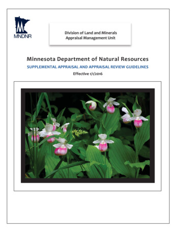
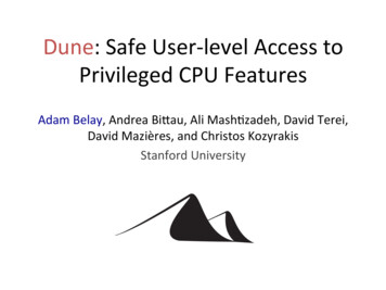
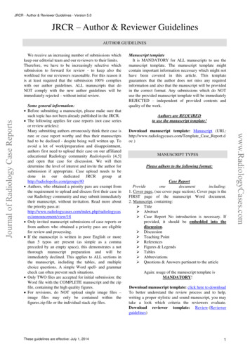
JRCR - Author & Reviewer Guidelines - Version 5.0Author Citations of Online-only ArticlesOnline-only article citations should include the authors’names, title of the article, journal or website title, year,volume, and issue of publication, the complete URL forthe document and the date the document was accessed orconsulted:1. Gossner J. Lissencephaly Type 1. PedRad [serialonline]. 2008; vol 8, no. 5. Available at:www.PedRad.info/?search 20080522125523. AccessedJune 10, 2008.Reference format is now required as following (example):Roentgen A, Hounsfield G, Fourier J. Title. RadiologyCase. 2011 Jan;5(1):30-31. PMID: 12345How to get the PubMed ID?Please go on http://www.pubmed.org and enter yourreference into the search box. If the reference is correct,the abstract you will then see the abstract of thatreference. The PubMed ID is located to the left bottom ofthe abstract and displayed in the format “PMID: #”, with“#” being the PubMed ID (Figure 1, red box and arrow).Figure 1. How to get the PubMed ID for an article: ThePubMed ID is located to the left bottom of the abstractand displayed in the format “PMID: #”, with “#” beingthe PubMed ID (red box and arrow). URL:www.pubmed.orgIf the reference is not entirely correct, PubMed willdisplay a list of similar articles found in their database.Each listed article has its PubMed ID displayed left belowits reference ID (Figure 2, red box and arrows). Pleasecopy this PubMed ID and add it to the end of yourreference as seen above. Of course, please provide thereference also correctly (no typos etc.)These guidelines are effective: July 1, 2014Figure 2. How to get the PubMed ID for an article: If thereference is not entirely correct, PubMed will display alist of similar articles found in their database. Each listedarticle has its PubMed ID displayed left below itsreference ID (red box and arrows). URL:www.pubmed.orgIf the reference is a book, the ISBN number has to beadded at the end of the reference. This could be either theISBN-10 or ISBN-13. An example:Haaga JR, Lanzieri CF, Sartoris DJ, Zerhouni EA.Neoplastic Diseases of the Lung. In: ComputedTomography And Magnetic Resonance Imaging Of TheWhole Body. 3rd ed. St. Louis; Mosby,1994; 724-729.ISBN: 1234567890FiguresAny imaging modality, which was mentioned in themanuscript, has to be provided as figure. Please providealso multiple views/planes of the same modality.Pathologic correlation is also necessary if it wasperformed (e.g. operative, macroscopic, microscopicimages). Figures should be either submitted as PNG oruncompressed JPG files. All figures need to be deidentified/anonymized. Figures embedded in PowerPointare not accepted. The figures should be initiallyimplemented into the Word file, but have to be uploadedaltogether in a zip file for the high quality final version.(Do not upload each figure as separate supplementaryfile) Images need to be also annotated to highlight thefindings (arrow, asterisk etc.). Annotations, created inWord and not saved as separate figure are not accepted.Reason for that is that the annotations might shift duringthe editing process for the review version. Please avoidtoo much black background by cropping the figuresappropriately.If the findings are not obvious, pleaseprovide in addition magnified (sub)figures. There is nolimitation of submitted images.Each provided image needs to be a separate figure andeach figure needs a figure legend. Subfigures need to becombined to one single image and labeled accordingly4www.RadiologyCases.comJournal of Radiology Case ReportsIn the past, some references were not provided in thecorrect format by some authors. This might have been dueto copying from an erroneous source or mistyping. Toguarantee a correct reference and linking for our readerswe now require from the authors to provide the PubMedID at the end of the reference.
JRCR - Author & Reviewer Guidelines - Version 5.0Annotated figuresALL figures need annotations (e.g. arrows, asterisk)and these need to be explained in the respective figurelegend. Figures HAVE to be annotated (arrows, asterisketc. – NO text) and saved in an external image editor –BEFORE embedding into the Word document. Creationof annotations on the images within the Word documentis not permitted. These annotations might shift during theeditorial process and distort the figure.Figure legends and stack legends need the followingformat: Age, gender and diagnosis in one sentence andFindings and Technique, each separated by a line breakand preceded with "Findings" or "Technique".Figure zipsThe zip file containing ALL figures provided in themanuscript have to be provided in HIGH quality and thebest resolution within ONE single zip file. This zip fileshould be labeled “Figures.zip” and uploaded as asupplemented file at the time of initial manuscriptsubmission.Figure legends & image stack legendsFigure legends need to be below the respectivefigure. Figure and image stack legends need to be splitinto three sections: “Age, gender, diagnosis”, Findingsand Technique. (An example can be seen below.)Figure/stack legends have to contain patient age,gender, diagnosis, and especially imaging techniqueused and a good description of the imaging findings.Figure/stack legends need detailed protocol informationabout the study. E.g. MRI: magnet strength, whatsequence (TR, TE), plane, contrast type and dose. Inwhich phase was the study obtained (arterial, venous,delayed etc.) Same applies to CT and Nuclear Medicinestudies (in addition: what radiopharmaceutical was given,which dose, at what time was imaging obtained.) E.g.:52 year old female with left internal carotid arterydissection.FINDINGS: Axial contrast enhanced CT of the neck inthe arterial phase demonstrates a dissection flap (arrow)in the left internal carotid artery.TECHNIQUE: Axial CT, mAs, kV, mm slicethickness, ml name contrast material) Please provide three-dimensional measurements formeasurable imaging findings.These guidelines are effective: July 1, 2014TablesRequired tables: summary table and differential table.These tables include condensed information found inthe manuscript discussion section. ALL informationprovided in the tables HAS to be included in thediscussion section of the manuscript. All tables need atable legend explaining what the respective table is about.Summary table: contains high yield information aboutthe reported entity. Some mandatory fields are: etiologyincidencegender ratioage predilectionrisk factorstreatmentprognosisfindings on imaging (can be adopted from thedifferential table)Differential table: contains differential diagnoses of thereported entity (including the entity itself). Eachdifferential diagnosis belongs in a separate rowrespectively and each imaging modality belongs in aseparate column, including pertinent imaging findings.Imaging modalities include: X-Ray US CT MRI - T1 MRI - T2 MRI - DWI Pattern of contrast enhancement (avid, none,homogeneous, heterogeneous etc.) Scintigraphy PETBoth tables need to be completed with up-to-dateknowledge found in the current literature. Do NOTsubmit these tables as lists or images – these need to beprovided as true tables.Table contents in discussionThe summary and differential tables provide a quickoverview about the presented entity and it’s differentialdiagnoses. However, ALL information provided in thetables HAS to be also included in the discussion sectionof the manuscript.AbbreviationsAny abbreviation used in the article should be writtenout. (e.g. HTN Hypertension). Abbreviations need to bespelled out the first time mentioned in the manuscript. Noabbreviations are permitted in the title, abstract orquestion & answer section.5www.RadiologyCases.comJournal of Radiology Case Reports(1a, 1b, 1c etc.). Otherwise they need to be listed asseparate figures (1, 2, 3 etc.) – including separate figurelegends. Otherwise these figures need to be separatelyand chronologically numerated (Figure 1, 2, 3 etc.)including their respective figure legend.
JRCR - Author & Reviewer Guidelines - Version 5.0QuestionsPlease provide 5 multiple choice questions - targeted tothe knowledge provided in the submitted manuscript.Each question should have 5 answer choices. The answersmay be either single best answer (only one correct) orcontain several correct answers (more than one correct).Only mark the choices that apply to the question with“(applies)” (not “true”, “wrong”, “false” etc. – it has to be“applies”). Furthermore, an explanation follows thequestion and answers, explaining why the answer choicesare correct (or wrong). The appropriate sentences in thearticle need to be cited in squared brackets “[]” to guidethe reader to the appropriate section in the article. Thequestions and answers should be understandable for thereader – even without having read the manuscript. Do notrepeat the answer choices in the explanation. Do notrepeat the same explanation per question (rather combineexplanations for several choices that share the sameexplanation).Question and Answer format:Question 1Answer choice 1Answer choice 2Answer choice 3Answer choice 4 (applies)Answer choice 5Explanation for question 1 (The appropriate sentence/s inthe article need to be cited in squared brackets “[]” toguide the reader to the appropriate section in thearticle.) this needs to be done five times ( 5 individualquestions)One example:Applies to article: Bryce Y, Wood B, Baron P, Gibbs L.Radiology Case. 2008 Oct; 2(4):18-23. An unusualcongenital hepatic cyst in an adolescent and review ofdifferential diagnoses of complex liver ologycases/article/view/55)Question: Which of the following answer choices is false?1. Simple hepatic cysts are congenital lesions.2. They measure plasma density on CT imaging.3. Hemorrhagic hepatic cysts are hypoechoic onultrasound. (applies)4. Complications of hepatic cysts might include ruptureinto the peritoneum and hemorrhage.5. Cysts demonstrate T1 hypointensity and T2hyperintensity.These guidelines are effective: July 1, 2014Explanation:1. [Simple hepatic cysts are congenital lesions, usuallylined with biliary-like epithelium, secreting a fluidsimilar to plasma.]2. [Simple hepatic cysts are congenital lesions, usuallylined with biliary-like epithelium, secreting a fluidsimilar to plasma.]3. [If there is hemorrhage within the cyst, typically theultrasound shows hyperechoic fluid.]4. [Rare complications of simple hepatic cysts are rightupper quadrant abdominal pain or discomfort, earlysatiety, hemorrhage within the cyst, infection,intraperitoneal rupture.]5. [On MRI, simple cysts are hypointense on T1-weightedimages and hyperintense on T2.]Image stacks for the Interactive Viewing ModeLatest after acceptance of the manuscript forpublication, the author has to provide for each crosssectional modality (CT, MRI, US - also PET, SPECT) astack of images – ideally in multiple planes for eachmodality. The stack of images should be numeratedchronologically, e.g. first image starts with "1", e.g. 1.jpg,2.jpg, 3.jpg, . n.jpg. (images have to be JPG files – NOTTIF or BMP files!) All images of an individual sequenceare collected in a separate zip-file. Also add a text orWord file, containing a description of the image stack(modality/sequence, plane, contrast or not, significantfindings). For sequential studies, such as fluoroscopy (IRor GI) or nuclear medicine studies (e.g. gastric emptying,HIDAA scan etc.) please provide also all images for theparticular sequence. These images should be alsocollected in a separate zip-file. An example of such animage stack zip file can be downloaded here k example.zip - Only parts of the abdominal CT are saved in thisexample zip to save you download time, however theauthor is required to submit the entire exam.) Pleaseinclude also the topogram/localizer image in case of CTor MR exams. The zip-file should be uploaded in thesubmission section as a supplementary file. Eachmodality and plane is ONE zip file! If g.Only case reports which provide stacks of images for theinteractive mode will receive a DOI number!Disclosures:Each submitted article needs written clarification about: Conflict-of-Interest (click here to read details) Informed Consent (click here to read details) Human and Animal Rights (click here to readdetails)These statements are provided in the cover page of themanuscript template.6www.RadiologyCases.comJournal of Radiology Case ReportsKeywords A minimum of 5 keywords related to the case reportis necessary Keywords should include the diagnosis itself,synonyms, eponyms and alternative names (somediagnoses run under several names), affected bodyregion, modality used, Mesh terms
JRCR - Author & Reviewer Guidelines - Version 5.0What to focus on when writing?To better understand the review process and to help,writing a proper stylistic and sound manuscript, you mayalso take a look which criteria the reviewers evaluate.Downloadreviewertemplate:Review (URL:http://www.radiologycases.com/Template Reviewer Case Report.doc )www.RadiologyCases.comJournal of Radiology Case ReportsThe reviewer guidelines can be found eviewerguidelines)These guidelines are effective: July 1, 20147
JRCR - Author & Reviewer Guidelines - Version 5.0REVIEWER GUIDELINESThe reviewer guidelines can be found eviewerguidelines)1.Is the manuscript written in proper and soundEnglish?2.Does the author adhere to the format according to theauthor guidelines? (e.g. Case report: Title, Abstract,Case Report, Discussion, Teaching Point,References, Figures, Tables, Abbreviations)3.This is a medical imaging focused journal. Are therepertinent and enough imaging modalities provided?4.Is any imaging modality, which has been mentionedin the article, available as figure?5.Do the figure legends contain patient age, gender,diagnosis, imaging technique used and a gooddescription of the imaging findings?6.Does this manuscript offer new knowledge aboutknown medical/radiological entities or reports new,unusual or unexpected findings?7.Does the manuscript describe a rare finding?(Applies to a general valence/incidence, risk factors, treatment,prognosis of the discussed entity AND differentialdiagnoses (DDs) and how to differentiate the entityfrom the DDs?9.Especially responding to the review request within therequired deadline AND providing the review within therequested deadline are of uppermost importance – inrespect to our authors who are eagerly waiting for adecision. Our reviewers are evaluated by an automaticrating system on a 5 point scale. Rating is determined bytimely response and quality of the review. One point (outof five) will be deducted if not responded by therequested deadline. If a reviewer does not respond 3times within one year he/she will be automaticallyremoved from our system.As an appreciation of their valuable contribution, eachreviewer who receives a total score of 3.5 or higher (outof 5), will be honored in our hall of fame and yearlyspecial journal issue.URL of this Published by EduRadLevel of interest and urgency10. Recommendation (e.g. accept, revisions required,decline)www.EduRad.org11. Other comments: (e.g. what can be done to improvethis manuscript?) This is mandatory!These guidelines are effective: July 1, 20148www.RadiologyCases.comJournal of Radiology Case ReportsReviewing CriteriaReviewer ratingWe appreciate the time and effort our reviewers donateto improve the quality of this journal. As an appreciation,we honor our reviewers who comply with our reviewguidelines in our hall of fame and our special yearlyissue. However, we require that all reviewers comply100% with our reviewer guidelines which are available ycases/about/editorialPolicies#peerReviewProcess.
These guidelines are effective: July 1, 2014 JRCR - Author & Reviewer Guidelines - Version 5.0 rts yCases.com preceded by an empty space), this thorough manuscript preparation and will be immediately declined. This applies check can often prevent such Word file with the COMPLETE manuscript and the zip w 1 AUTHOR GUIDELINES MANUSCRIPT TYPES