Transcription
JOURNAL OF NEUROTRAUMAVolume 21, Number 10, 2004 Mary Ann Liebert, Inc.Pp. 1457–1467Dietary Omega-3 Fatty Acids Normalize BDNF Levels,Reduce Oxidative Damage, and Counteract LearningDisability after Traumatic Brain Injury in RatsAIGUO WU,1 ZHE YING,1 and FERNANDO GOMEZ-PINILLA1,2ABSTRACTOmega-3 fatty acids (i.e., docosahexaenoic acid; DHA) regulate signal transduction and gene expression, and protect neurons from death. In this study we examined the capacity of dietary omega3 fatty acids supplementation to help the brain to cope with the effects of traumatic injury. Ratswere fed a regular diet or an experimental diet supplemented with omega-3 fatty acids, for 4 weeksbefore a mild fluid percussion injury (FPI) was performed. FPI increased oxidative stress, and impaired learning ability in the Morris water maze. This type of lesion also reduced levels of brainderived neurotrophic factor (BDNF), synapsin I, and cAMP responsive element-binding protein(CREB). It is known that BDNF facilitates synaptic transmission and learning ability by modulating synapsin I and CREB. Supplementation of omega-3 fatty acids in the diet counteracted all ofthe studied effects of FPI, that is, normalized levels of BDNF and associated synapsin I and CREB,reduced oxidative damage, and counteracted learning disability. The reduction of oxidative stressindicates a benevolent effect of this diet on mechanisms that maintain neuronal function and plasticity. These results imply that omega-3 enriched dietary supplements can provide protection againstreduced plasticity and impaired learning ability after traumatic brain injury.Key words: BDNF; fish oil; hippocampus; learning; traumatic brain injuryINTRODUCTIONT(TBI) is a major cause ofdisability such that a great concern exists to developmeans to decrease its short- and long-term effects. Dietary factors are emerging as an efficient means to modulate the capacity of the brain for plasticity (Mattson etal., 2003; Wu et al., 2004). Docosahexaenoic acid (DHA;C22: 6n-3), one of the major omega-3 polyunsaturatedfatty acids in the brain, has shown to be essential for norRAUMATIC BRAIN INJURYmal neurological development, maintenance of learningand memory, and neuronal plasticity (Green and Yavin,1998; Hashimoto et al., 2002; Salem et al., 2001). DHAcan affect neural function by enhancing synaptic membrane fluidity and function (Jump, 2002), regulating geneexpression (Duplus et al., 2000; Ikemoto et al., 2000; Kitajka et al., 2002; Puskas et al., 2003; Salem et al., 2001),mediating cell signaling (de Urquiza et al., 2000; Jump,2002; Vaidyanathan et al., 1994;), and enhancing longterm potentiation (LTP) (McGahon et al., 1999). Dietary1Department of Physiological Science, University of California at Los Angeles, and 2Division of Neurosurgery, UCLA BrainInjury Research Center, Los Angeles, California.1457
WU ET AL.DHA supplementation improves learning ability (Limand Suzuki, 2001; Suzuki et al., 1998) and enhances longterm memory in both young and old animals (Gamoh etal., 1999, 2001; Lim and Suzuki, 2000), and can reducecognitive decay during aging and Alzheimer’s disease(Hashimoto et al., 2002). Therefore, it is possible that select dietary components ingested at the appropriate timecan be used to overcome some of the deleterious effectsof TBI on brain function.It is well accepted that cognitive dysfunction is a prevalent consequence of TBI in humans (Borgaro et al., 2003;Piot-Grosjean et al., 2001) and animals (Fox et al., 1998;Smith et al., 1995; Wu et al., 2003). TBI is associatedwith a long-lasting decrement in the capacity of the brainto cope with future insults, and often with a reduced ability of individuals to maintain higher cognitive and intellectual function. Although cells may undergo differentialdegrees of degeneration, the large majority of neuronssurvive the primary insult (Hovda et al., 1995). It is,therefore, believed that neuronal death by itself cannotcompletely explain the chronic functional problems experienced by TBI patients (Lyeth et al., 1990; Scheff etal., 1997), and that these problems may reside in disrupted molecular mechanisms governing synaptic transmission.The BDNF system enhances the function and viability of select neuronal populations, and its action appearsto be crucial for maintaining molecular processes underlying cognitive function. BDNF promotes neuronal excitability (Bolton et al., 2000; Kafitz et al., 1999) and facilitates synaptic transmission (Kang and Schuman,1996; Levine et al., 1998; Sherwood and Lo, 1999; Tylerand Pozzo-Miller, 2001), and hippocampal BDNF seemsnecessary for the induction of LTP (Korte et al., 1995;Linnarsson et al., 1997; Patterson et al., 1996). BDNF issynthesized predominantly by neurons located in the hippocampus, a brain region intimately associated with theprocessing of cognitive function (Drapeau et al., 2003;Kelly et al., 2003; Steffenach et al., 2002; Sugaya et al.,1996). Synapsin I is a nerve terminal phospho-proteininvolved in neurotransmitter release, axonal elongationand maintenance of synaptic contacts (Brock andO’Callaghan, 1987; Wang et al., 1995), whose synthesis(Wang et al., 1995) as well as phosphorylation (Jovanovicet al., 1996) are affected by BDNF. CREB, a transcription factor involved in learning and memory, is an important modulator of gene expression induced by BDNF(Finkbeiner, 2000). It is possible that TBI can compromise the BDNF system, weakening the molecular substrates for maintaining neuronal function. We also hypothesize that enhanced function of the BDNF system byomega-3 fatty acids can be an effective therapy to reducecognitive impairment after TBI.MATERIALS AND METHODSExperimental Design and Tissue PreparationMale Sprague-Dawley rats (n 48, Charles RiverLaboratories, Inc., Wilmington, MA) weighing between200 and 240 g were housed in cages (two rats per cage)and maintained in environmentally controlled rooms(22–24 C) with a 12-h light/dark cycle. After acclimatization for 1 week on standard rat chow, the rats were randomly assigned to regular diet (RD; 0.9% DHA and 1%eicosapentaenoic acid (EPA). In this diet total fat content, 4.5%; total saturated fat, 1.5%; total monounsaturated, 1.58%) or diet containing 8% fish oil (FO; 12.4%DHA and 13.5% EPA. In this diet total fat content:10.4%; total saturated fat, 2.8%; total monounsaturated,2.29%) for 4 weeks. Omega-3 fatty acids from FO havebeen shown to provide beneficial effects on rodent brain(Kitajka et al., 2002; Puskas et al., 2003). The diets, fedad libitum, were provided in powder (TestDiet Inc., Richmond, IN) in a large bowl and contained a standard vitamin and mineral mix with all essential nutrients. After4 weeks of feeding with RD or FO diet, a subgroup ofrats was exposed to mild fluid percussion injury (FPI).After consumption of the same diet for 1 week post-injury, rats (n 6–8 within each group) were killed by decapitation. The brains were rapidly dissected and frozenon dry ice, and stored at 70 C until use for biochemical analyses including ELISA, western blot, and measurement of oxidized protein levels by Oxyblot. For immunohistochemistry, the rats (n 4 within each group)were transcardially perfused with 400 mL of 4%paraformaldehyde and 100 mL of 30% sucrose. The fixedbrains were then removed and stored at 70 C until use.All experiments were performed in accordance with theUnited States National Institute of Health Guide for theCare and Use of Laboratory Animals and were approvedby the University of California at Los Angeles Chancellor’s Animal Research Committee. The suffering andnumber of animals used were minimized.Fluid Percussion InjuryThe injury was performed as previously described (Wuet al., 2003). In brief, with the aid of a microscope (Wild,Heerburg, Switzerland) a 3.0-mm diameter craniotomywas made 3.0 mm posterior to bregma and 6.0 mm lateral (left) to the midline with a high-speed drill (Dremel,Racine, WI). A plastic injury cap was placed over thecraniotomy with silicone adhesive and dental cement.When the dental cement hardened, the cap was filled with0.9% saline solution. Anesthesia was discontinued andthe injury cap was attached to the fluid percussion device. At the first sign of hind-limb withdrawal to a paw1458
FISH OIL DIET NORMALIZES BDNF AND COGNITION IN TBIpinch, a mild fluid percussion pulse (1.5 atm) was administered. Sham animals underwent an identical preparation with the exception of the lesion. Immediately uponresponding to a paw pinch, anesthesia was restored andthe skull was sutured. Neomycin was applied on the suture and the rats were placed in a heated recovery chamber for approximately an hour before being returned totheir cages.Measurement of Oxidized ProteinsThe amounts of oxidized proteins containing carbonylgroups were measured by using an Oxyblot kit (Intergen,Purchase, NY). Briefly, the protein sample (10 g) fromhippocampal tissue was reacted with 1 dinitrophenylhydrazine (DNPH) for 15 min, followed by neutralization with a solution containing glycerol and -mercaptoethanol. These samples were electrophoresed on an 8%polyacrylamide gel and electrotransferred to a nitrocellulose membrane. After blocking, membranes were incubated overnight with a rabbit anti-DNPH antibody(1:150) at 4 C, followed by incubation in goat anti-rabbit (1:300) for 1 h at room temperature. After rinsing withbuffer, the immunocomplexes were visualized by chemiluminescence using the ECL kit (Amersham PharmaciaBiotech Inc., Piscataway, NJ) according to the manufacturer’s instructions. The Oxyblot bands were all groupedtogether in each group and then analyzed by NIH imagesoftware.Cognitive TestingThe cognitive testing was performed in a water mazeas described previously (Molteni et al., 2002; Wu et al.,2003) at day 5, 6, and 7 after surgery. The swimmingpool (130 cm diameter, 50 cm height) was divided intofour quadrants delimiting separate zones. The quadrantwhere the escape platform (12 cm diameter) was locatedin a fixed position with 2 cm under the water surface wasdefined as target zone; the other three quadrants were left,right and opposite zone. The water (22 2 C) was madeopaque with white nontoxic biodegradable dye to preventthe rats from seeing the platform. The rats were trainedin the water maze with 10 consecutive trials per day for3 days. The rats were placed into the tank facing the wallfrom one of the four equally spaced start locations thatwere randomly changed every trial. The spatial cues forreference around the pool were maintained constantthroughout the duration of the experiment. Each triallasted until the rat found the platform or for a max of 2min. If the rat failed to find the platform in the allocatedtime, it was gently placed on the platform. At the end ofeach trial, the animals were allowed to rest on the platform for 1 min. The behavioral variables including swim-ming distance (cm) and spent time (s) were recorded withthe computer-controlled Smart Video tracking system(San Diego Instruments, San Diego, CA).ELISAHippocampal tissue was homogenized in a lysis buffercontaining 137 mM NaCl, 20 mM Tris-HCl pH 8.0, 1%NP40, 10% glycerol, 1 mM PMSF, 10 g/mL aprotinin,0.1 mM benzethonium chloride, 0.5 mM sodium vanadate. The homogenates were then centrifuged, the supernatants were collected and total protein concentration wasdetermined according to MicroBCA procedure (Pierce,Rockford, IL), using bovine serum albumin as standard.BDNF protein was quantified using an enzyme-linkedimmunosorbent assay (ELISA) kit (BDNF Emax ImmunoAssay System kit, Promega Inc., Madison, WI) according to manufacturer’s protocol.Western BlotThe total proteins from hippocampal tissue were extracted as described above. Synapsin I and CREB wereanalyzed by western blot. Briefly, protein samples wereseparated by electrophoresis on an 8% polyacrylamidegel and electrotransferred to a nitrocellulose membrane.Non-specific binding sites were blocked in TBS,overnight at 4 C, with 2% BSA and 0.1% Tween-20.Membranes were rinsed for 10 min in buffer (0.1%Tween-20 in TBS) and then incubated with anti-actin,anti-synapsin I (1:2000, Santa Cruz Biotechnology, SantaCruz, CA), followed by anti-goat IgG horseradish peroxidase-conjugate (Santa Cruz Biotechnology); antiCREB (1:1000; Cell Signaling Technology, Beverly,MA), followed by anti-rabbit IgG horseradish peroxidase-conjugate (Santa Cruz Biotechnology). After rinsing with buffer, the immunocomplexes were visualizedby chemiluminescence using the ECL kit (AmershamPharmacia Biotech Inc., Piscataway, NJ) according to themanufacturer’s instructions. The film signals were digitally scanned and then quantified using NIH Image software. Actin was used as an internal control for westernblot such that data were standardized according to actinvalues.ImmunohistochemistrySerial coronal sections (25 m) were cut on a cryostat, mounted to gelatin-coated slides and processed forimmunohistochemistry, as previously described (GomezPinilla et al., 2001). A 1:1000 dilution was used for therabbit polyclonal anti-BDNF (Chemicon InternationalInc., Temecula, CA). Immunohistochemistry controlswere performed by omission of the primary antibody. The1459
WU ET AL.results of immunohistochemistry controls were negativeas no staining was observed in cell structures.Statistical AnalysisActin was employed as internal standard for westernblot. The RD rats with sham surgery were regarded asexperimental controls for comparisons with other experimental groups. For Western blot, the values were expressed as a ratio of actin value and then converted topercent of Sham-RD group as presented in bar figuresand represented the mean SEM. The data were analyzed by ANOVA followed by Fisher’s protected leastsignificance post hoc test. Statistical differences wereconsidered significant at p 0.05.RESULTSOmega-3 Fatty Acid SupplementationCompensates for Cognitive ImpairmentAssociated with TBIWe have previously shown that TBI impairs learningability in rats (Wu et al., 2003). To determine whetheromega-3 fatty acids in the diet can provide protectionagainst learning impairment after TBI, we assessed theeffects of FO supplementation on intact rats and rats undergoing FPI. The learning performance was assessed using the Morris water maze. The results demonstrated thatTBI-RD rats performed worse than Sham-RD rats as evidenced by longer escape latency to locate the platformin the Morris water maze (Fig. 1A). In contrast, TBI ratsfed the FO supplemented diet showed significant improvement in their learning ability with latency to findthe platform similar to that of Sham-RD animals (Fig.1A). There was no significant difference in swimmingspeed across the different experimental groups (Fig. 1B).Omega-3 Fatty Acid Supplementation NormalizesLevels of BDNF, Synapsin I, and CREB after TBIRobust evidence implicates BDNF in maintainingsynaptic plasticity and cognition (Bolton et al., 2000;Hariri et al., 2003; Kang and Schuman, 1996; Thoenen,1995). Therefore, we have focused on the action of BDNFas a manipulating factor to counteract the learning impairment observed after TBI. Levels of BDNF were measured in the hippocampus of four experimental groups:Sham-RD, TBI-RD, Sham-FO, and TBI-FO. We havepreviously shown that TBI significantly decreases hippocampal levels of BDNF in rats consuming a regulardiet (Wu et al., 2003). The current results showed thatomega-3 fatty acids supplementation normalizes BDNFlevels in the hippocampus of TBI rats (Fig. 2A). Immunohistochemistry showed relatively normal BDNFimmunostaining in the cornus amonus 3 (CA3) and thedentate gyrus (DG) of the hippocampal formation of TBIrats supplemented with FO (Fig. 2B).FIG. 1. Dietary omega-3 fatty acids supplementation provides protection against deficit in learning ability resulting from FPI.Learning performance was scored as average of escape latencies (s) to locate the platform in the Morris water maze. (A) The escape latency was significantly longer in FPI-RD rats compared with Sham-RD animals. FPI rats supplemented with omega-3fatty acids showed escape latencies similar to that of Sham-RD rats. *p 0.05. (B) There was no significant difference in swimming speed across the different experimental groups.1460
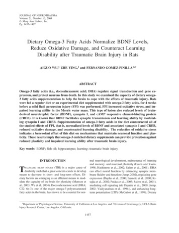
FISH OIL DIET NORMALIZES BDNF AND COGNITION IN TBIBDNF facilitates synaptic transmission and regulatesgene expression through activation of synapsin I andCREB (Finkbeiner, 2000; Jovanovic et al., 1996; Wanget al., 1995; Ying et al., 2002). Our previous report indicates that TBI may affect cognitive ability by compromising some of the action of BDNF on synaptic plasticity (Wu et al., 2003). To evaluate whether omega-3fatty acids supplemented in the diet could protect againstdisruption in molecular systems that maintain synapticplasticity after TBI, we measured the protein expressionof synapsin I and CREB in the hippocampus by Westernblot analysis. The results showed that dietary omega-3fatty acids supplementation normalized levels of synapsinI (Fig. 3A,B) and CREB (Fig. 4A,B) in the hippocampusof TBI rats.Omega-3 Fatty Acid Supplementation ReducesOxidative Damage in TBI AnimalsOxidative damage was assessed using Western blotanalysis of DNPH-derivatized carbonyl groups on oxidized proteins. A representative example of an Oxyblotgel is shown in Figure 5A. The oxidized protein levelswere significantly increased in TBI-RD animals relativeto Sham-RD rats (Fig. 5B). However, the TBI animalsfed the diet supplemented with FO had significant lowerlevels of oxidized proteins compared with TBI-RD,Sham-RD, and Sham-FO animals (Fig. 5B).DISCUSSIONOur results demonstrate that supplementation ofomega-3 fatty acids in the diet normalizes hippocampallevels of BDNF and its downstream effectors on synaptic plasticity synapsin I and CREB, after experimentalTBI. Restoration in levels of these molecular systems wasassociated with a normalized spatial learning ability inthe Morris water maze. These findings suggest thatomega-3 fatty acid–enriched dietary supplements can bea potent therapeutic agent for reducing the deleterious effects of TBI on synaptic plasticity and cognition. In addition, the finding that omega-3 fatty acids reduced oxidative damage as a result of TBI appear to reveal ageneral benevolent effect of omega-3 fatty acids onFIG. 2. Omega-3 fatty acids normalized BDNF levels in thehippocampus of FPI rats measured using ELISA. (A) FPI reduced BDNF levels, and this effect was reversed by FO dietarysupplementation. FO supplementation increased BDNF levelsin intact rats (Sham-FO). The values were converted to percentof Sham-RD (mean SEM). *p 0.05. (B) Immunohistochemistry shows that BDNF was predominantly distributedalong the mossy fiber system that runs between CA3 and thedentate gyrus (DG), and in the molecular layer of the DG ofthe hippocampal formation. The omega-3 fatty acids supplementation prevented the reduction in BDNF immunoreactivityin animals receiving FPI.1461
WU ET AL.and regulate gene expression through activation ofsynapsin I and CREB (Finkbeiner, 2000; Jovanovic et al.,1996; Wang et al., 1995; Ying et al., 2002). Our resultsshowed that omega-3 fatty acids normalize the proteinlevels of synapsin I and CREB in TBI rats. Since BDNFand its downstream effectors synapsin I and CREB areinvolved in learning and memory events, our findingssuggest that supplementation of omega-3 fatty acids inthe diet may provide protection against learning disability after TBI via upregulation of these molecular systems(Fig. 6).It is notable that FO supplementation increased BDNFbut did not affect cognitive function in intact rats. It ispossible that slight changes in BDNF may not significantly affect cognition under normal conditions. It seemslikely, however, that under pathological weakness smalldecreases in BDNF can be a factor to further deterioratecognitive function. This eventual possibility emphasizesthe necessity to use therapeutic means, such as dietaryFIG. 3. Omega-3 fatty acids reverse the FPI-elicited reduction of synapsin I in the hippocampus. (A) Western blot analysis using actin as standard control showed that FPI reducedsynapsin I levels, and that these effects of FPI were reversedby FO supplementation. FO supplementation did not affectsynapsin I levels in intact rats (Sham-FO). Protein values wereconverted to percent of sham (mean SEM). *p 0.05. (B)Representative immunoblot sample gels for synapsin I in eachexperimental group.mechanisms that maintain neuronal function and plasticity after TBI.Omega-3 Fatty Acids May Affect Cognition byModulating the BDNF SystemOur finding that supplementation of omega-3 fattyacids normalizes the protein levels of BDNF after TBIsuggests that BDNF mediates the beneficial effects ofomega-3 fatty acids on cognitive function. We have recently shown that TBI reduces BDNF, synapsin I, andCREB, with subsequent effects on cognitive function(Wu et al., 2003). The question is: How can omega-3fatty acids compensate for cognitive decline resultingfrom TBI? It is well accepted that BDNF modulatessynaptic plasticity (Bolton et al., 2000; Hariri et al., 2003;Kang and Schuman, 1996; Thoenen, 1995), and is required for normal learning in the Morris water Maze (Muet al., 1999). BDNF can facilitate synaptic transmissionFIG. 4. Omega-3 fatty acids dietary supplementation reversesthe FPI-elicited reduction of CREB in the hippocampus. (A)The level of CREB was determined by western blot analysisusing actin as a standard control. Results show that FPI decreased CREB, and that this effect was reversed by FO supplementation. FO supplementation did not affect CREB levelsin intact rats (Sham-FO). The values were converted to percentof sham-RD (mean SEM). *p 0.05. (B) Representative immunoblot gels for synapsin I in each experimental group.1462
FISH OIL DIET NORMALIZES BDNF AND COGNITION IN TBIFIG. 5. Measurements of oxidized protein levels in the hippocampus. The oxidized protein levels were determined by Oxyblot kit. (A) Representative sample of Oxyblot bands. (B) FPI resulted in higher oxidized protein levels compared with ShamRD animals, whereas FO feeding markedly reduced the FPI-induced elevation in protein carbonyl levels. FO supplementation did not affect oxidized protein levels in intact rats (Sham-FO). The values were converted to percent of Sham-RD(mean SEM). **p 0.01.supplementation of FO, to maintain normal levels ofBDNF under challenging conditions (Molteni et al., 2002,2004; Wu et al., 2003, 2004).Oxidative Stress May Be an Intermediate Step bywhich TBI and Dietary Factors ModulateSynaptic Plasticity and CognitionFIG. 6. Possible mechanisms underlying the effects ofomega-3 fatty acids supplementation on restoring neural plasticity after TBI. TBI results in degradation of membrane phospholipids, leading to cumulative oxidative stress, which in turnmay cause reduction in BDNF and its synaptic plasticity effectors synapsin I and CREB. These alterations may underliesynaptic dysfunction and cognitive impairment resulting fromTBI. Omega-3 fatty acids supplementation may prevent thedegradation of membrane phospholipids and subsequent cumulative oxidative stress. Omega-3 fatty acids can also contribute to normalized levels of BDNF, synapsin I, and CREB.The action of oxidative stress may also affect the regulation ofthe BDNF system and synaptic plasticity.We detected markedly elevated levels of protein carbonyl formation in TBI animals using a convenient Western blot analysis of DNPH derivatized carbonyls. Supplementation of omega-3 fatty acids in the diet, however,dramatically reduced the elevated protein carbonyls,which is consistent with the known antioxidant activityof DHA (Hashimoto et al., 2002; Hossain et al., 1999).For example, DHA has shown anti-oxidant capacity inthe aging brain with subsequent effects on cognition(Hashimoto et al., 2002; Hossain et al., 1999). It is possible that the potential anti-oxidant action of DHA in TBImay be achieved using mechanisms that maintain synaptic plasticity. It has been shown that TBI can result in cumulative ROS (Marklund et al., 2001; Paolin et al., 2002;Pratico et al., 2002), which may be associated with reduction of BDNF (Wu et al., 2003, 2004). Thus, DHAmay help to counteract elevated levels of ROS with subsequent effects on the action of BDNF on synaptic plasticity and cognition after TBI. Although the major com-1463
WU ET AL.ponents of FO in our study are DHA and EPA, it is likelythat the beneficial effects of FO supplementation are attributable to DHA. This possibility is supported by studies showing that (1) EPA is absent in normal brain (Kitajka et al., 2002); (2) EPA can be converted to DHA inthe body (Pawlosky et al., 2001); and (3) EPA cannotcross the blood–brain barrier. However, a direct link between DHA and the observed findings in our study needto be further investigated by using pure DHA in the diet.It is known that DHA affects LTP, one of the most impressive forms of synaptic plasticity (Bliss and Collingridge, 1993) and a biological substrate for learning andmemory (Moser et al, 1998). Emerging evidence indicates that dietary omega-3 fatty acids can restore theDHA in the neuronal membrane and reverse the age-related synaptic dysfunction such as impairment in LTP(McGahon et al., 1999). Indeed, DHA supplemented inthe diet can improve learning ability (Lim and Suzuki,2001; Suzuki et al., 1998) and enhance long-term memory in both young and old animals (Gamoh et al., 1999,2001; Lim and Suzuki, 2000), and can reduce cognitivedecline during aging and AD (Hashimoto et al., 2002).Our findings provide novel evidence suggesting that theaction of omega-3 fatty acids (i.e., DHA) in reducing cognitive impairment after TBI are associated with restoration of molecular systems that serve synaptic plasticity.Our results also suggest that this diet can exert its actionby diminishing cumulative ROS.Omega-3 Fatty Acids Can Help the TBI Brainby Restoring Membrane IntegrityEmerging evidence indicates that TBI can lead todegradation of membrane phospholipids (PLs), resultingin accumulation of reactive oxygen species. It is possible that these events can result in perturbations of synaptic plasticity (Fig. 6). It has been known that synapticmembranes phospholipids are preferentially enriched inomega-3 fatty acids, especially DHA. TBI can result inacute and long-lasting perturbation in brain phospholipidmetabolism (Homayoun et al., 1997; Marklund et al.,1997). Degradation of membrane PLs, a well-known phenomenon in acute brain injuries (Homayoun et al., 1997;Marklund et al., 1997), is thought to underlie the disturbance of cellular membrane functions, contributing tosecondary neuronal injury (Farooqui and Horrocks,1994). Synaptic terminals show a high activity in phospholipid-hydrolyzing enzymes (i.e. PLA2, PLC; Bazan etal., 1995), which can be intensified after TBI (Shohamiet al., 1989; Wei et al., 1982). Activation of PLA2 andPLC may hydrolyze PLs, resulting in cumulative oxidative stress and subsequent neuronal dysfunction. Thus,TBI-induced PL degradation and subsequent oxidativedamage may contribute to impairment in cognition andneuroplasticity (Fig. 6). The overall evidence suggeststhat supplementation of omega-3 fatty acids (i.e., DHA)in the diet may help the TBI brain preserve synapticmembrane integrity and fluidity, which are crucial factors for maintenance of vital cellular function.CONCLUSIONOur findings demonstrate that omega-3 fatty acids supplementation can restore cognitive function after TBI,and that this may be achieved by normalizing the actionof BDNF on synaptic plasticity using synapsin I andCREB. Results also show that this diet can provide protection against oxidative damage after TBI, which mayalso influence synaptic plasticity and cognition. These results suggest that dietary omega-3 fatty acids supplementation has a therapeutic potential to reduce the deleterious effects of TBI and perhaps other insults onsynaptic plasticity and cognitive function.ACKNOWLEDGMENTSWe would like to thank Dr. David Hovda for constructive advice related to the traumatic brain injury paradigm. This study was supported by NIH awards (NS39522 and 38978) and UCLA Brain Injury Research Center.REFERENCESBAZAN, N.G., RODRIGUEZ DE TURCO, E.B., and ALLAN,G. (1995). Mediators of injury in neurotrauma: intracellularsignal transduction and gene expression. J. Neurotrauma 12,791–814.BLISS, T.V., and COLLINGRIDGE, G.L. (1993). A synapticmodel of memory: long-term potentiation in the hippocampus. Nature 361, 31–39.BOLTON, M.M., LO, D.C., and SHERWOOD, N.T. (2000).Long-term regulation of excitatory and inhibitory synaptictransmission in hippocampal cultures by brain-derived neurotrophic factor. Prog. Brain Res. 128, 203–218.BORGARO, S.R., PRIGATANO, G.P., KWASNICA, C., andREXER, J.L. (2003). Cognitive and affective sequelae incomplicated and uncomplicated mild traumatic brain injury.Brain Inj. 17, 189–198.BROCK, T.O., and O’CALLAGHAN, J.P. (1987). Quantitative changes in the synaptic vesicle proteins synapsin I andp38 and the astrocyte-specific protein glial fibrillary acidic1464
FISH OIL DIET NORMALIZES BDNF AND COGNITION IN TBIprotein are associated with chemical-induced injury to the ratcentral nervous system. J. Neurosci. 7, 931–942.stem of aged hypercholesterolemic rats. J. Neurochem. 72,1133–1138.de URQUIZA, A.M., LIU, S., SJOBERG, M., et al. (2000). Docosahexaenoic acid, a ligand for the retinoid X receptor inmouse brain. Science 290, 2140–2144.HOVDA, D.A., LEE, S.M., SMITH, M.L., et al. (1995). Theneurochemical and metabolic cascade following brain injury:moving from animal models to man. J. Neurotrauma 12,903–906.DRAPEAU, E., MAYO, W., AUROUSSEAU, C., et al. (2003).Spatial memory performances of aged rats in the water mazepredict levels of hippocampal neurogenesis. Proc. Natl. Acad.Sci. USA 100, 14385–14390.DUPLUS, E., GLORIAN, M., and FOREST, C. (2000). Fattyacid regulation of gene transcription.
(CREB). It is known that BDNF facilitates synaptic transmission and learning ability by modulat-ing synapsin I and CREB. Supplementation of omega-3 fatty acids in the diet counteracted all of the studied effects of FPI, that is, normalized levels of BDNF and associated synapsin I and CREB, reduced oxidative damage, and counteracted learning .