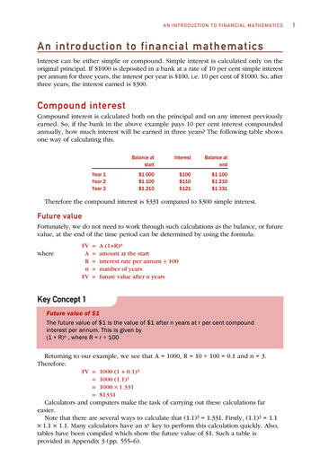
Transcription
Immunodiagnostic Value of Echinococcusgranulosus Recombinant B8/1 Subunit ofAntigen BAmir Savardashtaki1,2, Bahador Sarkari3,4, Farzane Arianfar2, Zohreh MostafaviPour1,2*1Department of Medical Biotechnology, School of Advanced Medical Sciences and Technologies,Recombinant Proteins Laboratory, Department of Biochemistry, 3Department of Parasitology andMycology, 4Basic Sciences in Infectious Diseases Research Center, School of Medicine, Shiraz Universityof Medical Sciences, Shiraz, Iran2ABSTRACTBackground: Cystic echinococcosis (CE), as a chronic parasitic disease, is a majorhealth problem in many countries. The performance of the currently availableserodiagnostic tests for the diagnosis of CE is unsatisfactory. Objective: The currentstudy aimed at sub-cloning a gene, encoding the B8/1 subunit of antigen B (AgB) fromEchinococcus granulosus, using gene optimization for the immunodiagnosis of humanCE. Methods: The coding sequence for AgB8/1 subunit of Echinococcus granulosuswas selected from GenBank and was gene-optimized. The sequence was synthesizedand inserted into pGEX-4T-1 vector. Purification was performed with GST tag affinitycolumn. Diagnostic performance of the produced recombinant antigen, native antigen Band a commercial ELISA kit were further evaluated in an ELISA system, using a panelof sera from CE patients and controls. Results: SDS-PAGE demonstrated that theprotein of interest had a high expression level and purity after GST tag affinitypurification. Western blotting verified the immunoreactivity of the producedrecombinant antigen with the sera of CE patients. In an ELISA system, the sensitivityand specificity (for human CE diagnosis) of the recombinant antigen, native antigen Band commercial kit were respectively 93% and 92%, 87% and 90% and 97% and 95%.Conclusion: The produced recombinant antigen showed a high diagnostic value whichcan be recommended for serodiagnosis of CE in Iran and other CE-endemic areas.Utilizing the combination of other subunits of AgB8 would improve the performancevalue of the introduced ELISA system.Savardashtaki A, et al. Iran J Immunol. 2017; 14(2):111-122.Keywords: Cloning, Recombinant antigen, Immunodiagnosis, Cystic ----------------------*Corresponding author: Dr. Zohreh Mostafavi-Pour, Professor of Biochemistry, School of Medicine, Shiraz University ofMedical Sciences, Shiraz, Iran, e-mail: zmostafavipour88@yahoo.co.ukIran.J.Immunol. VOL.14 NO.2 June 2017111
Savardashtaki A, et al.INTRODUCTIONEchinococcus granulosus, in its larval stage, is the causative agent of cysticechinococcosis (CE), a zoonotic infection affecting both human and livestock (1) and amajor health problem in different endemic areas of the world, including Iran (2-5),entailing heavy economic losses in the affected areas (3,6). Nearly 1.2 million peopleare affected by CE and the annual costs pertaining to the disease are estimated to bearound 3 billion US for patients’ management and damages to livestockproductiveness (7). An early and accurate diagnosis of CE is conducive both to itsmanagement and treatment. Diagnosis of CE is mainly based on a combination ofserological tests, along with imaging methods (6,8). Immunodiagnosis of CE has beenconsiderably improved over the past two decades, improvements that are mainly due tothe advancement in defining and synthesizing such immunodominant peptides asrecombinant antigens. However, the immunodiagnosis of CE is still problematic and theperformances of the available serological tests are not yet satisfactory (8-10).Purified recombinant antigens and synthetic peptides, mainly derived from two parasitemolecules, antigen B (AgB) and antigen 5 (Ag5), have been applied for the serologicaldiagnosis of CE with various accuracy performances where the 8 kDa subunit of AgBhas proven the most valuable antigen in native, recombinant or synthetic forms(8,11,12). During the last decades, recombinant DNA technology has been used for theheterologous expression of recombinant antigens in E. coli and their application to theimmunodiagnosis of infectious diseases (13-15). Owing to their low cost andreproducibility, recombinant proteins have drawn significant attention. However, a fewdrawbacks such as the differences in codon usage between species may result in thereduction of protein’s yield. Several studies have indicated that gene optimizationincrease the yield of protein expression (16,17). In the current study, the gene encodingthe B8/1 subunit of antigen B from E. granulosus genotypes 1 was sub-cloned in E.coli, using gene optimization for high-throughput protein expression. The producedrecombinant antigen was evaluated in an ELISA system for the immunodiagnosis ofhuman hydatid disease.MATERIALS AND METHODSGene optimization, synthesis and cloning. Sequence data was obtained from NCBIGenBank with the accession number DQ372074. Gene optimization of the AgB8/1,without signal sequence for heterologous expression in E. coli, was performed usingOptimum Gene Algorithm (Genscript, New Jersey, USA). Gene synthesis wasordered from Biomatik Company (Biomatik, Ontario, Canada). The optimized AgB8/1gene was cloned in pBluescript II SK( ) (Biomatik, Ontario, Canada) vector andexcised by restriction enzymes EcoRI and XhoI according to the manufacturer’sinstructions. So as to confirm the size of the fragments, the product was further checkedwith agarose gel electrophoresis. Subsequently, the band corresponding to the AgB8/1coding sequence was excised from the gel and purified through the use of gel extractionkit (Bioneer, Korea). Next, the purified fragment was cloned into pGEX-4T-1 (GEHealthcare, Illinois, US) expression vector. Plasmids transformation into the DH5α cellswas performed using standard CaCl2 method followed by plasmid extraction, employingQIAprep Spin Miniprep Kit (Qiagen, Hilden, Germany) according to the manufacturer’sIran.J.Immunol. VOL.14 NO.2 June 2017112
Immunodiagnosis of Human Cystic Echinococcosis by Recombinant AgB8/1 of Echinococcus granulosusguideline. After that, a new round of transformation was performed to transfer theplasmids into BL21 (DE3)pLysS cells for expression under the T7 promoter. CEAgB8/1 was expressed in fusion with N-terminus GST tag in BL21(DE3) pLysS E. colistrain by 0.5 mM IPTG (Invitrogen, USA) induction for 4 hours.Purification of the recombinant protein. The produced Echinococcus granulosusrecombinant AgB8/1 antigen (rEgAgB8/1) was purified via immunoaffinity column forGST-tag (Sigma-Aldrich, Germany), according to the manufacturer’s instructions as tothe native purification procedure. Digestion with thrombin was made for the removal ofthe tag. Purified protein was desalted overnight using a dialysis membrane in PBS (pH:7.5) (14). Native AgB was purified as previously described (18).SDS-PAGE and western blotting analysis of the recombinant protein. We carriedout the SDS-PAGE of the cleaved and uncleaved recombinant proteins and the nativeprotein (native antigen B purified from hydatid cyst fluid) in 18% (w/v) polyacrylamidegel containing 0.1% SDS. Protein samples (with and without GST tag) were mixed,with reducing sample buffers, and boiled in a water bath, for 5 minutes (14). Gel wasrun at a constant voltage of 200 V and transferred to nitrocellulose membranes (GEHealthcare) using a Mini Trans-blot Cell (Bio-Rad, Berkeley, California). Therecombinant proteins were evaluated by use of the pooled sera of CE patients at 1:1000dilution. Secondary horseradish peroxidase conjugated anti-human IgG (Sigma, USA),was diluted at 1:4000 dilution. Bound proteins were developed utilizingdiaminobanzidine (DAB) substrate.Serum samples. Serum samples of 30 pathologically-confirmed CE patients wereobtained from university affiliated hospitals in Shiraz, southern Iran. The cysts locationsin the patients were lungs, liver, lungs along liver, spleen (one case), or bladder alongpelvis (one case). Samples were also collected from 32 patients with parasitic diseasesother than hydatid cyst, including giardiasis (n 3), hymenolepiasis (n 3), toxocariasis(n 3), toxoplasmosis (n 3), fascioliasis (n 3), malaria (n 2), cryptosporidiasis (n 2),trichostrongyliasis (n 1), and patients with autoimmune diseases (n 9, including 3 withvitiligo, 3 with Behçet's disease and 3 with systemic lupus erythematosus). Laboratorydiagnosis of the parasitic diseases was based on the microscopic findings, with theexception of toxocariasis and toxoplasmosis which were based on serology, usingToxocara IgG ELISA (IBL International Gmbh, Hamburg, Germany) and ToxoplasmaIgG ELISA (Yekta Tajhiz, Iran). Moreover, sera were collected from 30 healthycontrols with no sign, symptoms or history of CE. Based on the tests, the healthycontrols were all negative for anti-hydatid cyst antibodies. The study was approved byEthics Committee of Shiraz University of Medical Sciences and informed consent wastaken from the subjects prior to collecting the samples.ELISA with native and recombinant antigens. ELISA was performed in flat-bottom96-well microplates (Guangzhou Jet Bio-Filtration, Guangzhou, China). The microplatewere coated with 100 μl/well of either rEgAgB8/1 or native antigen B (nAgB) with aconcentration of 5 µg/mL in 0.1 M carbonate/bicarbonate (pH 9.6) buffer through anovernight incubation at 4 C. Plates were blocked with 300 µl of 5% non-fat skimmedmilk in phosphate-buffered saline with 0.1% (V/V) Tween 20 for 2 hours at 37 C. Thewells were then washed 5 times with 300 µl of the washing buffer (PBST; PBScontaining 0.1% Tween 20). Serum samples were diluted (1:100) in PBST, applied tothe plates and incubated at 37 C over a period of 2 hours. The plate was washed asbefore and 100 μl aliquots of HRP-conjugated goat anti-human IgG (Sigma, USA) at a1:4000 dilution in PBST were added to the plates and incubated for 1 hour at roomIran.J.Immunol. VOL.14 NO.2 June 2017113
Savardashtaki A, et al.temperature. After washing, the plates were incubated with substrate (100 μl/well of 0.4mg/ml OPD, 0.3% H2O2 in 0.1 M citrate buffer, pH 5) for 20 minutes and the reactionwas stopped by 1 N H2SO4. The absorbance at 450 nm was monitored with a microplatereader (Bio-Tek, ELx800). The cutoff point was set at 2SD from the mean of controlsamples. The ELISA was also performed with tagless and also GST tag-containingrEgAgB8/1.ELISA with commercial kit. All the sera samples were further tested by a commercialELISA kit (Euroimmune, Germany) for the detection of anti-hydatid cyst antibodiesbased on the manufacturer’s instructions.Statistical analysis. The diagnostic accuracy of the native antigen B, rEgAgB/1 andcommercial kit were assessed by the area under the receiver operating characteristic(ROC) curve (AUC) using SPSS software (ver. 18). Cutoff values were set for the testaccording to Youden’s Index. Cohen's kappa statistic was used to find out the extent ofagreement among the ELISA systems, using different antigens and the commercial kit.RESULTSIn the present study, the gene encoding a B8/1 subunit of antigen B from E. granulosuswas sub-cloned in E. coli, with high-throughput protein expression. The producedrecombinant antigen was evaluated in an ELISA system for the serological diagnosis ofhuman CE.Expression and purification of rAgB8/1.A fragment of E. granulosus AgB8/1, without the signal sequence, was synthesized andcloned into pGEX-4T-1 expression vector, under the control of T7 promoter, andtransformed into the BL21 (DE3) pLysS expression host. The sequencing of the vectorconfirmed the correct reading frame and the insertion of the coding sequence in theGST-tag frame. REgAgB8/1 protein was expressed in LB as the culture medium.Furthermore, SDS-PAGE analysis confirmed the expression of the recombinant proteinwith the expected molecular weight of 34 kDa (Figure 1).Iran.J.Immunol. VOL.14 NO.2 June 2017114
Immunodiagnosis of Human Cystic Echinococcosis by Recombinant AgB8/1 of Echinococcus granulosusFigure 1. SDS-PAGE analysis of the rEgAgB8/1 protein expression and purification. Line1 Protein weight marker; Line 2 and 3, uninduced and induced cultures of E. coli by IPTG; Line4 prewash; Line 5, 6 and 7 consecutive washes; Line 8 final elution (GST-AgB8/1); Line 9cleaved.Western blot analysis of rEgAgB8/1 expression in E. coli.Western blotting analysis confirmed the reactivity of the produced recombinant antigenwith the sera of CE patients (Figure 2) in both the tagged and tagless types.Figure 2. Western blot analysis of rEgAgB8/1 expression in E. coli. Line 1: molecularweight marker; Lines 2, 3 and 4 native AgB with 20, 10 and 5 µg/well respectively; Lines 5, 6and 7 rEgAgB8/1; Line 8 rEGAgB8/1 fused with GST tag.Iran.J.Immunol. VOL.14 NO.2 June 2017115
Savardashtaki A, et al.Purification of rEgAgB8/1 by anti-GST-tag immunoaffinity column.The concentration of the purified rEgAgB8/1 prior to the cleavage of tag with thrombinwas 3 mg/mL; following the cleavage, on the other hand, the concentration was 1mg/mL. Appraising the purity of the purified proteins with SDS-PAGE, it became clearthat no unwanted proteins were present in the elutes, (Figure 1).Evaluation of rEgAgB8/1 for the diagnosis of human CE.Both pure rEgAgB8/1 (without tag) and GST-containing rEgAgB8/1 were employed inthe ELISA system for the detection of anti-hydatid cyst antibodies in the sera of CEpatients and controls. Using ELISA checkerboard titration method, the optimalconcentration of the rEgAgB8/1 antigen (both the tagless and GST-containing forms),serum and HRP-conjugated goat anti-human IgG were found to be 5 µg/ml, 1:100 and1:4000, respectively. Using rEgAgB8/1 in ELISA system, 28 out of 30 CE patients(93.3%) were found to be positive and in the controls, 5 cases (8.1%) were falsepositive. Therefore, the sensitivity and specificity of the produced rEgAgB8/1 antigenfor the diagnosis of human CE were 93% and 92%, respectively. Table 1 demonstratesthe performance of rEgAgB8/1, in the diagnosis of human CE.Table 1. Performance of ELISA, using recombinant Ag B8/1, in serodiagnosis ofCE.Type of serumNumberNumber of positive cases in rEgAgB8/1- ELISANumberPercentage (%)CE patients302893Vitiligo300Behcet's s100Normal30310Iran.J.Immunol. VOL.14 NO.2 June 2017116
Immunodiagnosis of Human Cystic Echinococcosis by Recombinant AgB8/1 of Echinococcus granulosusEvaluation of native antigen B for the diagnosis of human CE.Using native AgB, 26 out of 30 CE patients (86.7%) were found to be positive and 6cases (9.7%) in the controls were false positive. Accordingly, the sensitivity andspecificity of this antigen were 87 and 90%, respectively. Table 2 illustrates theperformance of native AgB regarding the diagnosis of human CE.Table 2. Performance of ELISA, using native antigen B, in serodiagnosis of CE.Type of serumNumberNumber of positive cases in native antigen B ELISANumberPercentage (%)CE patients302687Vitiligo300Behcet's sis100Normal3027Evaluation of commercial ELISA kit for the diagnosis of human CE.Using commercial ELISA kit, 29 out of 30 CE patients (96.7%) were found to bepositive and 3 control cases (4.8%) were false positive. In this regard, the sensitivity andspecificity of the commercial kit were respectively 97% and 95%. Table 3 shows theperformance of commercial ELISA kit and Table 4 compares the performances ofrEgAgB8/1, native AgB and commercial ELISA in the diagnosis of human CE.Iran.J.Immunol. VOL.14 NO.2 June 2017117
Savardashtaki A, et al.Table 3. Performance of commercial ELISA kit in serodiagnosis of CE.Type of serumNumberNumber of positive cases in commercial ELISAkitNumberPercentage (%)CE patients302997Vitiligo300Behcet's 0Malaria is100Normal3013Based on the statistical analysis of the data, there existed a good agreement(kappa 0.74) between the three systems using native AgB, rEgAgB8/1 and commercialkit (kappa 0.88 between rEgAgB8/1 and native AgB, kappa 0.76 between native AgBand commercial kit, and kappa 0.83 between rEgAgB8/1 and commercial kit).Iran.J.Immunol. VOL.14 NO.2 June 2017118
Immunodiagnosis of Human Cystic Echinococcosis by Recombinant AgB8/1 of Echinococcus granulosusTable 4. Comparative performances of recombinant AgB8/1, native AgB andcommercial kit in the serodiagnosis of CE.Type of antigensSensitivity (%)(95% CI)93(76-98)Specificity (%)(95% CI)92(81-96)PPV (%)(95% CI)85(67-94)NPV (%)(95% CI)96(87-99)Native AgB87(68-95)90(79-96)81(63-92)93(83-98)Commercial ntAgB8/1DISCUSSIONCE is known as a disease mostly remaining in an asymptomatic state during the primaryyears of infection (6). Since a late diagnosis entails severe implications with the severityof the outcomes, early diagnosis of the disease is crucial. Diagnosis of CE is mostlymade performed through such imaging modalities as ultrasonography, computedtomography (CT) and magnetic resonance imaging (MRI) (6,19). Further employed indiagnosing CE are serological tests with specific antigens of the parasite (8,10,20-22).Serological methods can be used for the confirmation of the results of imaging and alsoto diagnose the disease in subclinical stages. These methods suffer from not beingstandardized, resulting in poor specificity or sensitivity. Hence the developing a reliabletest with reasonable validity for routine laboratory diagnosis of CE is necessary. Recentstudies have been directed toward the use of recombinant antigens (11,12,23,24). Inorder to develop an immunodiagnostic assay in any regions, world health organizationhas recommended the use of antigens corresponding to the sequences obtained from thesame region. Accordingly, the current study was designed to produce a recombinantantigen of E. granulosus for serodiagnosis of CE in Iran. It has been reported thatAgB8/1 is one of the most appropriate antigens for the development of anyimmunoassay as far as diagnosing CE is concerned (8,12,19). Owing to the fact that thepresence of signal sequence residues of 17 to 81 of AgB8/1 affects the heterologousexpression of the protein, interferes with its collection in the cytoplasm and influencesthe immunoreactivity of the antigen, we removed these parts from the sequence tooptimize the heterologous expression of the antigen (24). In the present research, highlevels of heterologous AgB8/1 expression were achieved using several approaches.Firstly, gene optimization and gene synthesis were applied so as to surmount issuessuch as codon usage bias and differences between E. coli as a bacterium and E.granulosus as a eukaryote. The former problem poses challenges concerning theheterologous expression of recombinant proteins in biotechnology applications. Anumber of other studies have successfully used this approach to overexpress the proteinof interest in a heterologous host (16,17). In the present study, we were able to obtain arelatively high-throughput expression level of 3 mg/mL of purified protein. Withoutgene optimization, the yield of recombinant protein would be much lower incomparison to what we have achieved in the current study. Secondly, we used GST tagas a fusion partner in the amino tail of the protein which, by itself, may ameliorate theexpression of the protein (25). Beyond the use of this fusion partner as a proteinpurifier, it is always possible that fusing the N-terminus of recombinant proteins withIran.J.Immunol. VOL.14 NO.2 June 2017119
Savardashtaki A, et al.one of the abundant proteins of the host augment the expression of its gene, therebyaccelerating the translation (14). Therefore, it may positively induce the level of proteinexpression and improve the production yield of the recombinant protein. Furthermore, itconduces to the increase in the solubility of the recombinant protein, reducing theformation of inclusion bodies and, in the process, the toxicity of the recombinantproteins to the host cells. Also noteworthy is the facile purification of the recombinantprotein with GST-tag columns. In the current study, we followed the protocol for nativepurification, conducive to maintaining the native structure of a protein. Nativepurification pre-empts the denaturing of the protein, an event which may lead to the lossof certain important conformational epitopes of the antigen. The tag was successfullyused for the purification of recombinant antigens where we obtained a solubleexpression of recombinant proteins with high yield. In this method, the tag can be easilyremoved through enzymatic cleavage by thrombin enzyme.Sensitivity and specificity of the rEgAgB8/1-based ELISA in the current study were93% and 92%, respectively, which implies its dominance, regarding accuracy,comparisons with the commercial kits which, not infrequently, employ a combination ofseveral recombinant antigens. Similar performance have been reported byMohammadzadeh et al. in which sera samples from CE patients in Iran, Turkey, Chinaand Japan were tested by a recombinant antigen B8/1 (12). A study by Kalantari et al.reported the production and use of recombinant antigen B for the diagnosis of hydatidcysts in Iran. Using AgB8/4 subunit of antigen B, they observed 91.66% sensitivity and97.22% specificity (23). There exist, however, a number of differences between theirstudy and ours. Firstly, in the latter, AgB8/1, known as the most appropriate subunit ofantigen B for the diagnosis of CE, was produced and used while in the former AgB8/4subunit was generated and utilized. Secondly, we used gene optimization to enhance theheterologous expression of recombinant protein of interest. Another difference is that, inthe present study, we used GST-tag affinity columns for the native purification of theproduced recombinant antigen; in the other study, on the other hands, his-tag affinity (adenaturing protocol) was used for the purification (23).It is concluded that gene optimization enhances the heterologous expression of therEgAgB8/1 antigen in E. coli. GST-Tag fusion is a valuable option for the purificationof the produced antigen which in turn contributes to the high expression level of theantigen. The rEgAgB8/1 antigen have a higher diagnostic value, in comparison to thenative AgB and can be recommended for the serodiagnosis of CE in Iran and, perhaps,other CE-endemic areas. Combining other subunits of AgB8 with the currentrecombinant antigen would potentially improve the performance value of such antigen.ACKNOWLEDGEMENTSThis study was financially supported by the office of vice-chancellor for research ofShiraz University of Medical Sciences (Grant No. 93-7376). The results described inthis paper were part of PhD student thesis of Amir Savardashtaki.Iran.J.Immunol. VOL.14 NO.2 June 2017120
Immunodiagnosis of Human Cystic Echinococcosis by Recombinant AgB8/1 of Echinococcus 14.15.16.17.18.19.20.Romig T, Deplazes P, Jenkins D, Giraudoux P, Massolo A, Craig PS, et al. Ecology and life cyclepatterns of Echinococcus species. Adv Parasitol. 2017; 95:213-314.Deplazes P, Rinaldi L, Alvarez Rojas CA, Torgerson PR, Harandi MF, Romig T, et al. Globaldistribution of alveolar and cystic Echinococcosis. Adv Parasitol. 2017; 95:315-493.Fasihi Harandi M, Budke CM, Rostami S. The monetary burden of cystic Echinococcosis in Iran.PLoS Negl Trop Dis. 2012; 6:e1915.Sarkari B, Sadjjadi SM, Beheshtian MM, Aghaee M, Sedaghat F. Human cystic Echinococcosis inYasuj district in southwest of Iran: an epidemiological study of seroprevalence and surgical casesover a ten-year period. Zoonoses Public Health. 2010; 57:146-50.Sarkari B, Mansouri M, Khabisi SA, Mowlavi G. Molecular characterization and seroprevalence ofEchinococcus granulosus in wild boars (Sus scrofa) in south-western Iran. Ann Parasitol. 2015;61:269-73.Kern P, Menezes da Silva A, Akhan O, Mullhaupt B, Vizcaychipi KA, Budke C, et al. TheEchinococcoses: diagnosis, clinical management and burden of disease. Adv Parasitol. 2017;96:259-369.Budke CM, Deplazes P, Torgerson PR. Global socioeconomic impact of cystic Echinococcosis.Emerg Infect Dis. 2006; 12:296-303.Sarkari B, Rezaei Z. Immunodiagnosis of human hydatid disease: Where do we stand? World JMethodol. 2015; 5:185-95.Gottstein B, Soboslay P, Ortona E, Wang J, Siracusano A, Vuitton D. Immunology of alveolar andcystic Echinococcosis (AE and CE). Adv Parasitol. 2017; 96:1-54.Sadjjadi SM, Sedaghat F, Hosseini SV, Sarkari B. Serum antigen and antibody detection inEchinococcosis: application in serodiagnosis of human hydatidosis. Korean J Parasitol. 2009;47:153-7.Carmena D, Benito A, Eraso E. Antigens for the immunodiagnosis of Echinococcus granulosusinfection: An update. Acta Trop. 2006; 98:74-86.Mohammadzadeh T, Sako Y, Sadjjadi SM, Sarkari B, Ito A. Comparison of the usefulness ofhydatid cyst fluid, native antigen B and recombinant antigen B8/1 for serological diagnosis ofcystic Echinococcosis. Trans R Soc Trop Med Hyg. 2012; 106:371-5.Movahedi B, Mokarram P, Hemmati M, Mosavari N, Zare R, Safaee Ardekani L, Mostafavi-PourZ. IFN-γ and IL-2 responses to recombinant AlaDH against ESAT-6/CFP-10 fusion antigens in thediagnosis of latent versus active Tuberculosis infection. Iran J Med Sci; 2017; 42: 275-283.Hemmati M, Seghatoleslam A, Rasti M, Ebadat S, Mosavari N, Habibagahi M, et al. Expressionand purification of recombinant Mycobacterium Tuberculosis (TB) antigens, ESAT-6, CFP-10 andESAT- 6/CFP-10 and their diagnosis potential for detection of TB patients. Iran Red Crescent MedJ. 2011; 13:556-63.Savardashtaki A, Sharifi Z, Hamzehlou S, Farajollahi MM. Analysis of immumoreactivity ofheterologously expressed non-structural protein 4B (NS4B) from hepatitis C virus (HCV) genotype1a. Iranian J Biotechnol. 2016; 13:32-7.Bai J, Swartz DJ, Protasevich, II, Brouillette CG, Harrell PM, Hildebrandt E, et al. A geneoptimization strategy that enhances production of fully functional P-glycoprotein in Pichia pastoris.PLoS One. 2011; 6:e22577.Maertens B, Spriestersbach A, von Groll U, Roth U, Kubicek J, Gerrits M, et al. Gene optimizationmechanisms: a multi-gene study reveals a high success rate of full-length human proteinsexpressed in Escherichia coli. Protein Sci. 2010; 19:1312-26.Sarkari B, Sadjjadi S, Abidi H, Izadpanah A, Kazemian S, Rafati A. Application of westernblotting using native antigen B for serodiagnosis of human cystic echinococcosis. Iranian JParasitol. 2007; 2:7-12.Siles-Lucas M, Casulli A, Conraths FJ, Muller N. Laboratory diagnosis of Echinococcus spp. inhuman patients and infected animals. Adv Parasitol. 2017; 96:159-257.Sarkari B, Fatemie Sfedan A, Moshfe A, Abdolahi Khabisi S, Savardashtaki A, Hosseini F, et al.Clinical and molecular evaluation of a case of giant primary splenic hydatid cyst: A case report.Iran J Parasitol. 2016; 11:585-90.Iran.J.Immunol. VOL.14 NO.2 June 2017121
Savardashtaki A, et al.21. Sadjjadi SM, Abidi H, Sarkari B, Izadpanah A, Kazemian S. Evaluation of enzyme linkedimmunosorbent assay, utilizing native antigen B for serodiagnosis of human hydatidosis. Iran JImmunol. 2007; 4:167-72.22. Rahimi H, Sadjjadi S, Sarkari B. Performance of antigen B isolated from different hosts and cystlocations in diagnosis of cystic echinococcosis. Iranian J Parasitol. 2011; 6:12-9.23. Kalantari E, Bandehpour M, Pazoki R, Taghipoor-Lailabadi N, Khazan H, Mosaffa N, et al.Application of recombinant Echinococcus granulosus antigen B to ELISA kits for diagnosinghydatidosis. Parasitol Res. 2010; 106:847-51.24. Hernández-González A, Muro A, Barrera I, Ramos G, Orduña A, Siles-Lucas M. Usefulness offour different Echinococcus granulosus recombinant antigens for serodiagnosis of unilocularhydatid disease (UHD) and postsurgical follow-up of patients treated for UHD. Clin VaccineImmunol. 2008; 15:147-53.25. Kimple ME, Brill AL, Pasker RL. Overview of affinity tags for protein purification. Curr ProtocProtein Sci. 2013; 73:Unit9.9.Iran.J.Immunol. VOL.14 NO.2 June 2017122
Gene synthesis was ordered from Biomatik Company (Biomatik, Ontario, Canada). The optimized AgB8/1 gene was cloned in pBluescript II SK( ) (Biomatik, Ontario, Canada) vector and excised by restriction enzymes EcoRI and XhoI according to the manufacturer's instructions. So as to confirm the size of the fragments, the product was further checked