Transcription
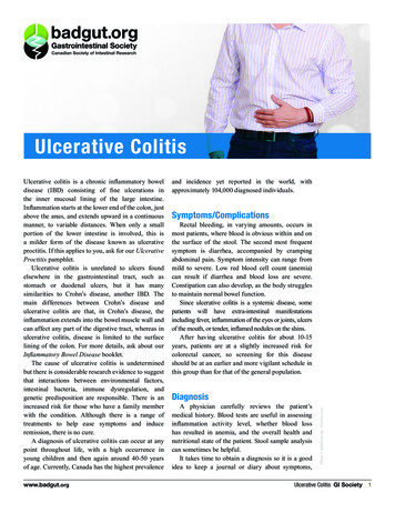
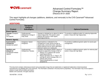
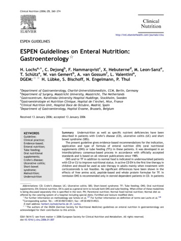
9Extraintestinal Manifestationsof Ulcerative ColitisBrian Huang, Lola Y. Kwan and David Q. ShihCedars Sinai Medical Center, Inflammatory Bowel and Immunobiology Institute (IB)USA1. IntroductionInflammatory bowel disease (IBD) consists predominantly of ulcerative colitis (UC) andCrohn’s disease (CD), which are clinically distinguished by intestinal localization, localfeatures of inflammation, a profile of complications, and familial aggregation. UC ischaracterized by recurring episodes of continuous inflammation limited to the mucosallayer of the colon and rectum, and approximately 42% of UC patients have extraintestinalcomplications (Ozdil et al., 2004). This chapter highlights key features of each involvedorgan system including association, diagnosis, and treatment options.2. Extraintestinal manifestations of Ulcerative ColitisThe extraintestinal manifestations (EIMs) of UC can affect any organ system ranging frommore common ones including the skin and hepatobiliary systems, to less common onesincluding the cardiovascular and renal systems (Das, 1999). Studies have shown that thedevelopment of one EIM can increase the risk of developing additional complications(Monsen et al., 1990).2.1 Skin and mucocutaneous complicationsThe skin is one of the most commonly affected organ systems in IBD patients. The two mostcommon skin disorders associated with UC, erythema nodosum (EN) and pyodermagangrenosum (PG), are reactive due to an immunologic response to UC. Non immunologiccauses can also occur secondary to nutritional deficiencies and medication side effects. (Das,1999; Timani & Mutasim, 2008). Skin disorders seen in UC caused by malnutrition includediseases such as pellagra and cheilitis. Aphthae are the most common complication of theoral mucosa, while fissures and fistulae are most common in the perianal mucosa.[FIGURES 1 AND 2] (Areias & Garcia E Silva, 1987). Standard medications used to treat UCinclude steroids and immunosuppressants which can cause skin disorders such ascushingoid features and drug eruptions (Timani & Mutasim, 2008). Other disorders, such asSweet’s syndrome and psoriasis have been associated with UC (Timani & Mutasim, 2008).2.1.1 Erythema NodosumEN affects about 3% of patients with UC (Areias & Garcia E Silva, 1987; Evans & Pardi, 2007;Mir-Madjlessi et al., 1985; Timani & Mutasim, 2008). Lesions affect females with UC morewww.intechopen.com
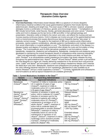
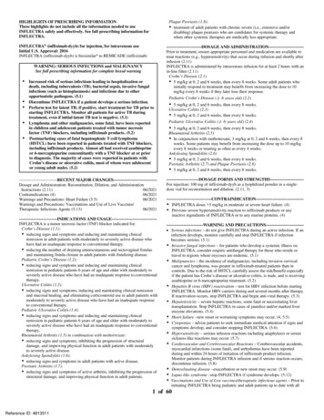
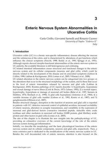
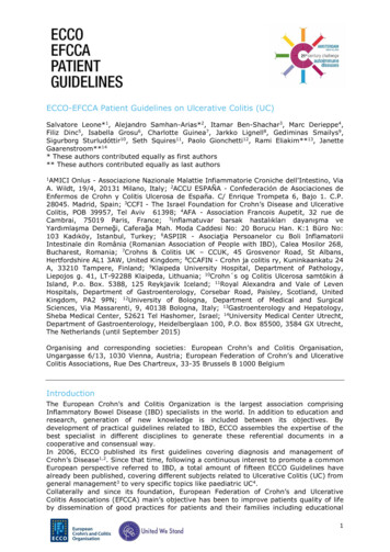
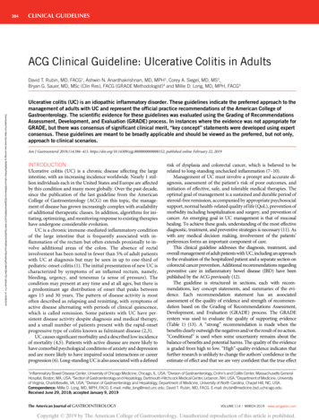
132Ulcerative Colitis – Epidemiology, Pathogenesis and ComplicationsFig. 1. Fistula with a setonFig. 2. UC patient with peri-anal fistula (left). Endoscopic view of fistula (right)frequently than men and EN rarely precedes the initial diagnosis of UC (Trost & Mcdonnell,2005; Weinstein et al., 2005). Typical EN lesions present as painful, raised subcutaneouslesions located on extensor surfaces of the extremities (Evans & Pardi, 2007; Timani &Mutasim, 2008). However, UC patients almost always have EN lesions on the anteriorsurface of the legs [FIGURE 3] (Mir-Madjlessi, Taylor et al., 1985). The skin nodules are nonulcerating and resemble a bruise on the skin (Timani & Mutasim, 2008). Unlike PG, ENlesions mirror UC disease activity and worsen with colonic flares (Timani & Mutasim, 2008).The average lag time between initial UC diagnosis and appearance of EN is 5 years (MirMadjlessi, Taylor et al., 1985).Fig. 3. EN affecting the lower extremities in a UC patientwww.intechopen.com
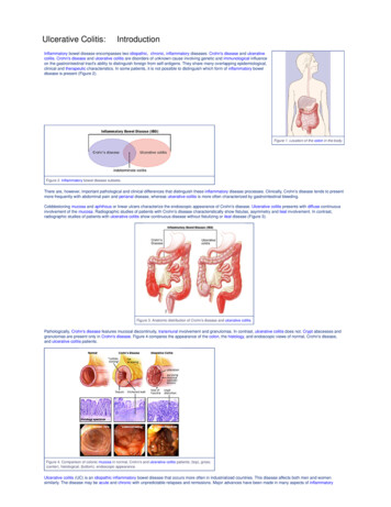
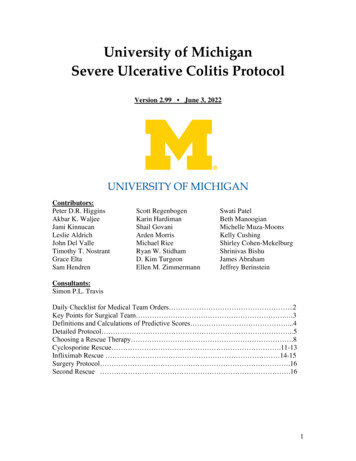
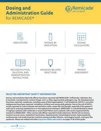
Extraintestinal Manifestations of Ulcerative Colitis133As EN lesions and UC disease activity usually parallel each other, treatment of theunderlying UC usually controls the EN lesions. Most lesions are self-limiting, thus aconservative approach to therapy is often practiced, such as leg elevation, rest, non-steroidalanti-inflammatory drugs (NSAIDs), and potassium iodide (Horio et al., 1981; Marshall &Irvine, 1997; Schulz & Whiting, 1976). In situations where lesions occur during quiescentphases of UC, treatment with oral steroids is effective. Studies have shown that time toremission in patients with EN is approximately 5 weeks which is significantly shorter thanthat seen in PG (Timani & Mutasim, 2008; Tromm et al., 2001).2.1.2 Pyoderma GangrenosumThe incidence of PG in patients with UC varies between 1.4 and 5% (Areias & Garcia E Silva,1987; Mccallum & Kinmont, 1968). PG is more commonly seen in patients with UC asopposed to CD, and as with EN, there is a slight female predilection (Bernstein et al., 2001b;Greenstein et al., 1976). PG was initially described in 1930 as necrotic ulcers with expandingborders of erythema (Newell & Malkinson, 1982). The onset of noninfectious pustules andnodules eventually expand outwards to develop painful shallow and deep ulcers (Callen,1998; Farhi & Wallach, 2008). PG usually occurs on the legs, but can also appear anywhereon the skin [FIGURE 4]. Pathergy, a phenomenon in which skin lesions develop secondaryto local trauma, has been reported in approximately 30% of cases of PG (Blitz & Rudikoff,2001; Callen, 1998). The average lag time between initial UC diagnosis and appearance of PGis 10 years (Mir-Madjlessi, Taylor et al., 1985). Diagnosis of PG is usually clinical, but skinbiopsy may be necessary for confirmation. PG is classified as a type of neutrophilicdermatosis in which the inflammatory infiltrate seen on microscopic examination showsdense dermal neutrophilic infiltrates without any evidence of infection (Cohen, 2009; Timani& Mutasim, 2008). Unlike EN, there is no temporal relationship between onset of UC flaresand the course of PG lesions (Thornton et al., 1980).Fig. 4. PG affecting the lower extremity (left) and face (right)In general, the PG lesions are more severe and refractory to therapy than EN. Treatment ofthe underlying UC activity does not always resolve the PG lesions and about 30% of patientsrequire additional treatment (Mir-Madjlessi, Taylor et al., 1985). The mainstay of treatingPG has been a combination of topical, intralesional, and systemic medications, but nospecific therapy has proven to be universally effective (Cohen, 2009; Timani & Mutasim,2008). Treatments with the best clinical evidence include systemic corticosteroids andwww.intechopen.com
134Ulcerative Colitis – Epidemiology, Pathogenesis and Complicationscyclosporine as maintenance therapy (Wollina, 2002), assuming there is no concurrentinfection. Initial doses of oral prednisone have ranged from 0.5 to 2 mg/kg/day and initialcyclosporine doses have ranged from 2 to 5 mg/kg/day (Timani & Mutasim, 2008; Wollina,2002). Maintaining target trough serum levels of 150-350 ng/ml for cyclosporine has shownto be effective in improving PG lesions (Cohen, 2009; Curley et al., 1985; Matis et al., 1992;Turner et al., 2010). In small lesions, intralesional steroid injections can be considered(Timani & Mutasim, 2008). Other agents including azathioprine, cyclophosphamide,methotrexate, high dose intravenous immunoglobulin, mycophenolate mofetil, minocycline,plasmapheresis, and hyperbaric oxygen treatment have been employed with variableefficacy (Cohen, 2009; Timani & Mutasim, 2008; Tutrone et al., 2007; Wasserteil et al., 1992).Biologics including infliximab and adalimumab have also been reported to improve PGlesions (Alkhouri et al., 2009; Brooklyn et al., 2006). Additionally, optical treatments such assteroids, tacrolimus, benzoyl peroxide, and hydrogen peroxide have shown positive results(Callen & Jackson, 2007). To avoid pathergy, unnecessary surgical interventions should beavoided. However, surgery can be considered if medical therapies are not successful. Propertiming of the surgery with immunosuppressants is essential for optimal long term woundstabilization (Rozen et al., 2001; Wittekindt et al., 2007; Wollina, 2002).2.1.3 Sweet’s syndromeSweet’s syndrome (SS), also known as acute febrile neutrophilic dermatosis, has a femalepredilection and classically affects women between the ages of 30 and 50 years (Timani &Mutasim, 2008). Although the pathogenesis is unclear, Sweet’s syndrome usually developsas a response to some type of underlying systemic disease, such as infection, malignancy,medications, or IBD (Vij et al., 2010) In fact, studies have shown that patients with SS haveunderlying disease in 50% of cases and an underlying malignancy in 20% of cases (Kemmett& Hunter, 1990; Souissi et al., 2007). UC and CD are the most common systemic diseasesassociated with SS (Timani & Mutasim, 2008). SS is characterized by an acute onset of fever,leukocytosis, and tender, erythematous plaques that can occur on the extremities, face, neck,and trunk (Burrall, 1999; Kemmett & Hunter, 1990; Souissi, Benmously et al., 2007). Whenthese lesions occur on the lower extremities, they often resemble EN and skin biopsy may benecessary to differentiate the two disease processes (Guhl & Garcia-Diez, 2008). Theselesions are burning-like and non-pruritic in quality. Other associated symptoms include, butare not limited to arthralgia, headache, fatigue, and other constitutional symptoms. Otherorgan systems such as the eye, kidney, liver, and pancreas can be involved as well (Cohen etal., 1988). As is also seen with PG, skin biopsy reveals neutrophilic infiltrates in the reticulardermis upon histopathological examination (Kemmett & Hunter, 1990; Timani & Mutasim,2008). The onset of symptoms from SS usually occur after the initial diagnosis of UC(Darvay, 1996).The mainstay of treatment for SS is steroids and multiple studies have shown dramaticimprovement with a 6 week course of systemic corticosteroid therapy (Cohen, Talpaz et al.,1988; Souissi, Benmously et al., 2007). Topical or intralesional steroids are effective forlocalized disease (Timani & Mutasim, 2008). Recurrence is common and has been reportedto affect approximately 1/3 of patients (Kemmett & Hunter, 1990). Untreated lesions havebeen reported to heal after variable periods of time, but can be associated with scarring(Kemmett & Hunter, 1990; Timani & Mutasim, 2008). Other alternative first line treatmentsinclude potassium iodide and colchicine. Second line agents including indomethacin andclofazimine have been used with successful results, but are not as effective ascorticosteroids, potassium iodide, and colchicine (Cohen, 2009; Cohen & Kurzrock, 2002).www.intechopen.com
Extraintestinal Manifestations of Ulcerative Colitis1352.1.4 Mucocutaneous manifestationsAphthous Stomatitis. About 4.3% of UC patients experience recurrent aphthous stomatitisand symptom onset often parallels UC disease activity (Areias & Garcia E Silva, 1987;Timani & Mutasim, 2008). Minor aphthous ulcers are small, round, painful, and heal within2 weeks without scarring, while major recurrent ulcers are larger, can last for 6 weeks, andfrequently scar [FIGURE 5] (Ship, 1996; Timani & Mutasim, 2008). A study showed that amajority of patients with multiple aphthous ulcers had underlying IBD (Letsinger et al.,2005). The pathogenesis of aphthous stomatitis and UC is still unclear; studies have not beensuccessful in proving that these ulcers are secondary to vitamin deficiencies as the lesionsdid not improve with vitamin therapy (Basu & Asquith, 1980). Treatment options includetreating the underlying UC, symptomatic relief with steroid elixirs, and systemic treatmentwith steroids and immunosuppressants (Basu & Asquith, 1980; Timani & Mutasim, 2008).Fig. 5. Aphthous stomatitis in a UC patientPyostomatitis Vegetans. Pyostomatitis vegetans, a rare disorder of the oral mucosa, hasbeen shown to be a specific marker for IBD, especially UC (Storwick et al., 1994; Timani &Mutasim, 2008). Lesions are hyperplastic folds of the mucosa with small abscesses anderosions and often manifest before the diagnosis of UC (Hansen et al., 1983).Pyostomatitis vegetans is usually resistant to treatments such as topical steroids, antibioticmouthwashes, or hydrogen peroxide. Systemic steroids and immunosuppressants havebeen employed with variable success, but were not always successful in maintainingremission (Timani & Mutasim, 2008). In one case report, topical fluocinonide gel resulted intemporary state of remission, but total colectomy was necessary to achieve completeremission (Calobrisi et al., 1995).2.1.5 Miscellaneous skin disordersStudies report an increased risk of psoriasis with UC. In one study, of 88 patients with UC,5.7% had psoriasis, compared to 1.5% in the control group, suggesting some type of geneticrelationship between the two (Yates et al., 1982). Another study found an associationbetween UC and hidradenitis suppurativa [FIGURE 6].www.intechopen.com
136Ulcerative Colitis – Epidemiology, Pathogenesis and ComplicationsFig. 6. UC patient with hidradenitis suppurativa2.2 Hepatopancreatobiliary complicationsThere are multiple types of hepatic, biliary, and pancreatic complications associated withUC. Pancreatic complications are less common than hepatobiliary complications, but will bediscussed in this section as well given its anatomic location.2.2.1 Primary Sclerosing Cholangitis (PSC)PSC is a chronic cholestatic liver disease that is caused by progressive inflammatorydestruction of intra- and extra-hepatic bile ducts, which leads to multifocal biliary strictures,resulting in liver cirrhosis and failure. PSC is believed to be the result of a combination ofgenetic and immunological factors, resulting in immune dysfunction and impropertargeting of the biliary system by lymphocytes and autoantibodies (Bergquist et al., 2008;Karlsen et al., 2007; Saarinen et al., 2000). Multiple autoantibodies including ANA, RF andatypical p-ANCA have been described in UC and PSC patients (Chapman et al., 2010;Terjung & Spengler, 2009). Atypical p-ANCA is detected by indirect immunofluorescencestaining on ethanol fixed neutrophils in a perinuclear or nuclear pattern whereas classicalANCA targets myeloperoxidase and proteinase 3 in a perinuclear pattern (Terjung et al.,1998). Atypical p-ANCA is detectable in approximately 70% of PSC patients with or withoutUC and is also described in autoimmune hepatitis in patients with IBD (Duerr et al., 1991;Saxon et al., 1990; Terjung & Spengler, 2009). Atypical p-ANCA recognizes both betaisoform 5 in human neutrophils and bacterial cell division protein FtsZ, which is highlyconserved across bacterial microflora in the gut (Terjung et al., 2010). This evidence supportsthe theory of a combined pathogenesis of PSC or autoimmune hepatitis in UC. It isproposed that an altered immune response to bacterial antigen in the gut lumen results in a"leaky gut” and stimulation of pattern recognition receptors (cross-recognition betweenbacterial antigen in the gut and host components, such as beta isoform 5 in neutrophils)which gives rise to autoimmunity phenomenon. (Terjung & Spengler, 2009).PSC typically affects young to middle aged males (male to female ratio of 2:1) and whileonly 5% of patients with UC will develop PSC, about 70% to 80% of cases of PSC occur inpatients with UC (Charatcharoenwitthaya & Lindor, 2006; Lundqvist & Broome, 1997). Theonset of PSC can precede or follow the onset of UC, and may not be related to UC diseaseactivity (Larsen et al., 2010). An isolated alkaline phosphatase level may be the only findingduring the early stages of the disease, but PSC usually manifests with chronic intermittentobstructive jaundice. As the disease progresses, serum bilirubin will rise (Lee & Kaplan,1995). In the latter course of the disease, the prothrombin time will be prolonged and serumalbumin level will decrease, signaling progression to hepatic failure. Other symptoms suchas right upper quadrant abdominal pain, pruritus, fatigue, fever, and weight loss arewww.intechopen.com
Extraintestinal Manifestations of Ulcerative Colitis137variably present. However, PSC should be suspected in asymptomatic patients with anisolated elevation of serum alkaline phosphatase (Larsen, Bendtzen et al., 2010).Symptomatic patients manifest with icterus, jaundice, hepatomegaly and splenomegaly(Chapman, Fevery et al., 2010; Farrant et al., 1991; Wiesner et al., 1989).Pericholangitis, described by studies as PSC of the small bile ducts, is another complicationof UC. A study of 107 patients with UC showed that 35% patients had “small duct” PSCcompared to 17% with “large duct” PSC, but histologic examinations were indistinguishablefrom each other (Wee & Ludwig, 1985). These findings imply that rather than being separatedisorders, PSC and pericholangitis merely affect different areas of the same disease process.Pericholangitis (or small-duct PSC) is now referred to patients with biochemical andhistologic characteristics of PSC, but with normal appearing cholangiograms (Wee &Ludwig, 1985). Small-duct PSC is more often associated with Crohn's disease whereas largeduct PSC (classical PSC) is more often associated with UC (Bjornsson et al., 2008; Loftus,1997; Rasmussen et al., 1997). Compared to large-duct PSC, small-duct PSC has a relativelyfavorable prognosis with longer transplant-free survival. Approximately 25% of patientswith small-duct PSC progress to large-duct PSC, but usually does not develop intocholangiocarcinoma unless it progresses to large-duct PSC (Bjornsson, Olsson et al., 2008).The gold standard for diagnosis is endoscopic retrograde cholangiopancreatography (ERCP)and typical radiographic findings include multifocal strictures and dilatation of the intraand/or extra-hepatic biliary tracts, producing a "beaded pattern" (Lee & Kaplan, 1995;Maccarty et al., 1983). Recently, magnetic resonance cholangiopancreatography (MRCP) hasemerged as a non-invasive alternative in diagnosing suspected PSC (Chapman, Fevery et al.,2010). ERCP may be more preferable over MRCP in diagnosing early stage PSC having aspecificity and sensitivity near 100% (Angulo et al., 2000; Berstad et al., 2006; Moff et al.,2006). However, ERCP does carry potential for significant risks and complications (Larsen,Bendtzen et al., 2010). Hence, there has been an increased use of MRCP as an initialdiagnostic tool, followed by ERCP as needed. If the diagnosis is in doubt, liver biopsy canassist in the confirmation of diagnosis as well as staging of disease (Charatcharoenwitthaya& Lindor, 2006). However, the classic pathognomonic finding of "onion-skin lesions" orperiductal concentric fibrosis is rarely seen (Chapman, Fevery et al., 2010). Liver biopsy canbe essential in the diagnosis of small-duct PSC, but is not required for the diagnosis of largeduct PSC (Burak et al., 2003; Chapman, Fevery et al., 2010).Multiple studies show that using ursodeoxycholic acid (UDCA) at moderate to high dosageshas been shown to improve liver histology and liver biochemistry (Smith & Befeler, 2007).Even though UDCA has not been demonstrated to improve either symptoms or mortality, ithas been shown to reduce the incidence of colonic dysplasia, colorectal carcinoma andcholangiocarcinoma (Pardi et al., 2003). Focus should be on starting these patients earlyenough to delay progression to cirrhosis, cholangiocarcinoma, and death. This is especiallyimportant as UDCA is not as effective in patients with end stage PSC. During treatmentwith UDCA, stenosis of bile ducts may occur and endoscopic intervention with dilatationhas been showed to be effective (Stiehl, 2004). At the present time, liver transplantation isthe only effective treatment especially for end-stage PSC or PSC with cholangiocarcinoma(Chapman, Fevery et al., 2010; Navaneethan & Shen, 2010). Orthotopic liver transplantationis the established treatment in PSC with 85% to 90% of 5-year survival rate (Gow &Chapman, 2000). However, 20 to 25% of transplanted patients develop recurrent PSC 5 to 10years after transplant (Alabraba et al., 2009; Campsen et al., 2008; Graziadei et al., 1999;Navaneethan & Shen, 2010).www.intechopen.com
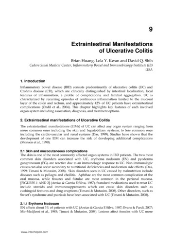
138Ulcerative Colitis – Epidemiology, Pathogenesis and Complications2.2.2 MalignanciesPSC has been shown to be complicated by malignancies including cholangiocarcinoma (CCA),bile duct carcinoma, gall bladder carcinoma, and hepatocellular carcinoma. Studies haveshown that patients with UC and PSC are more prone to develop cholangiocarcinoma,colorectal cancer, gallbladder cancer and hepatocellular carcinoma (Broome et al., 1995; Florinet al., 2004; Kitiyakara & Chapman, 2008). Typically CCA presents as an intraductal tumor,and at times can be quite difficult to differentiate from a benign PSC stricture [FIGURE 7].Evidence supporting diagnosis of CCA include high levels of CA 19-9, cross sectional liverimaging with long, confluent strictures and ERCP with brush biopsy of strictures, in additionto overall clinical presentation (Chapman, Fevery et al., 2010; Charatcharoenwitthaya et al.,2008; Levy et al., 2005). The relative risk of bile duct carcinoma in UC patients is 31.3 comparedto the general population and the prognosis is quite poor with a mean survival of less than oneyear. PSC and pericholangitis are common pre-existing lesions in UC patients with bile ductcarcinoma (Mir-Madjlessi et al., 1987). A case report recommended that in patients with UCand PSC with abnormal gallbladders, liver biopsy and cholecystectomy should be performed(Dorudi et al., 1991). Another case report documented an association between fibrolamellarhepatocellular carcinoma and UC complicated by PSC (Snook et al., 1989).Fig. 7. ERCP of a UC patient with PSC showing high grade strictures within the right andleft hepatic ducts with secondary pronounced intrahepatic ductal dilatation2.2.3 Primary Biliary Cirrhosis (PBC)PBC is autoimmune in nature with the unique presence of anti-mitochondrial antibodies(AMA) on laboratory testing. Studies have reported that in patients with UC, there is a 30fold risk of primary biliary cirrhosis compared to the general population (Koulentaki et al.,1999). UC patients with PBC, as compared to patients without UC, are younger and morelikely to be male. The mainstay treatment of PBC includes cholestyramine to relieve itchingby reducing the amount of bile acid in the blood, UDCA to increase bile flow to reduceinflammation in bile ducts, fat soluble vitamins for nutritional supplementation, and ERCPfor bile drainage. Ultimately, advanced PBC with liver failure and portal hypertensionrequires liver transplantation.www.intechopen.com
Extraintestinal Manifestations of Ulcerative Colitis1392.2.4 Autoimmune Hepatitis (AIH)Autoimmune chronic active hepatitis (CAH) has been observed in UC patients with andwithout PSC (Rabinovitz et al., 1992; Snook, Kelly et al., 1989). In addition to laboratory testsshowing the presence of autoantibodies, such ANA and anti-smooth muscle muscleantibody (ASMA) and abnormal liver function tests, liver biopsy may be necessary toconfirm the diagnosis (Alvarez et al., 1999). In patients with both UC and autoimmunehepatitis, up to 42% have abnormal cholangiographic findings indicating the coexistence oroverlap of PSC (Perdigoto et al., 1992). UC patients with both AIH and PSC respondrelatively poorly to immunosuppression and progress more rapidly to cirrhosis.Autoimmune CAH responds to steroids, but in the presence of PSC, dual therapy withsteroids and azathioprine is indicated (Perdigoto, Carpenter et al., 1992; Rabinovitz,Demetris et al., 1992). Thus, in patients with UC complicated by autoimmune CAH,cholangiography is necessary to rule out PSC to dictate further therapy.2.2.5 Other hepatic complicationsFatty liver or hepatic steatosis can be observed in UC patients and appears to correlate withdisease severity or duration and may contribute to abnormal liver function tests (Riegler etal., 1998). The ultrasound findings of fatty liver show hepatomegaly and a dysechogenicpattern (De Fazio et al., 1992). Causes of fatty liver include malnutrition, protein loss, andsteroid use. Patients are usually asymptomatic and treatment is geared towards treatment ofthe underlying UC and improving nutritional status (Navaneethan & Shen, 2010).Hepatic amyloidosis is an extremely rare complication of UC (0.07% vs. 0.9% in CD) andcontrol of gut inflammation can reduce amyloid deposition severity (Greenstein et al., 1992;Navaneethan & Shen, 2010; Wester et al., 2001). Because of the rarity and asymptomaticnature of this complication, the need for liver biopsy remains unclear and should be madeon a case-by-case basis.Hepatic and/or splenic abscesses are rare complications of IBD and even more rare in UC.They are diagnosed by ultrasound or CT scan showing non-enhancing hypodense lesions inthe liver and/or spleen. Clinical suspicion of hepatosplenic abscesses should be consideredin febrile patients with IBD whose presentations are inconsistent with IBD exacerbation. Thetreatments of hepatosplenic abscesses include drainage, broad-spectrum antibiotics, andtreatment of underlying IBD with sulfasalazine and/or steroids. The pathogenesis ofhepatosplenic abscess in UC is unclear at this time with possible mechanisms includinginfectious and immunologic etiologies (Navaneethan & Shen, 2010).2.2.6 Portal vein thrombosisPortal vein thrombosis is considered an EIM of UC and is thought to be secondary tocoagulation abnormalities caused by chronic bowel inflammation. Ulceration of the mucosabarrier can increase the chance of gut microbial translocation and thus portal veinthrombosis (Navaneethan & Shen, 2010). Portal vein thrombosis has also been observed inUC patients who were status post proctocolectomy (Navaneethan & Shen, 2010).2.2.7 Pancreatic complicationsPatients with UC have increased chances of developing both acute and chronic pancreatitis(Keljo & Sugerman, 1997). Causes include idiopathic, gallstones, PSC, and medication sideeffects from drugs such as 5-ASA, mesalamine, corticosteroids, azathioprine, and 6mercaptopurine. Medication induced pancreatitis does not lead to chronic pancreatitis andwww.intechopen.com
140Ulcerative Colitis – Epidemiology, Pathogenesis and Complicationstreatment involves cessation of the insulting drug (Bank & Wright, 1984). On the other hand,the etiology of chronic pancreatitis is unknown. Chronic pancreatitis with UC is usuallypainless, but is associated with pancreatic duct strictures and severe pancreatic exocrineinsufficiency (Pena et al., 2000).Autoimmune pancreatitis (AIP) is a less common complication of UC compared to druginduced or gallstone pancreatitis (Navaneethan & Shen, 2010). It affects elderly individualsand can present with obstructive jaundice. Steroids have become the standard treatment ofAIP for obstructive jaundice, abdominal pain, and prevention of future episodes ofpancreatitis (Finkelberg et al., 2006; Kamisawa et al., 2009). Failure to respond to steroidsraises the possibility of pancreatic cancer and warrants further work up for malignancy.2.3 Musculoskeletal complicationsMusculoskeletal complications are quite common and affect approximately 25% of all IBDpatients (Bourikas & Papadakis, 2009; Danese et al., 2005). The musculoskeletal involvementof UC patients can be divided into the following categories:1. Arthritis: peripheral arthritis, axial arthritis including ankylosing spondylitis (AS) andsacroiliitis2. Periarticular inflammation: enthesitis, tendonitis, dactylitis, clubbing, periostitis,myositis, fibromyalgia, and granulomatous lesions of the joint and bone3. Metabolic bone disorders: osteoporosis, osteopenia, osteonecrosis4. Localized myopathy: orbital myositis, gastrocnemius myalgia syndrome, polymyositis,dermatomyositis5. Impaired growth: seen in children and adolescents2.3.1 ArthritisPeripheral Arthritis: Arthritis, located peripherally or axially, can precede, occurconcurrently, or follow the diagnosis of UC (Bourikas & Papadakis, 2009). The type ofarticular involvement in UC is inflammatory and is associated with pain, heat, swelling, anddecreased joint mobility. Similar to rheumatoid arthritis (RA), the pain and stiffness ofarthritis in UC is worse in the morning and improves with physical activity. However, unlikeRA arthritis, UC arthritis is typically seronegative, non-deforming, and non-erosive. Theperipheral arthropathy seen with UC can be classified into two types: Type 1 and Type 2(Atzeni et al., 2009; Bourikas & Papadakis, 2009; Brakenhoff et al., 2010; Jose & Heyman, 2008).Type 1 is pauci-articular, asymmetrical in distribution, and typically affects less than 5 largejoints, such as the knees, elbows, and ankles. The risk of developing peripheral arthritis ishigher in UC patients and increases with colonic involvement (Bourikas & Papadakis, 2009;Jose & Heyman, 2008). The risk of developing type 1 peripheral arthritis is also increasedwith the presence of other EIMs, such as EN, PG, abscesses, stomatitis, and uveitis (Jose &Heyman, 2008). With the exception of small joints or type 2 polyarticular arthritis, thearthritis usually correlates with UC disease activity (Atzeni, Ardizzone et al., 2009). This typeof arthritis is sometimes referred to as colitic arthritis, affects up to 20% of UC patients, andusually presents as an acute self-limiting episode that lasts less than 10 weeks with a median of5 weeks (Bourikas & Papadakis, 2009; Das, 1999; Jose & Heyman, 2008). However, about 20%to 40% will experience recurrent episodes of arthritis.Type 2 arthropathy is typically polyarticular, involving 5 or more joints including the smalljoints of the hands, and most commonly affects the MCP joints of the hands. This type ofpolyarticular arthritis with IBD is associated with uveitis, but not with other EIMs of IBD.www.intechopen.com
Extraintestinal Manifestations of Ulcerative Colitis141Unlike type 1 arthritis, symptoms of type 2 arthritis are independent of bowel diseaseactivity and can persist for months to years with a median of 3 years (Bourikas & Papadakis,2009; Jose & Heyman, 2008). Genetic factors implicated in the association betweenperipheral arthritis and UC include: HLA-B27, HLA-B35, HLA-DR, and HLA-B44(Brakenhoff, Van Der Heijde et al., 2010).Axial Arthritis: Axial arthritis, referred as type 3 arthritis, includes both AS and isolatedsacroiliitis. This axial t
Ulcerative Colitis Epidemiology, Pathogenesis and Complications 134 cyclosporine as maintenance therapy (Wollina, 2002), assuming there is no concurrent infection. Initial doses of oral prednisone ha ve ranged from 0.5 to 2 mg/kg/day and initial cyclosporine doses have ranged from 2 to 5 mg/kg/day (Timani & Mu tasim, 2008; Wollina, 2002).