Transcription
Acta Biomaterialia 10 (2014) 11–25Contents lists available at ScienceDirectActa Biomaterialiajournal homepage: n scaffolds for tissue engineering of vascular graftsAnwarul Hasan a,b, Adnan Memic c, Nasim Annabi a,b, Monowar Hossain d, Arghya Paul a,b,Mehmet R. Dokmeci a,b, Fariba Dehghani e, Ali Khademhosseini a,b,f,g, aCenter for Biomedical Engineering, Department of Medicine, Brigham and Women’s Hospital, Harvard Medical School, Cambridge, MA 02139, USAHarvard–MIT Division of Health Sciences and Technology, Massachusetts Institute of Technology, Cambridge, MA 02139, USAcCenter of Nanotechnology, King Abdulaziz University, Jeddah 21589, Saudi ArabiadDepartment of Medicine, Lyell McEwin Hospital, University of Adelaide, SA 5112, AustraliaeSchool of Chemical and Biomolecular Engineering, The University of Sydney, Sydney 2006, AustraliafWyss Institute for Biologically Inspired Engineering, Harvard University, Boston, MA 02115, USAgWorld Premier International – Advanced Institute for Materials Research (WPI-AIMR), Tohoku University, Sendai 980-8577, Japanba r t i c l ei n f oArticle history:Received 11 April 2013Received in revised form 8 July 2013Accepted 13 August 2013Available online 22 August 2013Keywords:ElectrospinningTubular scaffoldsVascular graftsTissue engineeringMechanical propertiesa b s t r a c tThere is a growing demand for off-the-shelf tissue engineered vascular grafts (TEVGs) for the replacementor bypass of damaged arteries in various cardiovascular diseases. Scaffolds from the decellularized tissueskeletons to biopolymers and biodegradable synthetic polymers have been used for fabricating TEVGs.However, several issues have not yet been resolved, which include the inability to mimic the mechanicalproperties of native tissues, and the ability for long-term patency and growth required for in vivo function. Electrospinning is a popular technique for the production of scaffolds that has the potential toaddress these issues. However, its application to human TEVGs has not yet been achieved. This reviewprovides an overview of tubular scaffolds that have been prepared by electrospinning with potentialfor TEVG applications.Ó 2013 Acta Materialia Inc. Published by Elsevier Ltd. All rights reserved.1. IntroductionEach year, 1.4 million patients in the USA need arterial prostheses [1]. The available options for the replacement of vascular graftshave limited clinical success, with a cost of more than US 25 billion [1]. Particularly, the pathologies affecting small and medium-sized blood vessels are the primary cause of death [2,3].Atherosclerosis is a fatal disease that causes build-up of plaquesunder the intimal layer, reducing the cross-sectional area availablefor blood flow and thereby resulting in decreased flow of blood tothe tissues downstream to the plaque [4]. Eventually, cardiac andperipheral bypass surgeries become necessary, requiring thereplacement of a segment of blood vessels. In blue baby syndrome,only one of the two ventricles of an infant functions properly, and a‘‘Fontan operation’’ becomes necessary [5]. In ‘‘Fontan operations’’,an engineered blood vessel is required to connect the right pulmonary artery with the inferior vena cava so that the Corresponding author at: Center for Biomedical Engineering, Department ofMedicine, Brigham and Women’s Hospital, Harvard Medical School, Cambridge, MA02139, USA.E-mail address: alik@rics.bwh.harvard.edu (A. Khademhosseini).deoxygenated blood can bypass the heart and travel straight tothe lungs. Similarly coronary artery diseases and peripheral vascular diseases often require the replacement of diseased and damaged native blood vessels [6]. The currently available options forthese transplants are autologous grafts (e.g. coronary artery bypassgraft with autologous mammary arteries and saphenous veins),allografts (donor/cadaveric), xenografts (e.g. bovine or porcine pulmonary valve conduit), artificial prostheses or synthetic vasculargrafts made of expanded polytetrafluroethylene (ePTFE) and polyethylene terephthalate (PET) [7].The use of autografts and allografts is limited due to the lack oftissue donors, previous harvesting or anatomical variability [8,9].Besides, there are concerns about their long-term functionalitydue to the use of strong detergents and decellularizing agents.Xenografts suffer from their relatively shorter life span. As anexample, a bovine or porcine graft may last for up to 15 years,which is a major issue for pediatric patients, who will require anew implant replacement at intervals of 10–15 years. Other issuesinclude poor control over physical and mechanical properties,inflammation and calcification [10,11].Synthetic prosthetic grafts are rejected within a few months bythe immune system of the body if the diameter of the vessel issmaller than 6 mm. This rejection arises from the associated reocclusion problem caused by thrombosis, aneurysm and intimal1742-7061/ - see front matter Ó 2013 Acta Materialia Inc. Published by Elsevier Ltd. All rights 8.022
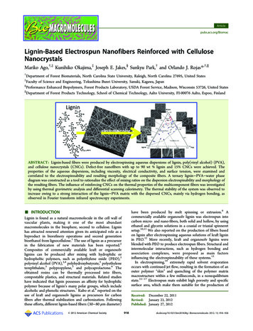
12A. Hasan et al. / Acta Biomaterialia 10 (2014) 11–25hyperplasia due to mismatch of compliance (compliance is theopposite of stiffness, measured as the strain/expansion or contraction of the graft with force) [8,10,12,13].Tissue engineering is an alternative approach for creating newvascular grafts. In this approach, cells are seeded or encapsulatedin scaffolds fabricated from a biodegradable polymer. In tissueengineering, it is anticipated that cells produce extracellular matrix(ECM) while the polymer is degraded, gradually creating the intended tissue. Extensive research has been conducted on tissueengineered vascular grafts (TEVGs) over the past few decades,and as a result significant progress has been made in terms ofachieving the remodeling of the tissue in the TEHV constructs similar to the native blood vessels [14–17], as depicted in Fig. 1. Several reviews have already been published that discuss thedifferent methods for tissue engineering of vascular grafts [3,18],the design of natural and artificial arteries as well as vascular networks [19] and the application of stem cells and other human cellsfor tissue engineering of blood vessels [20]. The main approachesfor fabrication of scaffolds include methods that are based onmolecular self-assembly, hydrogels, the solvent casting–particulate leaching technique, thermally induced phase separation andthe electrospinning process. The latter is a versatile technique forfabrication of nano/microscale fibers, which has a great potentialfor mimicking the microenvironment of natural ECM. The successof an implantable tissue engineered vascular graft is governed,among other factors, by the development of a scaffold that mimicsthe ECM [21]. It is well known that in natural tissues the ECM is athree-dimensional (3-D) network of 50–500 nm diameter structural protein and polysaccharide fibers. Electrospinning hasevolved to allow the fabrication of nanofiber scaffolds in this sizerange and beyond [13].Numerous review articles are available on the fundamentalsand applications of electrospinning, its historical developmentand various modifications [22–30]. These reviews illuminate thecapability of the electrospinning technique for the fabrication of fibers from a broad range of polymers, including purely natural, tosynthetic, to composite mixtures of ECM analogs and interstitialconstituents from organ-specific extracts [31–33]. These fiberswith nano/micron-sized pores and a large surface area have beenused as catalysts, filtration systems, protective clothing, drug delivery depots, optical wave guides, electronics and tissue engineeringscaffolds [31,32,34–39]. The versatility of this technique makes italso possible to process various categories of non-polymeric materials, including ceramics and their composite with polymer [34].Electrospinning is a flexible technique in which the mechanicaland biological properties of fibers can easily be tuned by varyingthe composition of a mixture which is not easily possible in otherscaffold fabrication methods.Due to these advantages, in recent years, the interest in theelectrospinning technique for the fabrication of tissue engineeringscaffolds has increased exponentially. In the current review wepresent a summary of the most recent application of electrospinning for the fabrication of tissue engineered vascular grafts. Thescope is limited to small and medium-sized blood vessels (diameter 6 5 mm), while large blood vessels (diameter 5 mm), microvasculature and capillary networks are beyond the scope of thisreview. The aspects that are critical for the design of TEVG suchas the structure of vascular grafts and function are also discussed.Fig. 1. Tissue engineered blood vessels. (A–C) the first clinically used sheet-based tissue engineered blood vessel tested on three human patients for application as highpressure arteries: (A) a tissue engineered graft was implanted between the axillary vein and the humeral artery as an arteriovenous shunt, (B) the vessel exhibited normalsuturing and surgical handling properties, (C) the shunt maintained high flow without signs of aneurysm or restenosis even after 6 months. Reprinted from Ref. [14] withpermission from Nature Publishing Group. (D–O) A comparative histological analysis of human pericyte cell-seeded TEVGs (D–I), unseeded scaffolds (J–L) and native rat aorta(M–O). The H&E (D, G, J and M), Masson’s trichrome (E, H, K and N) and Verhoeff–Van Gieson (F, I, L and O) stainings demonstrated remodeling of the tissue in the TEHVconstruct enriched with collagen and elastin similar to the native aorta. (G–I) are magnified images for the wall cross-section of (D–F). Arrows indicate the remodeled tissuewhile ES stands for electrospun scaffold layer. The remaining PEUU material was unspecifically stained in black by Verhoeff–Van Gieson stain in (I) and (L). Reprinted fromRef. [17] with permission from Elsevier Science.
A. Hasan et al. / Acta Biomaterialia 10 (2014) 11–252. Structure of vascular graftsThe native blood vessels in the human body have complexstructures with distinct features. The arterial wall is composed ofthree different layers: (i) intima, (ii) media and (iii) adventitia(Fig. 2). Intima, the innermost of the three concentric layers, consists of a continuous monolayer of endothelial cells (ECs) directlyattached to the basement membrane, which mainly comprisesconnective tissues. The media is the middle layer, comprisingdense populations of concentrically organized smooth muscle cells(SMCs) with fibers or bands of elastic tissues. Adventitia consists ofa collagenous ECM that mainly contains fibroblasts and perivascular nerve cells. In smaller arterioles and capillaries some of theselayers might be less obvious or absent [3]. An internal and anexternal elastic lamina separate the intima, media and adventitiafrom each other. Each layer serves a specific function: the collagenous adventitia functions to add rigidity while the elastic laminaprovides elasticity to the vessel walls.A confluent monolayer of EC in the lumen of the blood vessel iscritical for preventing the clotting of the blood, infection andinflammation of adjacent tissues. The EC monolayer is also important for regulating the gaseous and molecular (oxygen and nutrients) exchange. It also controls the signaling to the media,particularly to its muscular component [4]. The SMCs in the middlelayer (media) of the blood vessel walls have a unique contractilefunction. When the signals from EC or cytokines stimulate theSMCs, the latter cells dilate and contract in a coordinated manner.This leads to the dilation and contraction of the vessels as the pressure in the vessels changes. The concentric layer-wise structure ofECM, the spatial organization and alignment of the EC and SMCsand the interplay between the cells and ECM structures are therefore important factors that should be taken into account whendesigning tissue engineered blood vessels.3. Functional requirements for vascular graftsIt is crucial that TEVG functions, i.e. gets integrated to the adjacent blood vessels, sustains the load from blood pressure and allows blood flow without leakage, immediately after implantation.Biocompatibility and bioactivity are other primary requirementsfor engineering vascular grafts. In addition, the mechanical properties, adhesive ligands, growth factor presentation, transport anddegradation kinetics of the materials used for the scaffold shouldmimic the relevant ECM environment to a reasonable extent. It isfavorable to use cost-effective and cytocompatible materials withFig. 2. Schematic cross-sectional view of an artery. The arterial wall comprisesthree main layers: (i) adventitia, (ii) media and (iii) intima. A layer of endothelialcell covers the internal surface of the lumen while smooth muscle cells andfibroblast cells exist in outer layers.13tunable properties for a particular tissue [37]. An exact replicationof the native tissue structure is not always necessary, however, as afunctional construct, it is necessary that an engineered vasculargraft fulfills certain basic criteria [3,14]. For example, the burststrength above 260 kPa is required for a TEVG [6,14], to preventrupture due to variation in blood pressure. In addition, adequateelasticity is crucial to withstand cyclic loading in which no dilationoccurs in constructs within a month of in vitro cyclic loading within physiological ranges [14]. The engineered vascular graft shouldbe compatible with the adjacent host vessel and provide an antithrombotic lumen (autologous endothelium) [4]. The ability ofthe scaffold to provide initial mechanical function for the vasculargraft is another important factor, even though the structural andmechanical characteristics of the native vessels are expected tobe gradually acquired through remodeling, repair and growth uponimplantation. Finally, it is also important that the implanted vascular grafts minimize intimal hyperplasia and allow for regenerationof arterial tissues.The uniaxial tensile properties of several native human bloodvessels are presented in Table 1. These properties can be used astarget properties for tissue engineered vascular grafts. Some safetyfactors may also be incorporated in the design. There are someapparent discrepancies in the literature among the reported valuesof various mechanical properties for the same tissue by differentresearchers. For example, the uniaxial tensile values of 43 MPa[40], 4.2 MPa [41] and 2.25 MPa [54] are reported for humansaphenous vein in the circumferential direction. This discrepancyis due to the inconsistency in methods for extracting the resultsfrom the experimental load–displacement data. The values forthe elastic modulus in Donovan et al. [40], for example, were takenas the maximum linear slope of the curve before failure while thatin Stekelenburg et al. [41] was taken from the initial linear elasticregion of the curve. A reconciliation of literature data is thereforeimportant when comparing experimental results from differentgroups.In developing any tissue engineering product including TEVG itis also crucial that researchers take into account the requirementsbehind the Food and Drug Administration’s (FDA’s) approval process to ensure that the FDA’s approval can be obtained for releasingthe end product in the market and that it can be used in the clinic.Researchers need to contemplate the safety, efficacy, purity andidentity of biomaterials in the engineering and design of products.4. Application of electrospun scaffolds in tissue engineering ofvascular graftsElectrospinning offers the ability to fine-tune mechanical properties during the fabrication process, while also controlling thenecessary biocompatibility and structure of the tissue engineeredgrafts. The ability of the electrospinning technique to combinethe advantages of synthetic and natural materials makes it particularly attractive for TEVG, where a high mechanical durability, interms of high burst strength and compliance (strain per unit load),is required. In addition, the incorporation of natural polymers, withan abundance of cell binding sites, can promote the formation of acontinuous monolayer of EC in the lumen and proliferation of othercell types in the matrix of the graft’s wall. The electrospinningtechnique also offers precise control over the composition, dimension and alignment of fibers that have impact on the porosity, poresize distribution and architecture of scaffolds. This method allowsfor engineering of a wide range of tunable structural and mechanical properties as required for specific applications. Moreover,aligned nanofibers can be used for orienting cells in a specificdirection necessary to provide the anisotropy encountered incertain organs including blood vessels. Companies have recently
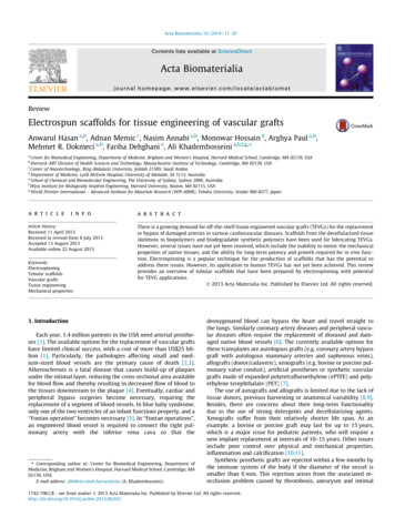
14A. Hasan et al. / Acta Biomaterialia 10 (2014) 11–25Table 1Uniaxial tensile mechanical properties (mean values): elastic modulus, ultimatetensile stress, tensile strain at failure and burst strength of some native human bloodvessels.Type of enous vein(circ.)Saphenous vein(long.)Saphenous vein(circ.)Saphenous vein(long.)Saphenous vein(circ.)Left internalmammaryartery (circ.)Left internalmammaryartery(long.)Femoral 63–764.1. Electrospinning process and parameters[40]1680–3900[41][41][41]NAcirc., circumferential; long., longitudinal; NA, not available.commenced the fabrication of electrospun grafts for transplantation of trachea, and other tissue engineered conduits. The world’sfirst tissue engineered tracheal transplant was successfully usedin a clinical trial in Sweden in June 2011 [42].[108,109]The electrospinning process is based on the stretching of a viscoelastic solution into nano/microfibers using a high electrostaticforce. In-depth reviews about the electrospinning process can befound elsewhere [15–23]. In brief, the material to be electrospunis loaded into a syringe and is pumped at a slow flow rate by a syringe pump (Fig. 3a). A high DC voltage is applied to the solution,causing a repulsive force within like charges in the liquid. Underthe large applied electric field, the tip of the liquid droplet makesa cone shape, also called the Taylor cone. When the applied voltageis high enough to overcome the surface forces acting on the Taylorcone, a narrow jet of liquid generates from the Taylor cone andtravels toward the collector. An electrode of either opposite polarity or neutral (grounded) charge is located nearby to attract andcollect the fibers. As the liquid jet travels through the ambient toward the collector, the solvent from the fiber jet evaporates and asolid fiber is deposited on the collector. Schematics of three different modified experimental setups of the electrospinning methodthat can be used for fabricating multilayer, composite and hybridFig. 3. Schematics of various experimental setups for the electrospinning process for the fabrication of tubular scaffolds. (A) A multilayer scaffold combining electrospinningand hydrogel fabrication. Adapted from Ref. [1] with permission from Elsevier. (B) Co-electrospinning of two polymer solutions concurrently. (C) Electrospinning withsimultaneous electrospraying. Adapted from Ref. [43] with permission from American Chemical Society.
A. Hasan et al. / Acta Biomaterialia 10 (2014) 11–25scaffolds are presented in Fig. 3. In one of these setups, a multilayered composite scaffold is prepared by forming an electrospuntubular scaffold using a rotating mandrel, as shown in Fig. 3a, followed by molding of a concentric layer of hydrogel around theelectrospun scaffold. It is also viable to prepare hybrid scaffoldsfrom two types of fibers collected simultaneously by electrospinning using the setup in Fig. 3b that contains a single mandrel. Amodified approach has also been developed in which a hydrogelprepolymer is electrosprayed concurrently with the electrospinning of fibers [1,43], as shown Fig. 3c. These modifications allowfabricating scaffolds with multilayer structures, enhancingmechanical and biological properties through the use of hybridand composite structures. The simultaneous electrospinning–electrospraying approach can be used to combine the advantages ofhydrogels and electrospinning, including uniform cell distributionthroughout the scaffold, enhanced cell attachment, spreading andproliferation, and improved mechanical properties resulting fromelectrospun fibers.The morphology of electrospun fibers is affected by variousparameters, including the density, viscosity, electrical conductivity, molecular weight, surface tension, applied voltage, flow rate,distance of the collector from the tip and environmental parameters such as humidity and temperature [32,44–46]. In brief, an increase in the concentration of solute increases the fiber diameter ina power law relationship, which in turn enhances the porosity. Asan example, micron-size fibers generate a more porous scaffoldcompared to nanofibers. Similar trends are observed for the effectof polymer molecular weight and viscosity; raising these parameters also increases the fiber diameter, and hence the pore size.However, prior to fiber formation it is critical to determine therange for each of these variables for the formation of uniform, continuous and stable fibers.An increase in the electrical conductivity of the solution generally decreases the fiber diameter. Contradicting results were reported for the effect of applied voltage [32]. While someresearchers reported no significant difference in fiber diameterwith variation of applied voltage [47], others reported an increaseor decrease of size with increase in applied voltage [48]. An increase in flow rate increases the fiber diameter. However, a lowerflow rate is commonly used to ensure that the solvents evaporatefrom fibers during the process. These data demonstrate that bycontrolling process parameters it is possible to tune the fiber characteristics, and hence the mechanical properties. Various studieshave been performed to create electrospun scaffolds with sufficientmodulus and strength for the engineering of vascular grafts, andthese are described in the following section.4.2. Electrospun tubular scaffoldsThe electrospinning technique has been widely used in the fabrication of scaffolds; however, with a few exceptions, the majorityof the investigations have been limited to in vitro characterizations. Tubular scaffolds have been electrospun using a broad rangeof materials as presented in Table 2. These materials include blendsof segmented polyurethane (SPU), styrenated (ST)-gelatin and typeI collagen, as well as SPU and polyethylene oxide (PEO) [49], andelastin, type I collagen and poly (D,L-lactide-co-glycolide) PLGApolymers [50]. The latter composite graft possesses superior biocompatibility, favorable tissue composition and attractive mechanical properties, particularly for applications in blood vessels [50].The poly e-caprolactone (PCL), collagen, PEO, gelatin and Heprasilgrafts have also been fabricated [43]; however, their mechanicalproperties have not been reported [43]. The addition of naturalpolymers significantly improves cellular attachment and infiltration. Zhu et al. [51] demonstrated that aligned PCL fibers coatedwith fibrin induce the alignment of human smooth muscle cells15(hSMCs) along the direction of these fibers. This scaffold promotedSMC modulation into a contractile phenotype and supported theirsurvival and biological function. The electrospun silk fibroningrafts coated with silk sponge [52] have also been utilized for vascular graft applications. A recent report on small-diameter polyurethane (PU) graft [53] is also noteworthy. Composite scaffoldsof poly(ester urethane) urea (PEUU) involving electrospinningand thermally induced phase separation (TIPS) [54–56], bilayerscaffolds of PCL-collagen blend [57], and bilayer scaffold of a stiffcircumferentially aligned fibrous PLA outer layer with a randomlyoriented elastic PCL inner layer scaffold [58] have also been fabricated using electrospinning. These studies demonstrate the feasibility of fabricating strong and pliable electrospun scaffolds witha multilayer structure similar to that of native blood vessels. Another work using chitosan–collagen–thermoplastic polyurethane(TPU) nanofiber scaffolds crosslinked by glutaraldehyde (GTA) vapor [59] showed flexible mechanical properties with a high degreeof tensile strength, and satisfactory biocompatibility for Schwanncells and endothelial cells. Finally, multi-layered (type I collagen,St-gelatin, segmented polyurethane and poly(ethylene oxide))scaffold [33] present interesting, novel and recent approaches fortissue engineered grafts. Besides, a series of studies also investigated the incorporation of various specialized cells such as macrophages [60] and mast cells [61] in electrospun scaffolds,particularly highlighting the bioresorbable properties of certainpolymers, and their applicability in the fabrication of vasculargrafts promoting the remodeling and regeneration of ECM. Apartfrom some exceptions, most of these constructs, however, havenot been tested in vivo to assess their performance in animalsand clinical settings.Acellular electrospun scaffolds have also been used in vivo byWei et al. [62], Wise et al. [8] and Soletti et al. [63]. Wei et al.[62] fabricated tubular poly-L-lactic acid-co-poly-e-caprolacton(PLL-CL) scaffolds of 3 mm diameter and implanted them in rabbitsfor a period of 7 weeks. The structural integrity of these scaffoldswas maintained and the grafts showed patency. They also incorporated ECs, obtained from human coronary artery, on the collagencoated scaffolds. Results showed that the employed ECs distributedevenly and spread well throughout the lumen of the scaffolds over1–10 days after seeding [62].Wise et al. [8] engineered a recombinant human tropoelastin/PCL conduit by electrospinning a 10% total concentration of tropoelastin and PCL solution in 1,1,1,3,3,3-hexafluro-2-propanol.They tailored the mechanical properties of the scaffolds to mimicthe elastic modulus, compliance, permeability and burst pressureof human internal mammary artery. Cellular attachment andproliferation behavior were investigated using human umbilicalvein endothelial cells (HUVECs). The acellular graft was also implanted in a rabbit model and explanted after 1 month followedby post-implant mechanical characterization. Enhanced vascularcompatibility with reduced platelet attachment and increasedendothelialization was observed for electrospun synthetic elastin/PCL conduits compared to elastin-free synthetic (PCL) graft.The results showed that the addition of tropoelastin significantlyenhanced EC attachment and proliferation. The addition of 5-9%tropoelastin to 5-1% PCL, maintaining a 10% (w/v) total solutionconcentration, allowed the development of TEVG that mimickedthe innate vascular graft properties including elastic modulus,compliance, permeability and burst pressure of those of humaninternal mammary artery.Soletti et al. [63] implanted acellular PEUU grafts in adult ratmodels as aortic interposition grafts for up to 24 weeks. The innerlining of the grafts was coated with a non-thrombogenic 2-methacryloyloxyethyl phosphorylcholine copolymer. The results showedthat the phospholipid coated grafts exhibit significantly decreasedplatelet adherence and improved patency as compared to the
16A. Hasan et al. / Acta Biomaterialia 10 (2014) 11–25Table 2A summary of conditions for fabrication of electrospun scaffold based TEVG, including the composition, molecular weight and concentration of solutes, the solvent composition,the mandrel specification and process parameters.Electrospun materialMandrelProcess erSpeed(rpm)Voltage(kV)Flow rate(ml h nds of rhTE (5–9%), PCL (1–5%)PLLACL (70:30)PEG-Sc12hydrogel 1518.5212.518[10]N/AHFP/TFA61046 CopperSSAL foil400018112–15N/A[59]100:0 and50:50 (v:v)N/AHFP65AL4[67]SSN/A641.01810, 253025101510N/A[51]PCL (PEUU inHFIP)Type I collagen,PEO, SPU andST-gelatin(mPCL/Col) withPEO, or gelatin,or 8N/A4.0 or 1.0250131.07N/A[54]N/AHFIP/THF/chloroform12.5, 10, 5.0SS310035.0–30.0N/A[49]N/AHFIP20N/AN/A1007.0 –30.0 P53, 84335,0008 m min12201820[53][52]HFP15440018.512.518[10]N/AHFP5, 8, 10, 13, 15,2010440.89 10 3cm min 110]PUSilk fibroin (fromB. Mori)prTEBiosyn67, 100,143 mg mlN/AN/A11HFP, 1,1,1,3,3,3-hexafluro-2-propanol; DCM, dichloromethane; DMF, N,N-dimethylformamide; DMAC, dimethylacetamide, TFA, trifluoroacetic acid; HFIP, 1,1,1,3,3,3-hexafluroisopropanol; THF, tetrahydrofuran; PCL, polycaprolactone; PLLACL, poly-L-lactic acid-co-poly-e-caprolacton; PEG, polyethylene glycol; PEU, poly(esterurethane); TPU,thermoplastic polyurethane; PDO, polydioxanone; PEUU, poly(esterurethane) urea; PEO, polyethylene oxide; PU, polyurethane; prTE, porcine recombinant tropoelastin; SS,stainless steel; AL, aluminum.uncoated PEUU grafts. The mechanical properties of the grafts werealso compatible with native arterial conduits.Boland et al. [64], and de Valence et al. [65] performed animalstudies on cell seeded electrospun scaffolds. Boland et al. [64] fabricated PGA (polyglycolic acid) scaffolds composed of small diameter fibers through electrospinning and attempted to improve theirbiocompatibility using acid pre-treatme
a Center for Biomedical Engineering, Department of Medicine, Brigham and Women's Hospital, Harvard Medical School, Cambridge, . new implant replacement at intervals of 10-15 years. Other issues include poor control over physical and mechanical properties, inflammation and calcification [10,11].