Transcription
http://www.bio-protocol.org/e1090Vol 4, Iss 7, Apr 05, 2014Flow Cytometric Analysis of Autophagic Activity with Cyto-ID Stainingin Primary CellsMetodi Stankov1*, Diana Panayotova-Dimitrova2, Martin Leverkus2, Jan-Henning Klusmann3 andGeorg Behrens11Department of Clinical Immunology and Rheumatology, Hannover Medical School, Hannover,Germany; 2Section of Molecular Dermatology, Department of Dermatology, Venerology andAllergology, Medical Faculty Mannheim, University of Heidelberg, Mannheim, Germany;3Department of Pediatric Hematology and Oncology, Hannover Medical School, Hannover,Germany*For correspondence: metodi stankov@yahoo.com[Abstract] Flow cytometry allows very sensitive and reliable high-throughput analysis ofautophagic flux. This methodology permits to screen cells in flow and capture multi-componentimages. Using this technology autophagic flux may be analysed accurately in both suspension ofhowheterogeneoustheautophagosomal content might be. The method is based on Cyto-ID staining of autophagiccompartments (pre-autophagosomes, autophagosomes, and autophagolysosomes) in live cellsusing Cyto-ID Autophagy Detection Kit. Autophagic compartments are intermediate constituentsof a dynamic lysosomal degradation process and their intracellular abundance at a particular timepoint is a function of the established equilibrium between their generation and degradation.Determination of autophagic flux facilitates the discrimination between early induction ofautophagosome formation and late inhibition of autophagosome maturation as both results in anultimate increase in autophagosomal presence. Cyto-ID assay is based on the usage of a specificdye that selectively stains autophagic compartments and therefore allows determination ofautophagic flux as accumulation of stained compartments in basic or activated conditions[rapamycin (1-5 µmol/L), PP242 (1-5 µmol/L) or Hanks’ Balanced Salt Solution containing 6mmol/L glucose (starvation medium)] after blockage of autophagolysosomal degradation usinglysosomotropic compounds such as ammonium chloride (NH4Cl) (10–20 mmol/L) or chloroquine(CQ) (5–10 µmol/L). ΔMFI Cyto-ID MFI Cyto-ID ( CQ/NH4Cl) - MFI Cyto-ID (-CQ/NH4Cl).Materials and Reagents1. Cells lines of interest (HepG2, HUH7, CMK, K562 etc.) or primary cells [for example:murine bone marrow-derived dendritic cells (BMDCs)]Copyright 2014 The Authors; exclusive licensee Bio-protocol LLC.1
http://www.bio-protocol.org/e1090Vol 4, Iss 7, Apr 05, 20142. Cyto-ID Autophagy Detection Kit (Enzo Life Sciences, catalog number: ENZ-51031K200)3. Eagle's minimal essential medium (EMEM) (ATCC, catalog number: 30-2003) containing10% fetal bovine serum (FBS) (Biochrom, catalog number: S0615) with 100 U/100 μg/mlpenicillin/streptomycin (Life Technologies, Gibco , catalog number: 15140-122)4. RPMI 1640 with L-glutamine (Lonza, catalog number: BE12-702F) containing 10% FBSwith 100 U/100 μg/ml penicillin/streptomycin5. DMEM (low glucose, pyruvate, no glutamine, no phenol red) (Life Technologies, Gibco ,catalog number: 11880-028)6. Dulbecco’s Phosphate Buffered Saline (DPBS) (Biochrom, catalog number: L1825)7. 1x 0.05% Trypsin-EDTA (phenol red) (Life Technologies, catalog number: 25300)8. Hanks Balanced Salt Solution (HBSS) (Life Technologies, Gibco , catalog number:14025) containing 6 mM glucose (starvation medium)9. Rapamycin from Streptomyces hygroscopicus (1-5 µmol/L) (Sigma-Aldrich, catalognumber: R0395)10. PP242 hydrate (1-5 µmol/L) (Sigma-Aldrich, catalog number: P0037)11. 3-methyladenine (3-MA) (3-10 mmol/L) (Sigma-Aldrich, catalog number: M9281)12. Wortmannin (30-100 nmol/L) (Sigma-Aldrich, catalog number: W3144)13. LY294002 (7-20 µmol/L) (Sigma-Aldrich, catalog number: L9908)14. Nocodazole (12-50 µmol/L) (Sigma-Aldrich, catalog number: M1404)15. Vinblastine (12-50 µmol/L) (Sigma-Aldrich, catalog number: V1377)16. Ammonium chloride (NH4Cl) (10-20 mmol/L) (Sigma-Aldrich, catalog number: A0171)17. Hydrohychloroquine sulphate (HCQ) (5-10 µmol/L) (Sigma-Aldrich, catalog number:H0915)18. Chloroquine (CQ) (5-10 µmol/L) (Sigma-Aldrich, catalog number: C6628)19. Dimethyl sulfoxide DMSO (Sigma-Aldrich, catalog number: D8418)Equipment1. 37 C, 5% CO2 humidified incubator2. Centrifuge3. FACSCalibur, LSR II (BD) or analogous equipmentProcedure1. Maintain cells under standard tissue culture conditions at 37 ºC, 5% CO2 in a humidifiedincubator. Keep cell density below 1 x 10⁶/ml.Copyright 2014 The Authors; exclusive licensee Bio-protocol LLC.2
http://www.bio-protocol.org/e1090Vol 4, Iss 7, Apr 05, 2014Caution: Prior to analysis cell should be kept for several hours (min 12 h) in fresh mediumto avoid potential activation of autophagy due to nutrients exhaustion. Generally culturemedium contains autophagy affecting substances: amino acids, glucose, growth factors,hormones etc. Take care when comparing autophagic flux in different conditions tonormalize for all the necessary factors. Normalize also for the solvent used whenanalysing the effect of different substances on autophagy – for example DMSO, ethanoletc. might affect autopagic flux.2. Incubate the cells for the desired time and under the conditions of interest. Negativecontrol subjected to vehicle treatment should be included.Caution: When analysing prolonged periods of time and/or under conditions potentiallyaffecting cells numbers or viability, differences in nutrient consumption and thereforeabundance might occur and influence your results as autophagic activity is highly relatedto the nutritional status.3. Positive control [rapamycin (1-5 µmol/L), PP242 (1-5 µmol/L), Hanks’ Balanced SaltSolution containing 6 mmol/L glucose (starvation medium)] and negative control [3methyladenine (3-MA) (3-10 mmol/L), wortmannin (30-100 nmol/L), LY294002 (7-20µmol/L), nocodazole (12-50 µmol/L), vinblastine (12-50 µmol/L), ammonium chloride(NH4Cl) (10-20 mmol/L), hydrohychloroquine (HCQ) or chloroquine (CQ) (5-10 µmol/L)]may be also included.4. Upon completion of treatment period, wash the cells with DPBS. For suspension cellsuse centrifugation, for adherent cells wash as they are adherent (do not trypsinize).5. Resuspend suspension cells, or add to adherent cells Cyto-ID Green containing indicatorfree cell culture medium, containing 5% FBS. Suggested cell density for staining stage is105 to 106 cells/ml. Suggested Cyto-ID Green concentration is 1 μl of Cyto-ID GreenDetection Reagent in 1 ml cell culture medium.6. Mix well and incubate for 30 min under standard tissue culture conditions at 37 ºC, 5%CO2 in the dark.7. At the end of staining procedure, wash away the Cyto-ID containing medium. Wash oncemore with DPBS and resuspend suspension cells or trypsinize, wash and resuspendadherent cells in ice cold DPBS containing 2% FBS.Note: Caution upon completion of staining step whenever possible keep always on iceand in the dark.8. Analyze samples using flow cytometer and plot data of cell counts as FITC (FL1)fluorescence intensity. Table 1 may be used to facilitate the interpretation of the results.Copyright 2014 The Authors; exclusive licensee Bio-protocol LLC.3
http://www.bio-protocol.org/e1090Vol 4, Iss 7, Apr 05, 2014Table 1. Analysis of unknown substance XX plusMediumActivatorI II III or Notes:a. : ΔMFI Cyto-ID MFI Cyto-ID ( CQ/NH4Cl) - MFI Cyto-ID (-CQ/NH4Cl).CQ/NH4Cl stays for either CQ or NH4Cl.Caution: The calculation of ΔMFI Cyto-ID requires both CQ/NH4Cl and -CQ/NH4Clcondition for each individual sample!b.Suggested incubation with ammonium chloride (NH4Cl) (10–20 mmol/L) orchloroquine (CQ) (5–10 µmol/L) is 5 h.c.(Activator): autophagy induction [rapamycin (1-5 µmol/L), PP242 (1-5 µmol/L) orstarvation medium]. Suggested incubation 5 h. (Caution: The concentration of theactivator should be established for the particular cell type.)d.Analysis of unknown substance X should include co incubation with one or moreactivators. Table 1 illustrates three possible scenarios. a) Scenario (I) unknownsubstance X does not impact cellular autophagic activity; b) Scenario (II) X is anactivator; c) Scenario (III) X is an inhibitor of autophagy.e.Caution: Although considered the most accurate sensitive and reliable method foranalysis of autophygic flux, flow cytometry determination of Cyto-ID stained cellsshould be combined with alternative methods with non overlapping limitations suchas electron microscopy (EM) fluorescence microscopy (FM), western blotting (WB)etc.f.Example data:Copyright 2014 The Authors; exclusive licensee Bio-protocol LLC.4
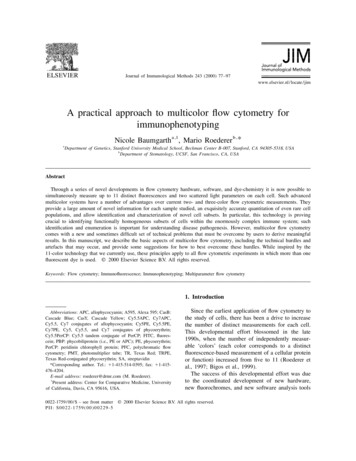
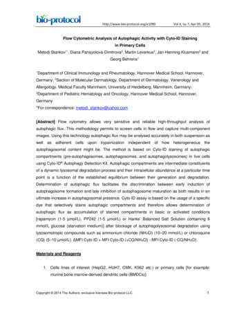
http://www.bio-protocol.org/e1090Vol 4, Iss 7, Apr 05, 2014Figure 1. Flow cytometry analysis of autophagic flux in BMDC incubated withautophagy inhibitor 3MA (10 mM) for 10 h in the presence or absence of autophagyactivator PP242 for the last 5 h (left) with representative histograms (right)AcknowledgmentsStankov, M and Behrens, G were supported by the DFG (BE-2089/2-1 and EXC62/1).Klusmann, J was supported by a grant from the Wilhelm Sander-Foundation (2011.057.1),the German Research Foundation (DFG; KL-2374/1-1). Klusmann, J is a fellow of the EmmyNoether-Programme from the DFG (KL-2374/2-1). Leverkus, M by the DFG (LE-953/8-1 andLE-953/6-1) and the Mildred-Scheel-Stiftung (#109891).References1. Cyto-ID /51031-K200.pdf.2. Mizushima, N., Yoshimori, T. and Levine, B. (2010). Methods in mammalian autophagyresearch. Cell 140(3): 313-326.Copyright 2014 The Authors; exclusive licensee Bio-protocol LLC.5
http://www.bio-protocol.org/e1090Vol 4, Iss 7, Apr 05, 20143. Stankov, M. V., El Khatib, M., Kumar Thakur, B., Heitmann, K., Panayotova-Dimitrova, D.,Schoening, J., Bourquin, J. P., Schweitzer, N., Leverkus, M., Welte, K., Reinhardt, D., Li,Z., Orkin, S. H., Behrens, G. M. and Klusmann, J. H. (2013). Histone deacetylaseinhibitors induce apoptosis in myeloid leukemia by suppressing autophagy. Leukemia.4. Sinicrope, F. A., Sirko, A., Siu, P. M. et al. (2012). Guidelines for the use andinterpretation of assays for monitoring autophagy. Autophagy 8(4): 445-544.Copyright 2014 The Authors; exclusive licensee Bio-protocol LLC.6
FACSCalibur, LSR II (BD) or analogous equipment . . Analyze samples using flow cytometer and plot data of cell counts as FITC (FL1) . Please cite this article as: Metodi et. al., (2014). Flow Cytometric Analysis of Autophagic Activity with Cyto-ID Staining in Primary Cells, Bio-protocol 4 (7): e1090. DOI: 10.21769/BioProtoc.1090.