Transcription
Jiang et al. BMC Infectious Diseases (2018) ARCH ARTICLEOpen AccessIL-10 NK and TGF-β NK cells playnegative regulatory roles in HIV infectionYongjun Jiang1, Mei Yang1, Xiaojuan Sun1,3, Xi Chen1, Meichen Ma1, Xiaowan Yin1, Shi Qian1, Zining Zhang1,2,Yajing Fu1, Jing Liu1, Xiaoxu Han1,2, Junjie Xu1,2 and Hong Shang1,2*AbstractBackground: Natural killer (NK) cells play cytotoxic roles by targeting tumor cells or virus infected cells, they alsoplay regulatory roles by secreting cytokines and chemokines. Transforming growth factor (TGF)-β and interleukin(IL)-10 are important immunosuppressive cytokines potentially related to the immune dysregulation that occurs inthe infection of human immunodeficiency virus (HIV). NK cells are an important source of TGF-β and a main earlyproducer of IL-10 in response to viral infection. Here, we evaluated the percentages of IL-10 and TGF-β NK cells inHIV-infected patients relative to healthy controls (HCs).Methods: Study participants (n 63) included 31 antiretroviral treatment (ART)-naïve HIV-infected patients, 17ART-treated HIV-infected patients, and 15 HIV-negative HCs. Expression of IL-10 or TGF-β in NK cells was examined byflow cytometry, and the influences of recombinant IL-10 (rIL-10) or recombinant TGF-β (rTGF-β) on NK cell functionwere investigated in vitro.Results: Compared with HCs, ART-naïve HIV-infected patients had increased percentages of IL-10 (2.0% vs. 0.4%, p 0.015) and TGF-β (4.5% vs. 2.1%, p 0.022) NK cells, and ART-treated patients also had a higher percentage of IL-10 NKcells (2.5% vs. 0.4%, p 0.002). The percentages of IL-10 and TGF-β NK cells were positively correlated (r 0.388; p 0.010). The results of in vitro experiments demonstrated that rIL-10 and rTGF-β inhibited NK cell CD107a expression (p 0.037 and p 0.024, respectively), IFN-γ secretion (p 0.006, p 0.016, respectively), and granzyme B release afterstimulation (p 0.014, p 0.040, respectively).Conclusions: Our data suggest that the percentages of IL-10 or TGF-β NK cells are increased in HIV-infected patients,and that rIL-10 and/or rTGF-β can inhibit NK cell functions in vitro, providing a potential therapeutic target for strategiesaimed at combating HIV infection.Keywords: HIV, IL-10, TGF-β, NK, Antiretroviral treatment, IFN-γ, Immune regulationBackgroundNatural killer (NK) cells serve as the first line of immunedefense in host protection against viruses and tumors [1]. Inhumans, NK cells account for 2%–18% of the lymphocytesin peripheral blood and express various inhibitory andactivating receptors, including C-type lectin-like, naturalcytotoxicity, and killer cell immunoglobulin-like receptors* Correspondence: hongshang100@hotmail.com1Key Laboratory of AIDS Immunology of National Health and Family PlanningCommission, Department of Laboratory Medicine, The First AffiliatedHospital, China Medical University, No. 155, Nanjingbei Street, Heping District,Shenyang, Liaoning Province 110001, China2Collaborative Innovation Center for Diagnosis and Treatment of InfectiousDiseases, 79 Qingchun Street, Hangzhou, ChinaFull list of author information is available at the end of the article[2, 3]. NK cell functions include killing target cells, cytokineproduction, and antibody-dependent cellular cytotoxicity(ADCC) [2]. Moreover, NK cells are critical effectors mediating cytotoxicity, and regulators modulating the activationand development of other immune response components[1]. NK cells are identified via their lack of CD3 and expression of CD56 cell surface markers, and they can be furtherdivided into CD56dim and CD56bright subsets [3]. Generally,CD56dim NK cells release perforin or granzymes, which playa key role in killing target cells, whereas CD56bright NK cellssecrete interleukin (IL)-10, interferon (IFN)-γ, transforminggrowth factor (TGF)-β and other cytokines, to exertimmunomodulatory effects [4–6]. The Author(s). 2018 Open Access This article is distributed under the terms of the Creative Commons Attribution 4.0International License (http://creativecommons.org/licenses/by/4.0/), which permits unrestricted use, distribution, andreproduction in any medium, provided you give appropriate credit to the original author(s) and the source, provide a link tothe Creative Commons license, and indicate if changes were made. The Creative Commons Public Domain Dedication o/1.0/) applies to the data made available in this article, unless otherwise stated.
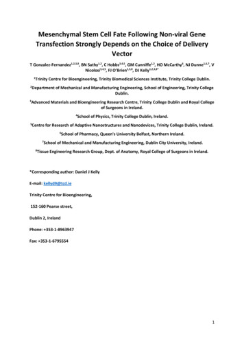
Jiang et al. BMC Infectious Diseases (2018) 18:80IL-10 and TGF-β are important immunoregulatorycytokines in vivo [7, 8], which suppress adaptive andinnate immunity [9]. IL-10 is produced by multiple celltypes, including T cells, NK cells, monocytes, and B cells;NK cells are a major early source of this cytokine inresponse to viral infection [10–13]. IL-10 is involved inthe impairment of T cell function during persistent viralinfections, and blockage of the IL-10 pathway alone issufficient to restore T cell activities and increase viralcontrol [14]. TGF-β is also secreted by various cell types,particularly NK cells, which are the only lymphocytepopulation that constitutively produces this cytokine [15].TGF-β plays important roles in immunomodulation, inflammation, and tissue repair [16], and can inhibit T cellproliferation and cytotoxicity [17]. IL-10 is reported tocause harmful effects during human immunodeficiencyvirus (HIV) infection by reducing IL-2 and IL-12 production, thereby inhibiting antigen-presentation and cellularimmune responses [18–20]. HIV-infected CD4 T cellscan produce IL-10, leading to persistent viral infection[11]. High levels of TGF-β in the plasma were reported inHIV-infected patients compared with healthy controls(HCs) [21]; however, the cell types producing TGF-β inthis context remain to be determined.IL-10 NK cells play significant modulatory roles in various viral, bacterial, and parasitic infections [12, 22–24].TGF-β NK cells have been reported to serve as an important co-stimulatory signal to induce suppressive T cells[15]. In HIV infection, multiple cells can produce IL-10and TGF-β. The majority of research has focused only onT cells, rather than NK cells, which are a major source ofthese cytokines and play important roles during acute HIVinfection. The percentage of IL-10 or TGF-β NK cells inHIV-infected patients and the regulatory effect of IL-10and TGF-β have yet to be elucidated.In the present study, we determined the percentagesof IL-10 and TGF-β NK cells in HIV-infected patientsand healthy controls (HCs). We also explored the immunomodulatory effects of recombinant IL-10 (rIL-10) andrecombinant TGF-β (rTGF-β) on NK cell functions,including the expression of lysosomal-associatedmembrane glycoprotein-1 (LAMP1; also known asCD107a), and IFN-γ secretion. The results indicated thatIL-10 and TGF-β NK cells may be risk factors for HIVdisease progression, and are potential therapeutic targetsin combating HIV infection.MethodsPage 2 of 10cells/μl (interquartile range [IQR] 277–421 cells/μl) andmedian viral load of 24,760 copies/ml (range,374–388,000 copies/ml). ART-naïve patients were furthergrouped according to CD4 T cell counts (CD4 T or 350 cells/μl groups) or viral load (viral load 104 or 104copies/ml groups). The 17 ART-treated patients hadreceived ART for 2 years, and had undetectable viralloads (HIV RNA, 20 copies/ml); the median duration oftheir virologic suppression at the time of sampling was33 months (IQR: 24–60 months), and they had a medianCD4 T cell count of 586 cells/μl (IQR: 453–684 cells/μl).HCs were HIV-negative, anti-hepatitis C antibodynegative, and hepatitis B surface antigen-negative, withnormal liver and kidney functions, and without immunesystem diseases.Detection of IL-10 or TGF-β NK cells by flow cytometryPeripheral blood mononuclear cells (PBMCs) were isolatedfrom HIV-infected patients and HCs by Ficoll densitygradient centrifugation. PBMCs were cultured for 6 h withrecombinant IL-12 (rIL-12, 10 ng/ml) and recombinantIL-15 (rIL-15, 50 ng/ml) in a 5% CO2 incubator at 37 C.Total NK cells and NK cell subsets were defined by theirexpression of CD3 and CD56, detected using the anti-CD3PerCP and anti-CD56 PE-Cy7 antibodies (BD Biosciences,San Jose, CA, USA), respectively. Cells were stained iolegend, San Diego, CA, USA) and fixed in 1% paraformaldehyde before detection using a flow cytometer (LSRII; Becton Dickinson, Franklin Lakes, NJ, USA) andanalysis with FACSDiva software (Becton Dickinson); thegating strategy is shown in Fig. 1.Determination of NK cell CD107a expression and IFN-γsecretionPBMCs were co-cultured with different concentrationsof rIL-10 and/or rTGF-β for 5 h, then stimulated withrIL-12 (10 ng/ml, R&D) and rIL-15 (50 ng/ml, R&D) for24 h at 37 C, and anti-CD107a-PE antibody (Biolegend,San Diego, CA, USA) added simultaneously. Cells wereco-cultured in GolgiStop ((Becton Dickinson) for 5 h before the end of stimulation. PBMCs were collected, andanti-CD3-Percp and anti-CD56-PE-cy7 were used for surface staining. Cells were then stained for intracellular IFN-γwith anti-IFN-γ-APC antibody (BD Biosciences, San Jose,CA, USA), washed with phosphate-buffered saline, andfixed in 1% paraformaldehyde, followed by analysis usingthe LSR II flow cytometer.Study participantsSixty-three individuals participated in this study, including31 antiretroviral treatment-naïve HIV-infected patients(ART-naïve), 17 ART-treated HIV-infected patients(ART-treated), and 15 HIV-negative HCs. The 31 ART-naïve patients had a median CD4 T cell count of 363Determination of NK cell release of granzyme B andperforinPBMCs were co-cultured with different concentrations ofrIL-10 and/or rTGF-β for 5 h. Then, cells were stimulatedwith PMA (10 ng/μL) and ionomycin (1 μg/μL) (Sigma
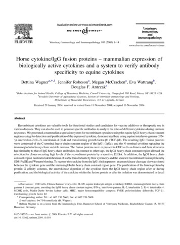
Jiang et al. BMC Infectious Diseases (2018) 18:80Page 3 of 10Fig. 1 Gating strategy for identification of NK cells and their subsetsP1 represents lymphocytes identified using a side and forward scatter dotplot. P2 represents CD56bright NK cells; gating, CD3 CD56bright. P3 represents CD56dim NK cells; gating, CD3 CD56dim. P2 P3 represents total NKcells. P4 represents IL-10 NK cells. P5 represents TGF-β NK cellsChemical St. Louis, MO, USA) for 6 h at 37 C. PBMCswere collected, and surface-stained with anti-CD3-PerCPand anti-CD56-PE-Cy7. Anti-granzyme B-PE and osciences, San Jose, CA, USA) were used for intracellular staining, and cells detected by flow cytometry (LSR II).test v2.0 (Roche Diagnostic Systems, Basel, Switzerland).The detection range of this method is 20–10,000,000copies/ml. The number of HIV RNA copies wascalculated according to the manufacturer’s referencestandard.Statistical analysesDetection of CD4 T cell countCD4 T cell counts were determined using a flow cytometer (FACS Calibur; Becton Dickinson). High, medium,and low Trucount Control Beads (BD Biosciences, SanJose, CA, USA) were used to confirm the accuracy andquality of CD4 T cell counts. A single-platform lyse-nowash procedure was conducted with TriTEST anti-CD3PerCP/CD4-FITC/CD8-PE reagents and Trucount tubes(BD Biosciences, San Jose, CA, USA) according to themanufacturer’s instructions.The non-parametric Mann–Whitney U-test was used forevaluation of differences between two groups, and theKruskall–Wallis test employed for comparisons of multiple groups. The Friedman’s test was used for multiplepairwise comparisons. Correlation analysis was performed using the Spearman’s rank correlation test. Allanalyses were undertaken using SPSS v19.0 (IBM,Armonk, NY, USA). p 0.05 was considered significant.ResultsDetermination of HIV viral loadPercentages of total NK cells and NK subsets in HIV-infectedpatients and HCsPlasma HIV RNA levels were determined with by reverse transcription-polymerase chain reaction (RT-PCR)using the COBAS AmpliPrep /COBAS TaqMan HIVThe clinical characteristics of ART-naïve and ARTtreated patients, and HCs, are presented in Table 1. Thesex and age of HIV-infected patients were matched with
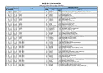
Jiang et al. BMC Infectious Diseases (2018) 18:80Page 4 of 10Table 1 Clinical characteristics of study subjectsParametersSexHC(n 15)ART-naive patients(n 31)ART-treated patients(n e(years)27 (25–33)25 (21–37)30 (27.5–38)CD4 T cells (cells/μl)ND363 (277–421)586 (453–684)HIV Viral Load(copies/ml) Log10NA4.19 (3.87–4.86)Undetectable ( 1.30)Months since infectionNA33 (19–68)67 (40.5–86.5)Months of ART treatmentNANA42 (30–66)NOTE: ND: no data; NA: not applicablethose of HCs. The percentages of total NK cells and NKsubsets among lymphocytes in peripheral blood samplesfrom the three groups were measured by flow cytometry.The percentages of total NK cells in ART-naïve andART-treated patient samples were lower than that inHCs (p 0.014, p 0.024) (Fig. 2a). In addition, the percentages of CD56dim NK cells in ART-naïve patientswere lower than those in HCs (p 0.006) (Fig. 2b) andthere was a tendency towards lower percentages ofCD56bright NK cells in ART-naïve patients comparedwith those of HCs (p 0.067) (Fig. 2c).Percentages of IL-10 or TGF-β NK cells in HIV-infectedpatientsPBMCs obtained from HCs, and ART-naïve, and ARTtreated patients, were stimulated with rIL-12 and rIL-15.The percentage of IL-10 or TGF-β NK cells was examined by intracellular staining and multi-color flow cytometry. Representative flow cytometry plots of IL-10 orTGF-β expression in NK cells from each participantgroup are shown in Fig. 3a. The percentage of IL-10 NK cells in ART-naïve patients was higher than that inHCs (p 0.015); moreover, the percentage of IL-10 NKcells was also higher in the ART-treated HIV patientgroup than that in HCs (p 0.002) (Fig. 3b). The percentage of TGF-β NK cells in ART-naïve patients waselevated compared with that of HCs (p 0.022), while inART-treated patients, the percentage of TGF-β NKcells still exhibited a tendency to be higher than that ofHCs (p 0.060) (Fig. 3c). There was no difference in thepercentage of IL-10 CD56dim NK cells among the threegroups (Fig. 3d), whereas the percentage of TGF-β CD56dim NK cells in ART-naïve patients was higher thanthat in HCs (p 0.024) (Fig. 3e). Moreover, there wereno differences in the percentages of IL-10 CD56brightand TGF-β CD56bright NK cells among the three groups(Fig. 3f, g). In ART-naïve patients, the percentage ofIL-10 NK cells was positively correlated with that ofTGF-β NK cells (r 0.388, p 0.010) (Fig. 3h) and thepercentage of IL-10 CD56dim NK cells correlated positively with that of TGF-β CD56dim NK cells (r 0.438,p 0.003) (Fig. 3i).Comparison of the percentages of IL-10 and TGF-β NKcells in different groups according to CD4 T cell count orHIV viral loadCD4 T cell counts and viral loads are importantmarkers for HIV disease progression. We analyzed thecorrelation between the percentage of IL-10 or TGF-β NK cells and CD4 T cell counts and viral loads and foundno significant correlations between them. Furthermore,Fig. 2 Percentages of total NK cells and NK cell subsets in HIV-infected patients and HCs The percentages of total NK cells among lymphocytesfrom HCs, ART-naïve, and ART-treated patients (a). The percentages of NK cell subsets (CD56dim and CD56bright) in lymphocytes from HCs, ART-naïve, and ART-treated patients (b, c)
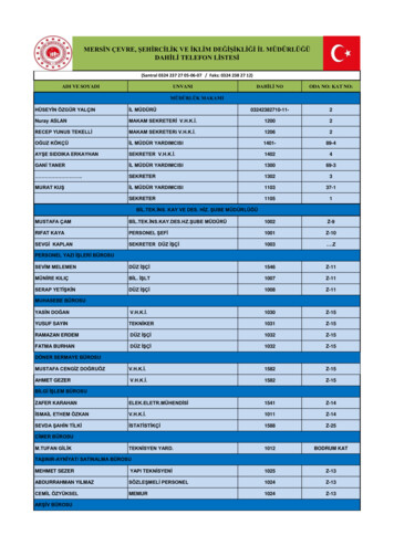
Jiang et al. BMC Infectious Diseases (2018) 18:80Page 5 of 10Fig. 3 Comparison of the percentages of IL-10 and TGF-β NK cells in HIV-infected patients and HCsPBMCs from HCs, ART- naïve, and ART-treated patients were stimulated using rIL-12 (10 ng/ml) and rIL-15 (50 ng/ml). IL-10 and TGF-β NK cells were examined by multi-color flow cytometry, with gates defined according to isotype controls. Representative flow-cytometric plots of IL-10 and TGF-β NK cells in HIV-infectedpatients and HCs (a). Comparison of the percentages of IL-10 NK cells (b), TGF-β NK cells (c), IL-10 CD56dim NK cells (d), TGF-β CD56dim NKcells (e), IL-10 CD56bright NK cells (f), and TGF-β CD56bright NK cells (g), among the three groups. Correlation between the percentage of IL-10 NK cells and TGF-β NK cells (h). Correlation between the percentage of IL-10 CD56dim NK cells and TGF-β CD56dim NK cells (i)we analyzed groups subdivided by CD4 T cell count orviral load. The percentage of IL-10 NK cells in the CD4 T 350 cells/μl group of ART-naïve patients was higherthan that of the HC group (p 0.032), and there was anincreasing trend in the CD4 T 350 cells/μl group ofART-naïve patients compared with HCs (p 0.075)(Fig. 4a). There was no difference in the percentages ofTGF-β NK cells among groups (Fig. 4b). When grouped
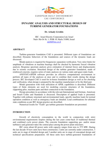
Jiang et al. BMC Infectious Diseases (2018) 18:80Page 6 of 10Fig. 4 Percentage of IL-10 or TGF-β NK cells in different groupsThe percentage of IL-10 or TGF-β NK cells in different groups according toCD4 T cell counts (a, b). The percentage of IL-10 or TGF-β NK cells in different groups according to viral load (c, d)according to the level of viral load, the percentage ofIL-10 NK cells in ART-naïve patients with viral load 104 copies/ml was higher than that in the HC group (p 0.023) (Fig. 4c), and that in the group with viral load 104 copies/ml, while the percentage of TGF-β NKcells was higher than that in the HC group (p 0.027) (Fig. 4d).Effect of rIL-10 and rTGF-β on the NK cell CD107a expressionand IFN-γ secretionThe expression of CD107a and IFN-γ secretion can beused to assess the functional anti-viral ability of NKcells. The inhibitory effects of rIL-10 or rTGF-β on NKcell functions were evaluated, and the percentages ofIFN-γ and CD107a NK cells determined. Representative flow cytometry plots, demonstrating the effects ofrIL-10 or rTGF-β on CD107a expression by NK cells arepresented in Fig. 5a. CD107a expression in NK cells wasinhibited by high concentrations of rIL-10 or rTGF-β invitro (p 0.037, p 0.024), and the rate of inhibition increased gradually with increasing concentrations of thesecytokines (Fig. 5b, c). Moreover, the effect of rIL-10 andrTGF-β on CD107a expression in NK cells was synergistic, demonstrating inhibitory effects much greater thanthose of either cytokine individually (p 0.006) (Fig. 5d).Representative flow-cytometric plots demonstrating theeffects of rIL-10 or rTGF-β on IFN-γ secretion from NKcells are shown in Fig. 6a. IFN-γ secretion by NKcells was also inhibited by rIL-10 or rTGF-β (p 0.006, p 0.016), and the rate of inhibition increasedgradually with their concentrations (Fig. 6b, c). Again,the effect of both IL-10 and TGF-β was synergistic(p 0.006) (Fig. 6d).Effects of rIL-10 and rTGF-β on granzyme B and perforinrelease by NK cellsGranzyme B and perforin are important functionalmarkers for NK cells, which indicate the killing capability of NK cells. The percentages of IL-10 and TGF-β NK cells were elevated in HIV infected patients; however, their influence on the release of granzyme B andperforin by NK cells is not known. Our results demonstrate that a low concentration of rIL-10 or rTGF-β didnot inhibit the release of granzyme B, whereas high concentrations of either cytokine did inhibit the release ofthis factor (p 0.014, p 0.040) (Fig. 7a, b). A synergisticeffect of rIL-10 and rTGF-β on inhibition of the releaseof granzyme B was also noted (p 0.037) (Fig. 7c). Similarly, rIL-10 and rTGF-β inhibited perforin release byNK cells (Fig. 8a–c).
Jiang et al. BMC Infectious Diseases (2018) 18:80Page 7 of 10Fig. 5 Effect of rIL-10 and rTGF-β on CD107a expression in NK cellsPBMCs were incubated with rIL-10 or rTGF-β and CD107a expression by NKcells evaluated. Representative flow-cytometry plots demonstrated the effect of rIL-10 or rTGF-β on CD107a expression in NK cells (a). CD107aexpression by NK cells was inhibited by different concentrations of rIL-10 (b) or rTGF-β (c). Fold-change in CD107a expression by NK cells wassynergistically inhibited by rIL-10 and rTGF-β (d). Fold-change of CD107a expression (percentage CD107a expression in untreated controls percentage of CD107a expression after treatment with rIL-10 and/or rTGF-β)/percentage of CD107a expression in untreated controlsDiscussionRegulation of the immune system has attracted considerable attention over the last decade, and the cytokines,IL-10 and TGF-β, have important roles in controllingimmune processes. The suppression of anti-cancer immunity by TGF-β and IL-10 has been reported in severalstudies [25–28]. In breast cancer, circulating levels ofIL-10 and TGF-β NK cells are increased, which inhibits the production of the NK cell effectors, IFN-γ andCD107a, and promotes the migration and invasion ofbreast-cancer cells [9].Brockman et al. reported that IL-10 mRNA levels wereincreased in several PBMC subpopulations in HIVinfected patients compared with HCs, particularly Tcells, B cells, monocytes, and NK cells, and that theinhibition of the IL-10 pathway enhanced the proliferative capacity of T cells [29]. Based on these findings regarding the levels of IL-10 mRNA in NK cells, wefurther explored the intracellular secretion of IL-10 andTGF-β proteins in NK cells (total and subsets) and investigated the relationship between IL-10 /TGF-β NKcells and CD4 T cell counts/viral loads. Our data elucidate the effects of IL-10 and TGF-β on NK cell functions, including IFN-γ secretion, CD107a expression,and granzyme B or perforin release.We found that ART-naïve patients had elevatedpercentages of IL-10 NK cells relative to HCs; however,patients who had received ART, also had higher percentages of IL-10 NK cells than those of HCs. Comparisonsof TGF-β NK cell percentages among study groups
Jiang et al. BMC Infectious Diseases (2018) 18:80Page 8 of 10Fig. 6 Effect of rIL-10 and rTGF-β on IFN-γ secretion by NK cellsPBMCs were co-cultured with rIL-10 and/or rTGF-β for 5 h, then stimulated withrIL-12 and rIL-15 for 24 h. Representative flow-cytometry plots demonstrated the effect of rIL-10 or rTGF-β on IFN-γ secretion by NK cells (a). IFN-γsecretion by NK cells was inhibited by different concentrations of rIL-10 (b) or rTGF-β (c) and synergistically by treatment with both rIL-10 andrTGF-β (d)Fig. 7 Effects of rIL-10 and rTGF-β on granzyme-B release by NK cellsPBMCs were co-cultured with rIL-10 and/or rTGF-β for 5 h, and then stimulatedwith PMA and ionomycin for 6 h. The inhibition of granzyme-B release by NK cells using different concentrations of rIL-10 (a) or rTGF-β (b) wasdetermined. The effect of treatment with both rIL-10 and rTGF-β on granzyme-B release by NK cells was synergistic (c)
Jiang et al. BMC Infectious Diseases (2018) 18:80Page 9 of 10Fig. 8 Effects of rIL-10 and rTGF-β on perforin release by NK cellsPBMCs were co-cultured with rIL-10 and/or rTGF-β for 5 h, and then stimulatedwith PMA and ionomycin for 6 h. The inhibition of perforin release by NK cells by different concentrations of rIL-10 (a) or rTGF-β (b) was determined.The effects of treatment with both rIL-10 and rTGF-β on perforin release by NK cells were synergistic (c)generated similar results to those for IL-10 NK cells.These data indicate that ART is insufficient to inducecomplete recovery of immune functions, and thatspecific immune therapies are required to overcome thenegative regulation. Our previous study showed thatHIV infection can result in impaired NK cell functions,including diminished levels of perforin and IFN-γ production [30, 31], which may be related to negative regulation by cytokines. In the present study, the results ofour in vitro experiments demonstrate that the expression of CD107a and secretion of IFN-γ by NK cells canbe inhibited by rIL-10 or rTGF-β. High concentrationsof rIL-10 or rTGF-β could also inhibit the release ofgranzyme B and perforin; therefore, we suspect that theincrease in the percentage of IL-10 and TGF-β NKcells may be related to impaired NK cell cytotoxicfunction during HIV infection.The mechanisms underlying the elevated percentageof IL-10 and TGF-β NK cells during HIV infection remain unclear; however, it was reported that the HIV Envand Tat peptides are responsible for IL-10 production byCD4 T cells [32–34], and the mitogen-activated proteinkinase/extracellular signal-regulated kinase pathway wasreported as involved in this process [35, 36]. Further, invitro experiments have indicated that HIV Tat caninduce TGF-β production by NK cells [21], and thatTGF-β may suppress NK cell functions by repressing themTOR pathway [37], or inhibit IFN-γ secretion and theADCC response of NK cells via SMAD3-mediatedsignaling [38].HIV patients also have a high percentage of IL-10 NKcells. We also show that rIL-10 and/or rTGF-β caninhibit the functions of NK cell effectors. Our researchprovides a potential therapeutic target for combatingHIV infection, along with information useful forresearch into the pathogenesis and immunotherapy ofHIV infection.ConclusionsThe results of our study demonstrate that the percentages of IL-10 and TGF-β NK cells are elevated inART-naïve HIV-infected patients, and that ART-treatedEthics approval and consent to participateThe Medical Science Research Ethics Committee of the First AffiliatedHospital of China Medical University (Shenyang, China) approved the studyprotocol (KELUNSHEN [2011] number 36). Our study was conductedaccording to the principles enshrined in the Declaration of Helsinki. WrittenAbbreviationsADCC: Antibody-dependent cellular cytotoxicity; ART: Antiretroviraltreatment; HCs: Healthy controls; HIV: Human immunodeficiency virus;IFN: Interferon; IL: Interleukin; NK: Natural killer; PBMCs: Peripheral bloodmononuclear cells; rIL: Recombinant interleukin; rTGF-β: Recombinant TGF-β;TGF-β: Transforming growth factor-βAcknowledgmentsThe authors express their gratitude for the generosity of the patients whoparticipated in this study.FundingFunding for this study was provided by the Mega Projects of NationalScience Research for the 12th Five-Year Plan (2014ZX10001002001004), andthe Platform Project for Close Combination of Basic and Clinical Medicine(YIDA FAZI [2013] no. 5). The funding sources had no role in the studydesign, data collection, data analyses, preparation of the manuscript, ordecision to publish.Availability of data and materialsAll data generated or analyzed during this study are included in thismanuscript.Authors’ contributionsYJ and HS designed the experiments, interpreted the data, and wrote themanuscript. MY, XS, XC, MM, XY, SQ, and YJ carried out the NK cellexperiments. ZZ and YF carried out CD4 T cell counts. XH participated inmeasurement of HIV viral load. JX and JL carried out the epidemiologicalstudy and helped to recruit study participants. All authors have read andapproved the manuscript.
Jiang et al. BMC Infectious Diseases (2018) 18:80informed consent to take part in the study was obtained from allparticipants.Consent for publicationNot applicable.Competing interestsThe authors declare that they have no competing interests.Publisher’s NoteSpringer Nature remains neutral with regard to jurisdictional claims inpublished maps and institutional affiliations.Author details1Key Laboratory of AIDS Immunology of National Health and Family PlanningCommission, Department of Laboratory Medicine, The First AffiliatedHospital, China Medical University, No. 155, Nanjingbei Street, Heping District,Shenyang, Liaoning Province 110001, China. 2Collaborative Innovation Centerfor Diagnosis and Treatment of Infectious Diseases, 79 Qingchun Street,Hangzhou, China. 3Clinical Laboratory, Shenyang Women and Children’sHospital, Shenyang, China.Received: 6 December 2016 Accepted: 7 February 2018References1. Cacalano NA. Regulation of natural killer cell function by STAT3. FrontImmunol. 2016;7:128.2. Littwitz-Salomon E, Dittmer U, Sutter K. Insufficient natural killer cellresponses against retroviruses: how to improve NK cell killing of retrovirusinfected cells. Retrovirology. 2016;13(1):77.3. Chester C, Fritsch K, Natural KHE. Killer cell immunomodulation: targetingactivating, inhibitory, and co-stimulatory receptor signaling for cancerimmunotherapy. Front Immunol. 2015;6:601.4. Caligiuri MA. Human natural killer cells. Blood. 2008;112(3):461–9.5. Cooper MA, Fehniger TA, Turner SC, Chen KS, Ghaheri BA, Ghayur T, et al.Human natural killer cells: a unique innate immunoregulatory role for theCD56(bright) subset. Blood. 2001;97(10):3146–51.6. Cooper MA, Fehniger TA, Caligiuri MA. The biology of human natural killercell subsets. Trends Immunol. 2001;22(11):633–40.7. Laouar Y, Sutterwala FS, Gorelik L, Transforming FRA. Growth factor-betacontrols T helper type 1 cell development through regulation of naturalkiller cell interferon-gamma. Nat Immunol. 2005;6(6):600–7.8. Eriksson M, Meadows SK, Wira CR, Unique SCL. Phenotype of human uterineNK cells and their regulation by endogenous TGF-beta. J Leukoc Biol. 2004;76(3):667–75.9. Ostapchuk YO, Cetin EA, Perfilyeva YV, et al. Peripheral blood NK cellsexpressing HLA-G, IL-10 and TGF-beta in healthy donors and breast cancerpatients. Cell Immunol. 2015;298(1–2):37–46.10. Elrefaei M, Burke CM, Baker CA, et al. TGF-beta and IL-10 production by HIVspecific CD8 T cells is regulated by CTLA-4 signaling on CD4 T cells. PLoSOne. 2009;4(12):e8194.11. Shete A, Suryawanshi P, Godbole S, Pawar J, Paranjape R, Thakar M. HIVinfected CD4 T cells use T-bet-dependent pathway for production of IL-10upon antigen recognition. Scand J Immunol. 2016;83(4):288–96.12. Wagage S, John B, Krock BL, et al. The aryl hydrocarbon receptor promotesIL-10 production by NK cells. J Immunol. 2014;192(4):1661–70.13. Perona-Wright G, Mohrs K, Szaba FM, et al. Systemic but not local infectionselicit immunosuppressive IL-10 production by natural killer cells. Cell HostMicrobe. 2009;6(6):5
Chemical St. Louis, MO, USA) for 6 h at 37 C. PBMCs were collected, and surface-stained with anti-CD3-PerCP and anti-CD56-PE-Cy7. Anti-granzyme B-PE and anti--perforin-fluorescein isothiocyanate (FITC) (BD Biosciences, San Jose, CA, USA) were used for intracellu-lar staining, and cells detected by flow cytometry (LSR II). Detection of CD4 T .