Transcription
Rustenhoven et al. Journal of Neuroinflammation (2016) 13:37DOI 10.1186/s12974-016-0503-0RESEARCHOpen AccessTGF-beta1 regulates human brain pericyteinflammatory processes involved inneurovasculature functionJustin Rustenhoven1,3, Miranda Aalderink1,3, Emma L. Scotter1,3, Robyn L. Oldfield4, Peter S. Bergin3,5,Edward W. Mee3,5, E. Scott Graham1,3, Richard L. M. Faull2,3, Maurice A. Curtis2,3, Thomas I-H. Park1,2,3and Mike Dragunow1,3*AbstractBackground: Transforming growth factor beta 1 (TGFβ1) is strongly induced following brain injury and polarisesmicroglia to an anti-inflammatory phenotype. Augmentation of TGFβ1 responses may therefore be beneficial inpreventing inflammation in neurological disorders including stroke and neurodegenerative diseases. However,several other cell types display immunogenic potential and identifying the effect of TGFβ1 on these cells is requiredto more fully understand its effects on brain inflammation. Pericytes are multifunctional cells which ensheath thebrain vasculature and have garnered recent attention with respect to their immunomodulatory potential. Here, wesought to investigate the inflammatory phenotype adopted by TGFβ1-stimulated human brain pericytes.Methods: Microarray analysis was performed to examine transcriptome-wide changes in TGFβ1-stimulatedpericytes, and results were validated by qRT-PCR and cytometric bead arrays. Flow cytometry,immunocytochemistry and LDH/Alamar Blue viability assays were utilised to examine phagocytic capacity ofhuman brain pericytes, transcription factor modulation and pericyte health.Results: TGFβ1 treatment of primary human brain pericytes induced the expression of several inflammatory-relatedgenes (NOX4, COX2, IL6 and MMP2) and attenuated others (IL8, CX3CL1, MCP1 and VCAM1). A synergistic induction ofIL-6 was seen with IL-1β/TGFβ1 treatment whilst TGFβ1 attenuated the IL-1β-induced expression of CX3CL1, MCP-1and sVCAM-1. TGFβ1 was found to signal through SMAD2/3 transcription factors but did not modify nuclear factorkappa-light-chain-enhancer of activated B cells (NF-kB) translocation. Furthermore, TGFβ1 attenuated the phagocyticability of pericytes, possibly through downregulation of the scavenger receptors CD36, CD47 and CD68. WhilstTGFβ did decrease pericyte number, this was due to a reduction in proliferation, not apoptotic death orcompromised cell viability.Conclusions: TGFβ1 attenuated pericyte expression of key chemokines and adhesion molecules involved in CNSleukocyte trafficking and the modulation of microglial function, as well as reduced the phagocytic ability ofpericytes. However, TGFβ1 also enhanced the expression of classical pro-inflammatory cytokines and enzymeswhich can disrupt BBB functioning, suggesting that pericytes adopt a phenotype which is neither solely pro- noranti-inflammatory. Whilst the effects of pericyte modulation by TGFβ1 in vivo are difficult to infer, the reduction inpericyte proliferation together with the elevated IL-6, MMP-2 and NOX4 and reduced phagocytosis suggests adetrimental action of TGFβ1 on neurovasculature.Keywords: Phagocytosis, Inflammation, Cytokine, Chemokine, BBB, Alzheimers, NOX4, MMP-2, IL-6, SMAD2/3* Correspondence: m.dragunow@auckland.ac.nz1Department of Pharmacology and Clinical Pharmacology, The University ofAuckland, Auckland 1023, New Zealand3Centre for Brain Research, The University of Auckland, Auckland 1023, NewZealandFull list of author information is available at the end of the article 2016 Rustenhoven et al. Open Access This article is distributed under the terms of the Creative Commons Attribution 4.0International License (http://creativecommons.org/licenses/by/4.0/), which permits unrestricted use, distribution, andreproduction in any medium, provided you give appropriate credit to the original author(s) and the source, provide a link tothe Creative Commons license, and indicate if changes were made. The Creative Commons Public Domain Dedication o/1.0/) applies to the data made available in this article, unless otherwise stated.
Rustenhoven et al. Journal of Neuroinflammation (2016) 13:37BackgroundNeuroinflammation contributes to the development andprogression of epilepsy [1], traumatic brain injuries [2],stroke [3] and many neurodegenerative diseases [4]. Inflammatory cytokines are key mediators of the brain’simmune response and associated neurodegeneration [5].Transforming growth factor beta (TGFβ) is a pleiotropiccytokine in the brain with roles in regulating cell proliferation, differentiation, survival and scar formation [6–9].Furthermore, TGFβ orchestrates both pro- and antiinflammatory responses in a cell and context-dependentmanner [10–12]. TGFβ exists in three isoforms (TGFβ1–3),of which TGFB1 was the first identified and is the mostwidely studied. TGFβ exerts its actions through the serine/threonine kinase receptors, transforming growth factor betareceptor 1 (TGFBR1), TGFBR2 and TGFBR3. Activation ofTGFBRs by TGFβ ligand binding initiates a signal transduction pathway predominantly through SMAD transcriptionfactors [13].Several cell types in the central nervous system (CNS)are capable of producing TGFβ1. Endothelial cells secretethis growth factor, and this is enhanced by co-culture withpericytes [14, 15]. Following brain injury, microglia and astrocytes produce large concentrations of TGFβ1 [8, 16, 17],whilst glioblastoma multiforme enhances TGFβ1 expression, possibly due to the increased and abnormal angiogenesis [18–20]. Furthermore, TGFβ1 is elevated in severalinflammatory conditions including Alzheimer’s disease,type 1 diabetes mellitus and acute brain injuries such as ischaemic stroke [21–24].A number of immunologically active brain cells expressTGFBRs and can therefore respond to secretions of thisgrowth factor. Microglia are the brain’s predominant immune cell and the most widely studied with respect to theirinflammatory response [25]. TGFβ1 promotes a stronganti-inflammatory phenotype in microglia through attenuated cytokine, chemokine, adhesion molecule and reactiveoxidative species (ROS) production; however, it has no effect on human leukocyte antigen (HLA) antigen presentation complexes [10, 26–28]. Furthermore, TGFβ1 enhancesmicroglial phagocytosis of amyloid beta, thereby reducingplaque burden [29]. Astrocytes show a more complex regulation of immune responses by TGFβ1 stimulation, with reports of both enhancement and inhibition of chemokineand cytokine production [11, 12, 30, 31]. Other cells, including those derived from the choroid plexus and leptomeninges, also display attenuated inflammatory responseswith TGFβ1 treatment [27, 32].Previous work in our lab using human brain pericytesdemonstrated a reduction in interferon gamma (IFNγ)-induced HLA-DR, HLA-DP and HLA-DQ expression withTGFβ1 treatment, suggesting an anti-inflammatory role[27]. Pericytes are multifunctional cells that surround endothelial cells and contact numerous parenchymal brain cellsPage 2 of 15including astrocytes, neurons and microglia to form a neurovascular unit [33]. Pericyte coverage of brain vasculatureis vital with respect to blood-brain barrier (BBB) formationand maintenance [34–36]. Several labs, including our own,have shown that pericytes can contribute to the brain’s immune response through cytokine, chemokine and adhesionmolecule expression and that they display phagocytic potential [37–42].The function of TGFβ1 on pericytes has been brieflystudied, however, largely with respect to differentiation,proliferation or angiogenesis [43, 44]. Little is knownabout the role of TGFβ1 in the context of human brainpericyte immune function. We therefore sought to investigate how TGFβ1 modifies the human brain pericyteinflammatory response.MethodsTissue sourceHuman middle temporal gyrus brain tissue was obtained,with informed consent, from surgeries of patients withdrug-resistant temporal lobe epilepsy. All specimens werecollected with written patient consent. All protocols usedin this study were approved by the Northern RegionalEthics Committee (New Zealand), and all methods werecarried out in accordance with the approved guidelines.Mixed glial cultures isolated from human brain tissueMixed glial cultures containing astrocytes, pericytes andmicroglia were isolated from adult human brain tissue withminor modifications as described previously [45]. As per[42], cells were grown until passage five before use in orderto dilute out non-proliferating microglia and astrocytes andobtain a pure pericyte population. Pericyte cultures at passages five-eight were used for all experiments. Detailedcharacterisation of pericyte cultures has been performedpreviously [37, 42].Cytokine and drug treatmentsTo examine the effect of cytokines on pericyte responses,cells were treated with 0.1–10 ng/mL TGFβ1 (PeproTech,NJ, USA; vehicle, 1 mM citric acid, pH 3 with 0.1 % bovineserum albumin (BSA)) or interleukin 1 beta (IL-1β; Peprotech, NJ, USA; vehicle, 0.1 % BSA in phosphate buffered saline (PBS)) for 0–72 h. Details for each experiment arenoted in appropriate figure legends. To induce apoptosis asa positive control for cleaved caspase 3 (CC3) staining, pericytes were treated with 50 nM okadaic acid (Sigma-Aldrich,MO, USA) for 24 h. Cells were treated with drugs/cytokinesby addition of a 1:100 dilution of a 100 stock.Collection of conditioned media and cytokinemeasurement by cytometric bead arrayConditioned media was collected from cells grown in a96-well plate. Media was spun at 160 g for 5 min to
Rustenhoven et al. Journal of Neuroinflammation (2016) 13:37collect any detached cells or debris. Supernatant wasobtained and stored at 20 C. The concentration of cytokines was measured using a cytometric bead array(CBA; BD Biosciences, CA, USA) as per manufacturer’sinstructions. CBA samples were run on an Accuri C6flow cytometer (BD Biosciences, CA, USA). Data wasanalysed using FCAP-array software (version 3.1; BDBiosciences, CA, USA) to convert fluorescent intensityvalues to concentrations using a ten-point standardcurve (0–5000 pg/mL) as described previously [46].ImmunocytochemistryCells were fixed in 4 % paraformaldehyde (PFA) for15 min and washed in PBS with 0.1 % triton X-100(PBS-T). Cells were incubated with primary antibodies(Additional file 1: Table S1) overnight at 4 C in immunobuffer containing 1 % goat serum, 0.2 % Triton X-100and 0.04 % thimerosal in PBS. Cells were washed inPBS-T and incubated with appropriate anti-species fluorescently conjugated secondary antibodies overnight at4 C. Cells were washed again and incubated withHoechst 33258 (Sigma-Aldrich, MO, USA) for 20 min.Images were acquired at 10 magnification using the automated fluorescence microscope ImageXpress MicroXLS (version 5.3.0.1, Molecular Devices, CA, USA).Quantitative analysis of intensity measures and positivelystained cells was performed using the Cell Scoring andShow Region Statistics analysis modules on MetaXpress software (version 5.3.0.1, Molecular Devices, CA, USA).Phagocytosis assaysTo evaluate phagocytosis by microscopy, cells were treatedwith 0–10 ng/mL TGFβ1 for 24 h, followed by a further 24h incubation with Fluoresbrite YG carboxylate microspheres of 1 μm diameter (Polysciences Inc, PA, USA;1:1000 dilution) at 37 C, 5 % CO2. At completion, cellswere washed twice with PBS to remove un-phagocytosedbeads and fixed in 4 % PFA as per immunocytochemistry.Nuclear staining was visualised by a 30-min incubationwith the DNA-specific dye DRAQ5 (BioStatus, UK). Imageswere obtained using the ImageXpress Micro XLS microscope and the percentage of phagocytic cells determinedusing the Cell Scoring module on MetaXpress software.To evaluate phagocytosis by flow cytometry, cells weretreated with 0–10 ng/mL TGFβ1 for 24 h, followed by afurther 2-h incubation with Fluoresbrite YG carboxylatemicrospheres of 1-μm diameter (1:1000 dilution) at 37 C,5 % CO2. At completion, cells were washed twice withPBS, and 0.25 % trypsin-ethylenediaminetetraacetic acid(EDTA) was added to remove beads bound to the cell surface and bring cells into suspension. Selected sampleswere incubated for 10 min with 7-aminoactinomycin D(7-AAD; BD Biosciences, CA, USA) to assess viability.Samples were run on an Accuri C6 flow cytometer andPage 3 of 15viable cells gated based on forward scatter and side scatter. Mean fluorescent intensity (MFI) of the live cells wasdetected, indicative of the quantity of beads internalised.Confocal laser scanning microscopyCells destined for confocal microscopy were plated at 5000cells/well on 8-mm #1.5 glass coverslips (Menzel Gläser,Germany) within a 48-well plate. Fluoresbrite YG carboxylate microspheres of 1-μm diameter (1:10,000 dilution)were added to cells for 24 h at 37 C, 5 % CO2 and at completion washed twice in PBS to remove un-phagocytosedbeads. Cells were fixed in 4 % PFA and immunostained forplatelet-derived growth factor receptor beta (PDGFRβ) asper immunocytochemistry, with the exception of dilutingprimary and secondary antibodies in donkey immunobuffer(1 % donkey serum, 0.2 % Triton X-100 and 0.04 % thimerosal in PBS). Coverslips were mounted onto glass slidesusing fluorescent mounting medium (DAKO, Denmark).Confocal images were acquired using an oil immersion lens( 63 magnification, 1.4NA) in a Z-series with a gap of0.8 μm using a Zeiss LSM 710 inverted confocal microscope (Biomedical Imaging Research Unit, University ofAuckland) with ZEN 2010 software (Carl Zeiss, Germany).EdU proliferation assay5-Ethynyl-2′-deoxyuridine (EdU; 10 μM) was added topericyte cultures 24 h prior to completion of experiment.Cells were fixed in 4 % PFA for 15 min and EdU visualisedas per manufacturer’s instructions (Life Technologies, CA,USA). Cell nuclei were counterstained with Hoechst33258 as per immunocytochemistry.Alamar Blue viability assayTo determine cell viability, Alamar Blue (1:10 final dilution in culture medium; Life Technologies, CA, USA) wasadded to cells in a 96-well plate for 2 h at 37 C, 5 % CO2.Fluorescence (Ex 544/ Em 590) was read on a FLUOStarOptima plate reader (BMG Labtech, Germany). Background fluorescence of wells with media but no cells wassubtracted from all values, and fluorescence intensityvalues were normalised to cell number using imaging ofHoechst positive cells. Fluorescence intensity values werethen normalised to vehicle control.LDH cytotoxicity assayTo determine cellular toxicity, extracellular lactate dehydrogenase (LDH) was detected as per manufacturer’sinstructions (Roche, Switzerland). Briefly, a positive control was first prepared by lysing cells through addition ofDMEM/F12 media containing 1 % Triton X-100. Theculture media from all samples was then transferred to anew plate and centrifuged at 160 g for 10 min at roomtemperature. To a new 96-well plate, 50 μL of centrifuged media was transferred and 50 μL of LDH reaction
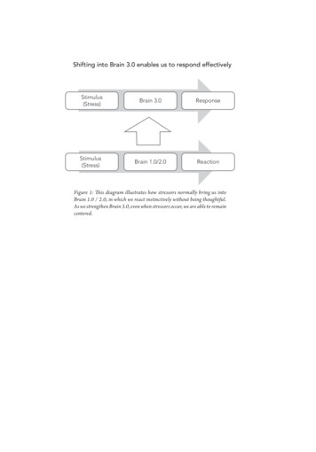
Rustenhoven et al. Journal of Neuroinflammation (2016) 13:37mixture was added. The plate was incubated at roomtemperature for 30 min and absorbance measured at540 nm on a FLUOStar Optima plate reader. Absorbance of culture media containing no cells was subtractedfrom all values and the absorbance normalised to that ofthe positive control (100 % cytotoxicity).Quantitative real-time reverse transcriptase PCRCells destined for quantitative real-time reverse transcriptase PCR (qRT-PCR) were plated at 100,000 cells/well in a six-well plate. At completion of the experiment, cells were washed in PBS and RNA extraction,and purification was performed using the RNeasy minikit (Qiagen, Netherlands) as per manufacturer’s instructions from duplicate wells. RNA was treated withDNase I (1 μg DNase I/1 μg RNA) using the RQ1RNase-free DNAse kit (Promega, WI, USA) and cDNAmade using the Superscript III First-Strand Synthesiskit (Life Technologies, CA, USA). Quantitative realtime PCR was performed using Platinum SYBR GreenqPCR SuperMix-UDG with Rox (Life Technologies,CA, USA) on a 7900HT Fast Real-Time PCR system(Applied Biosystems, Life Technologies, CA, USA).Standard curves were run for all primers, and efficiencies were all 100 10 % (Additional file 2: Table S2).Relative gene expression analysis was performed usingthe 2 ΔΔCt method to the housekeeping gene GAPDH.MicroarrayMicroarray analysis of TGFβ1-treated pericytes was conducted on technical triplicates of a single patient donor.Cells destined for microarray were treated for 72 h withvehicle (1 mM citric acid, pH 3 with 0.1 % BSA) or10 ng/mL TGFβ1. Cells were washed in PBS and lysed in1 mL of TRIzol reagent (Life Technologies, CA, USA).To this, 200 μL of chloroform was added, and sampleswere vigorously shaken and centrifuged at 12,000 g for15 min at 4 C. The upper aqueous phase was transferred to a new tube and an equal volume of 70 % ethanol added. RNA purification was performed using theRNeasy mini kit as per qRT-PCR experiments. RNAquality was analysed using a 2100 Bioanalyzer (AgilentTechnologies, CA, USA) with all samples having a RINvalue of 10. RNA was labelled and hybridised to Affymetrix Genechip Primeview Human Gene ExpressionArrays (Santa Clara, CA, USA) according to manufacturer’s instructions. Microarray was performed and analysed by New Zealand Genomics Limited (NZGL).Candidate microarray hits for qRT-PCR and CBA validation were selected based on the magnitude of changeand prior evidence for involvement in neuro-immune responses but do not include all modified inflammatorygenes.Page 4 of 15Statistical analysisUnless otherwise stated, all experiments were performedat least three independent times on tissue from threedifferent donors. Statistical analysis was carried outusing an unpaired Student’s t test, one-way analysis ofvariance (ANOVA) with Dunnett’s multiple comparisontest to compare treatments versus vehicle control,ANOVA with Newman-Keuls multiple comparison testto compare all conditions, or a two-way ANOVA tocompare the effect of both time and cytokine stimulation (Graphpad Prism 5.02). For statistical analysis ofqRT-PCR data, ΔCt values were used. Statistical analysisof microarray data was performed as described previously [37].ResultsTGFβ1 modifies inflammatory gene expression in humanbrain pericytesThe role of TGFβ1 in modifying pericyte phenotype andassociated functional responses including scarring, proliferation and differentiation has been previously studied[43, 44]. However, its effect on pericyte-mediatedimmune responses has not been well characterised. Previous work from our lab has identified a potential antiinflammatory role for TGFβ1 in pericytes throughprevention of IFNγ-induced HLA-DR expression [27]. Inorder to further understand the inflammatory phenotypeadopted by TGFβ1-stimulated brain pericytes, microarrayanalysis was undertaken. As expected, several inflammatorygenes showed attenuated expression, including interleukin8 (IL8), p 8.06 4; CX3CL1, p 3.09 6; monocyte chemoattractant protein 1 (MCP1), p 2.98 6; and vascular celladhesion molecule 1 (VCAM1), p 5.57 7 (Fig. 1a). Surprisingly, however, a number of inflammatory-related geneswere also found to be induced by TGFβ1 treatment, including nicotinamide adenine dinucleotide phosphate-oxidase 4(NOX4), p 1.12 6; cyclooxygenase 2 (COX2), p 1.35 6;matrix metalloproteinase 2 (MMP2), p 1.09 4; and interleukin 6 (IL6), p 2.03 4 (Fig. 1a). These changes were confirmed by qRT-PCR with TGFβ1-induced up-regulation ofNOX4 (p 0.001; Fig. 1b), COX2 (p 0.001; Fig. 1c), MMP2(p 0.05; Fig. 1d) and IL6 (p 0.05; Fig. 1e) and attenuationof IL8 (p 0.05; Fig. 1f), CX3CL1 (p 0.01; Fig. 1g), MCP1(p 0.001; Fig. 1h) and VCAM1 (p 0.001; Fig. 1i) at 72 h.Furthermore, several changes were apparent as early as24 h after TGFβ1 treatment (NOX4, p 0.001, Fig. 1b;COX2, p 0.001, Fig. 1c; IL8, p 0.01, Fig. 1f; MCP1, p 0.001, Fig. 1h and VCAM1, p 0.001, Fig. 1i).TGFβ1 modifies inflammatory protein secretion in humanbrain pericytesHaving identified a role for TGFβ1 in modifying pericyteinflammatory gene expression, we sought to ascertainwhether this translated to a functional change in protein
Rustenhoven et al. Journal of Neuroinflammation (2016) 13:37Page 5 of 15Fig. 1 TGFβ1 modifies inflammatory gene expression in human brain pericytes. Primary human brain pericytes were treated with vehicle (1 mMcitric acid, pH 3 with 0.1 % BSA) or 10 ng/mL TGFβ1 for 72 h. RNA was extracted and expression of inflammation-related genes determined bymicroarray (a). Changes in gene expression (NOX4 (b), COX2 (c), MMP2 (d), IL6 (e), IL8 (f), CX3CL1 (g), MCP1 (h) and VCAM1 (i)) were confirmed byqRT-PCR from three independent experiments with 0–72 h treatments with 10 ng/mL TGFβ1. *p 0.05, **p 0.01, ***p 0.001 versus 0 h timepoint (ANOVA)levels. Several of the genes affected by TGFβ1 are secretedcytokines, chemokines or cleavable/secreted adhesionmolecules. We therefore determined the concentration ofthese proteins in conditioned media from TGFβ1-treatedpericytes. All tested inflammatory mediators were foundto be secreted basally by pericytes and, consistent withmicroarray and qRT-PCR results, a 72-h treatment withTGFβ1 attenuated this secretion of soluble VCAM-1(sVCAM-1; p 0.001; Fig. 2a), CX3CL1 (p 0.001; Fig. 2b)and MCP-1 (p 0.001; Fig. 2c). Interestingly, the expression of IL-8 was found to be unchanged (p 0.05, Fig. 2d).Furthermore, IL-6 secretion was significantly enhanced byTGFβ1 treatment (p 0.001; Fig. 2e). Secretions ofsVCAM-1 (p 0.001; Fig. 2a) and CX3CL1 (p 0.05;Fig. 2b) were significantly decreased after a 24-h treatmentwith TGFβ1, whilst MCP-1 attenuation (p 0.001; Fig. 2c)and IL-6 induction (p 0.001; Fig. 2e) was apparent at48 h.Synergistic and preventative effects of TGFβ1 on IL-1βinduced inflammatory protein secretionAs TGFβ1 appears to be a critical modifier of the pericyte immune phenotype, we sought to investigate itsability to attenuate or synergize with IL-1β, another pro-
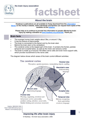
Rustenhoven et al. Journal of Neuroinflammation (2016) 13:37Page 6 of 15Fig. 2 TGFβ1 modifies inflammatory protein secretion in human brain pericytes. Primary human brain pericytes were treated with vehicle (1 mMcitric acid, pH 3 with 0.1 % BSA) or 10 ng/mL TGFβ1 for 0–72 h, and conditioned media was collected. Concentration of secreted proteins(sVCAM-1 (a), CX3CL1 (b), MCP-1 (c), IL-8 (d) and IL-6 (e)) was determined by a multiplex cytometric bead array. One representative experiment(N 4 wells) from three independent experiments is shown. *p 0.05, **p 0.01, ***p 0.001 versus vehicle control at each time point(two-way ANOVA)inflammatory cytokine. IL-1β alone significantly enhancedthe basal secretion of sVCAM-1 (p 0.001; Fig. 3a),CX3CL1 (p 0.001; Fig. 3b), MCP-1 (p 0.001; Fig. 3c),IL-8 (p 0.001; Fig. 3d) and IL-6 (p 0.001; Fig. 3e).TGFβ1 was found to significantly reduce IL-1β-inducedsVCAM-1 (p 0.001; Fig. 3a), CX3CL1 (p 0.001; Fig. 3b)and MCP-1 (p 0.001; Fig. 3c) secretions; however, it hadno effect on IL-8 expression (p 0.05, Fig. 3d). Furthermore, co-stimulation of pericytes with IL-1β and TGFβ1showed a synergistic release of IL-6, well above that seenwith IL-1β treatment alone (p 0.001, Fig. 3e).TGFβ1 signals through SMAD2/3 transcription factors butdoes not affect NF-kB translocationInflammatory protein expression is largely controlled bytranscription factor-mediated gene transcription. In orderto further understand how TGFβ1 modifies the pericyteimmune response, we investigated the ability of this cytokine to modify the localisation of two transcription factors,SMAD2/3 and nuclear factor kappa-light-chain-enhancerof activated B cells (NF-kB). Whilst SMAD2/3 showed alow level of basal nuclear localisation, TGFβ1 treatmentsignificantly enhanced this at 2 h (p 0.001; Fig. 4a, b),and remained slightly elevated (albeit not significantly) forup to 48 h (p 0.05; Fig. 4a, b). IL-1β was found to haveno effect on SMAD2/3 nuclear localisation at 2, 24 or48 h (p 0.05) alone and did not alter TGFβ1-induced nuclear localisation at any time point (p 0.05; Fig. 4a, b).IL-1β significantly enhanced nuclear translocation of NFkB at 2 h (p 0.001) but not 24 h (p 0.05) or 48 h (p 0.05), whilst TGFβ1 has no effect at any time point (p 0.05; Fig. 4a, c). Furthermore, TGFβ1 did not modify IL-
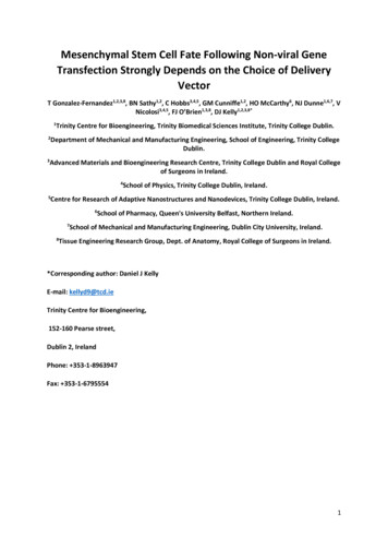
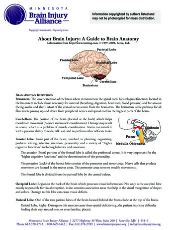
Rustenhoven et al. Journal of Neuroinflammation (2016) 13:37Page 7 of 15Fig. 3 Synergistic and preventative effects of TGFβ1 on IL-1β-induced inflammatory protein secretion. Primary human brain pericytes were treatedwith vehicle (1 mM citric acid, pH 3 with 0.1 % BSA) or 10 ng/mL TGFβ1 for 24 h followed by a further 24 h in the presence of 10 ng/mL IL-1β orvehicle (0.1 % BSA in PBS). Conditioned media was collected and the concentration of secreted proteins (sVCAM-1 (a), CX3CL1 (b), MCP-1 (c), IL-8(d) and IL-6 (e)) was determined by a multiplex cytometric bead array. One representative experiment (N 4 wells) from three independentexperiments is shown. ***p 0.001 (ANOVA)1β-induced NF-kB translocation at 2 h (p 0.05), 24 h(p 0.05) or 48 h (p 0.05; Fig. 4a, c).TGFβ1 attenuates phagocytosis by pericytesPhagocytosis is a critical function of specific immune cellsand is necessary to clear pathogenic substances from thebrain [47]. Inflammatory cytokines, including TGFβ1, havepreviously been shown to modify the phagocytic ability ofparenchymal brain cells, particularly microglia and astrocytes. Given the ability of TGFβ1 to modify pericyte inflammatory responses, we queried whether it couldinfluence phagocytosis by these cells. TGFβ1 significantlyattenuated pericyte phagocytosis of fluorescent latex beadsat 1 ng/mL (p 0.01) or 10 ng/mL (p 0.001) as determined by an automated imaging-based assay (Fig. 5a, b).To confirm that beads were internalised and not simplybound to the cell membrane confocal microscopy wasperformed (Fig. 5c). A flow cytometry based assay was alsoemployed to measure phagocytosis which again revealed asignificant reduction in phagocytic ability by pericytes following a 10 ng/mL TGFβ1 treatment (p 0.05, Fig. 5d, e)whilst lower concentrations trended towards a reduction.TGFβ1 reduces gene expression of scavenger receptorsScavenger receptors have a key role in recognising and coordinating the uptake of a wide range of macromolecules,including fluorescent beads and disease related proteinssuch as amyloid beta [48, 49]. As TGFβ1 was found to inhibit the phagocytic ability of pericytes, we investigated itsability to modify the expression of receptors involved inscavenging macromolecules. A 24-h treatment with TGFβ1was found to significantly attenuate pericyte expression, as
Rustenhoven et al. Journal of Neuroinflammation (2016) 13:37Page 8 of 15Fig. 4 TGFβ1 signals through SMAD2/3 transcription factors but does not affect NF-kB translocation. Primary human brain pericytes were treatedfor 0–48 h with combinations of vehicle (1 mM citric acid, pH 3 with 0.1 % BSA), 10 ng/mL TGFβ1 and 10 ng/mL IL-1β. Cells were fixed andimmunostained for NF-kB p65 and SMAD2/3. Representative images at 2 h are shown (a). The intensity of nuclear NF-kB (b) andnuclear SMAD2/3 (c) was determined by automated image analysis from four independent experiments. ***p 0.001 versus vehiclecontrol at each time point (two-way ANOVA). Scale bar 50 μmdetermined by qRT-PCR, of the receptors CD36 (p 0.001,Fig. 6a), CD68 (p 0.05, Fig. 6b) and CD47 (p 0.05,Fig. 6c), highlighting a possible mechanism behind TGFβ1reduced phagocytic ability.TGFβ1 reduces pericyte proliferation but does not affecttheir viabilityFunctional responses of cells, including phagocytosis,can be influenced by general changes in cell viability. In
Rustenhoven et al. Journal of Neuroinflammation (2016) 13:37Page 9 of 15Fig. 5 TGFβ1 attenuates phagocytosis by pericytes. Pericytes were treated for 24 h with 0–10 ng/mL TGFβ1 in 1 mM citric acid, pH 3 with 0.1 %BSA, followed by a further 24 h in the presence or absence of a 1:1000 dilution of fluorescent latex beads. Cells were fixed and nucleicounterstained with DRAQ5. Representative images for vehicle and 10 ng/mL TGFβ1 treatments are shown (a). The percentage of phagocyticpericytes was determined by automated image analysis from five independent experiments (b). Confocal microscopy of pericytes plated on glasscoverslips, incubated for 24 h with a 1:10,000 dilution of fluorescent beads and immunostained with PDGFRβ confirmed internalisation of beads(c). Pericytes were treated for 24 h with 0–10 ng/mL TGFβ1 in 1 mM citric acid, pH 3 with 0.1 % BSA. For an additional two hours cells wereincubated in the presence or absence of a 1:1000 dilution of fluorescent latex beads. Phagocytosis was determined by flow cytometry. Onerepresentative plot is shown (d) and the mean fluorescent intensity (MFI) of five independent experiments was determined (e). *p 0.05, **p 0.01, ***p 0.001 versus vehicle control (ANOVA). Scale bar 50 μmorder to exclude the possibility that TGFβ1 modifiesphagocytosis through compromising pericyte viability,we undertook a range of assays with respect to examining cell health. TGFβ1 was unable to stimulate pericyteapoptosis, as determined by antibody labelling and imaging of the early apoptotic marker cleaved caspase 3(CC3) at any time point (p 0.05; Fig. 7a, b). Due to thelow level of basal pericyte apoptosis, validation of theCC3 antibody was determined by okadaic acid-inducedapoptosis (Fig. 7a, b). TGFβ1 treatment did result in asignificant reduction of EdU positive cells, indicative of reduced cell proliferation, at 24, 48 and 72 h (p 0.001;
Rustenhoven et al. Journal of Neuroinflammation (2016) 13:37Page 10 of 15Fig. 6 TGFβ1 reduces gene expression of scavenger receptors. Primary human brain pericytes were treated with vehicle (1 mM citric acid, pH 3with 0.1 % BSA) or 10 ng/mL TGFβ1 for 24 h. RNA was extracted and expression of CD36 (a), CD68 (b) and CD47 (c) and determined by qRT-PCRfro
instructions. CBA samples were run on an Accuri C6 flow cytometer (BD Biosciences, CA, USA). Data was analysed using FCAP-array software (version 3.1; BD Biosciences, CA, USA) to convert fluorescent intensity values to concentrations using a ten-point standard curve (0-5000 pg/mL) as described previously [46]. Immunocytochemistry