Transcription
HindawiStem Cells InternationalVolume 2020, Article ID 8811963, 13 pageshttps://doi.org/10.1155/2020/8811963Research ArticleAmygdalin Promotes Fracture Healing through TGF-β/SmadSignaling in Mesenchymal Stem CellsJun Ying,1,2 Qinwen Ge,3 Songfeng Hu,4 Cheng Luo,5 Fengyi Lu,3 Yikang Yu,3 Taotao Xu,1,2Shuaijie Lv,1,2 Lei Zhang,6 Jie Shen,7 Di Chen,8 Peijian Tong,1,2 Luwei Xiao,2 Ju Li ,1,2Hongting Jin ,1,2 and Pinger Wang 1,21Department of Orthopaedic Surgery, The First Affiliated Hospital of Zhejiang Chinese Medical University, Hangzhou,310006 Zhejiang Province, China2Institute of Orthopaedics and Traumatology, The First Affiliated Hospital of Zhejiang Chinese Medical University, Hangzhou,310053 Zhejiang Province, China3First Clinical College of Zhejiang Chinese Medical University, Hangzhou, 310053 Zhejiang Province, China4Department of Orthopaedics, Shaoxing Hospital of Traditional Chinese Medicine, Affiliated with Zhejiang ChineseMedical University, Shaoxing, 312000 Zhejiang Province, China5Department of Orthopaedic Surgery, Fuyang Orthopaedics and Traumatology Affiliated Hospital of Zhejiang ChineseMedical University, Hangzhou, Zhejiang, China6Department of Orthopedics, Xiaoshan District Hospital of Traditional Chinese Medicine of Hangzhou, Hangzhou,311201 Zhejiang Province, China7Department of Orthopaedic Surgery, School of Medicine, Washington University, St. Louis, MO 63110, USA8Research Center for Human Tissues and Organs Degeneration, Shenzhen Institutes of Advanced Technology, Chinese Academyof Sciences, Shenzhen 518055, ChinaCorrespondence should be addressed to Ju Li; juli@zcmu.edu.cn, Hongting Jin; hongtingjin@163.com,and Pinger Wang; pingerwang@zcmu.edu.cnReceived 15 June 2020; Revised 13 August 2020; Accepted 21 August 2020; Published 7 September 2020Academic Editor: Darius WideraCopyright 2020 Jun Ying et al. This is an open access article distributed under the Creative Commons Attribution License, whichpermits unrestricted use, distribution, and reproduction in any medium, provided the original work is properly cited.Chondrogenesis and subsequent osteogenesis of mesenchymal stem cells (MSCs) and angiogenesis at injured sites are crucial forbone fracture healing. Amygdalin, a cyanogenic glycoside compound derived from bitter apricot kernel, has been reported toinhibit IL-1β-induced chondrocyte degeneration and to stimulate blood circulation, suggesting a promising role of amygdalinin fracture healing. In this study, tibial fractures in C57BL/6 mice were treated with amygdalin. Fracture calluses were thenharvested and subjected to radiographic, histological, and biomechanical testing, as well as angiography and gene expressionanalyses to evaluate fracture healing. The results showed that amygdalin treatment promoted bone fracture healing. Furtherexperiments using MSC-specific transforming growth factor- (TGF-) β receptor 2 conditional knockout (KO) mice(Tgfbr2Gli1-Cre) and C3H10 T1/2 murine mesenchymal progenitor cells showed that this effect was mediated through TGFβ/Smad signaling. We conclude that amygdalin could be used as an alternative treatment for bone fractures.1. IntroductionBone fractures, mainly caused by traumatic incidents andmedical conditions, including osteoporosis, are a growingglobal health burden currently affecting millions of people[1, 2]. Even with treatment, fractures are associated withgreat economic burden, loss of independence, and high ratesof morbidity and mortality [3]. In clinical practice, fracturescan be stabilized using various fixation methods, such asbraces and internal, external, and intramedullary fixation[4–6]. However, fracture nonunion is observed in 5-10% ofpatients annually [7]. In addition, bedfast patients receiving
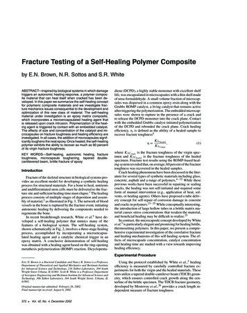
2Stem Cells International4d7d10 d21 d14 dControl⁎OOHOOHHOHOOOOHRadiographic union scoreAmygdalinOHHO⁎5⁎4321CN0Amygdalin : C20H27NO1147101421Days after fractureControlAmygdalin7d10 d14 d21 dAmygdalinBone volume (mm2)Control4d(b)BV/TV (%)(a)50403020100⁎⁎47101421Days after fracture⁎4⁎321047101421Days after fractureControlAmygdalin(c)(d)Figure 1: Amygdalin accelerated bone fracture healing in mice. (a) The chemical structure of amygdalin. (b) X-ray images and radiographicunion scores showed clear fracture lines at days 4-21 in mice of the control group, but only at days 4-10 in the amygdalin-treated group. Redarrows point to clear fracture lines. (c). μCT three-dimensional images of fractured tibiae from both groups at days 4, 7, 10, 14, and 21.Scale bar 500 μm. (d) μCT analysis of BV/TV. Data are presented as mean standard deviation (SD). P 0:05, n 6.conservative treatment have higher risks of pressure ulcers,hypostatic pneumonia, lower extremity venous thromboembolism, and death.Bone fracture healing is a complicated biological processthat involves specific regenerative patterns and diverse geneexpression changes [8, 9]. Secondary healing is the mostcommon form of bone healing, which normally includesendochondral and intramembranous bone healing that canbe separated into three consecutive stages: inflammation,bone repair, and remodeling [9]. The inflammatory phase ischaracterized by the formation of a hematoma and theimmediate release of soluble inflammatory mediators thatinduce immune cell infiltration [10].Mesenchymal stem cells (MSCs) can migrate to the fracturesite and differentiate into chondrocytes, which form a cartilaginous soft callus. This primary callus is subsequently surroundedby new bone material produced by osteoblasts in the perichondrium and permeated with blood vessels. The cartilaginouscallus then undergoes remodeling to replace the cartilage withnew bone. Thus, the promotion of chondrogenesis/osteogenesisand angiogenesis can accelerate fracture healing. The origin ofMSCs is not fully understood, but the recruitment, proliferation,and differentiation of these cells are indispensable for fracturehealing [11]. Successful bone repair and remodeling are alsodependent on an adequate blood supply and revascularizationof the injured area [12].
Stem Cells InternationalAmygdalin (D-mandelonitrile-β-gentiobioside) is derivedfrom bitter apricot kernel and has been used clinically for thetreatment of asthma, aplastic anemia, tumors, and alloxaninduced diabetes [13–15]. Amygdalin has also been reportedto inhibit IL-1β-induced chondrocyte degeneration inendplates and to both improve microcirculation and relieveblood stasis [16, 17]. Amygdalin has been implicated in theregulation of transforming growth factor- (TGF-) β/Smadsignaling [18], which has a fundamental regulatory functionfor bone homeostasis [19, 20]. Therefore, we hypothesizedthat amygdalin can promote bone fracture healing throughTGF-β/Smad signaling.2. Materials and Methods2.1. Preparation and Administration of Amygdalin. Amygdalin (CAS NO. 29883-15-6, purity 97:0%, Sigma, St. Louis,MO, USA) (Figure 1(a)) used in this study was purchasedfrom the National Institutes for Food and Drug Control ofChina and dissolved in sterile normal saline (NS)(0.05 mg/mL). In the amygdalin-treated group, mice wereintraperitoneally injected with 0.5 mg/kg of amygdalin dailyfrom day 1 postoperation. Mice in the control group wereintraperitoneally injected with an equal amount ofphosphate-buffered saline (PBS).2.2. Experimental Animals. This study was approved by theAnimal Experimentation Ethics Committee of Zhejiang Chinese Medical University. The animal center of the ZhejiangChinese Medical University provided C57BL/6 mice (SCXK,Shanghai, 2012-0002). Gli1-CreER transgenic mice can efficiently target MSCs by injecting tamoxifen (TM) to inducethe formation of Cre (CreERT2) from the endogenous Gli1locus [21]. The Gli1-CreER transgenic mice were crossedwith Tgfβr2flox/flox mice [22] to specifically knock out theTGF-β receptor 2 (Tgfbr2) in MSCs. Tamoxifen was administered (5 mg, dissolved in corn oil) once daily for 5 consecutive days by intraperitoneal injection.A DNA extraction kit (Sigma, USA) was used to determine mouse genotyping. The sequences of primers used forgenotyping were as follows: Cre, Fw: 5 ′ -ATT GCT GTCACT TGG TCG TGGC-3 ′ ; Rv: 5 ′ -GAA AAT GCT TCTGTC CGT TTGC-3 ′ ; Tgfbr2 loxP, Fw: 5 ′ -TAA ACA AGGTCC GGA GCC CA-3 ′ ; Rv: 5 ′ -ACT TCT GCA AGA GGTCCC CT-3 ′ (wild-type, 420-base-pair PCR product; homozygotic type, 540-base-pair PCR product). Cre-negativelittermates were used as controls.2.3. Cell Culture. C3H10 T1/2 murine mesenchymal progenitor cells (ATCC, Manassas, VA, USA) were cultured in Dulbecco’s modified Eagle medium (DMEM) (Hyclone, Logan,Utah, USA) supplemented with 10% fetal bovine serum(FBS), 100 U/mL penicillin and 100 mg/mL streptomycin(Gibco, Grand Island, NY, USA) in a 5% CO2 humidifiedincubator at 37 C. After reaching 80% confluence, cells werecollected and divided into the control group, the TGF-β1group (treated with 10 ng/mL recombinant TGF-β1), theamygdalin group (treated with 10 μM amygdalin), the TGFβ1 plus TGF-β/Smad pathway inhibitor-SB525334 group3Table 1: Primer name and sequences for PCR analysis.Primer nameSequencesβ-Actin forward5 ′ -GGAGATTACTGCCCTGGCTCCTA-3 ′β-Actin reverse5 ′ -GACTCATCGTACTCCTGCTTGCTG-3 ′Sox9 forward5 ′ -GAGGCCACGGAACAGACTCA-3 ′Sox9 reverse5 ′ -CAGCGCCTTGAAGATAGCATT-3 ′Col2a1 forward5 ′ -TGGTCCTCTGGGCATCTCAGGC-3 ′Col2a1 reverse5 ′ -GGTGAACCTGCTGTTGCCCTCA-3 ′Col10a1 forward5 ′ -ACCCCAAGGACCTAAAGGAA-3 ′Col10a1 reverse5 ′ -CCCCAGGATACCCTGTTTTT-3 ′Runx2 forward5 ′ -GAGGGCACAAGTTCTATCTGGA-3 ′Runx2 reverse5 ′ -GGTGGTCCGCGATGATCTC-3 ′Osteocalcin forward5 ′ -AGGGAGGATCAAGTCCCG-3 ′Osteocalcin reverse5 ′ -GAACAGACTCCGGCGCTA-3 ′VEGF forward5 ′ -GCACATAGAGAGAATGAGCTTCC-3 ′VEGF reverse5 ′ -CTCCGCTCTGAACAAGGCT-3 ′(treated with 10 ng/mL recombinant TGF-β1 and 10 μMSB525334), the amygdalin plus SB525334 group (treated with10 μM amygdalin and 10 μM SB525334), and the SB525334group (treated with 10 μM SB525334). After 4 hours of treatment, the cells were collected, and changes in the levels ofphosphorylated-Smad2/3 were analyzed. In addition, relativechondrogenic (Col2a1, Sox9) gene expressions were analyzedfollowing three days in culture.2.4. Tibial Fracture Model. Right transverse tibial fractureswere performed and fixed with an intramedullary needle[23]. Ten-week-old male C57BL/6, Tgfβr2f/f, andTgfβr2Gli1Cre mice were anesthetized with ketamine(60 mg/kg) by intraperitoneal injection. After local disinfection, a 1.0 cm long cut in the anteromedial skin of the tibiawas created. A 26-gauge syringe needle was then inserted intothe bone marrow cavity through the tibial plateau at themedial of patellar ligament. The needle was then removed,and the tibia was transected using a No.11 surgical blade.The 26-gauge syringe needle was reinserted into tibia to simulate intramedullary fixation. A 5-0 silk suture was selected toclose the incision, and buprenorphine administration (indrinking water) was used to reduce pain during the first threedays following surgery. Immediately following surgery, X-raytests (Carestream, FX Pro, USA) were performed on the rightlower limb in the anterior-posterior and lateral directions toconfirm the correctness of osteotomy and the alignment ofthe bone. The mice were then divided into groups foranalysis: C57BL/6 mice treated with amygdalin or PBS, andTgfβr2Gli1Cre and Cre-negative controls both treated withamygdalin.2.5. X-Ray and μCT Analyses. At days 4, 7, 10, 14, and 21postsurgery, mice were euthanized, and tibiae were collectedfor X-ray and microcomputed tomography (μCT) (Skyscan
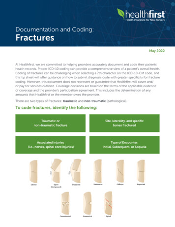
⁎25⁎2015⁎1050101421Days after fractureModulus of elasticity (MPa)Stem Cells InternationalMaximum loading (N)4⁎400⁎300200⁎1000101421Days after fractureControlAmygdalin(a)(b)Figure 2: Biomechanical testing was performed in bone samples harvested at days 10, 14, and 21 after surgery. (a) Maximum loading and (b)modulus of elasticity were measured in the amygdalin-treated and control groups. Data are presented as mean standard deviation (SD). P 0:05, n 6.1176; Bruker μCT, Kontich, Belgium) analyses to examinefracture healing quality. At each time point, harvested specimens were scanned using μCT at 8.73-micron isotropic resolution, with settings of 42 kV and 555 μA and 786 msintegration time. Followed by radiographic union scale(RUST) in the tibial fracture score system, semiquantitativeanalyses of the X-ray were performed. Callus bone volume(BV) and callus mineralized volume fraction (BV/TV) (%)parameters were then measured.ing day, a biotinylated anti-rabbit secondary antibody and anenzyme conjugate (Histostain-Plus Kit, Invitrogen, USA)were separately incubated on sections. IHC signals weredeveloped using DAB chromogen (Histostain-Plus Kit) andcounterstained for nuclei using Tacha’s CAT Hematoxylin.2.6. Biomechanical Testing. Full-length tibiae were harvestedand removed from their surrounding soft tissues (n 6 atdays 4, 7, 10, 14, and 21). Both ends of each tibia were thenfixed in bone cement to ensure that the fracture site wasexposed. Specimens were installed on an Endura Tec TestBench TM system (200 N mm torque cell; Bose Corporation,Minnetonka, MN, USA) to apply torsion at a rate of 1 /s untilfailure to determine the modulus of elasticity and maximumloading of the fracture callus.2.8. Quantitative Real-Time Polymerase Chain Reaction. Thecallus was cut with a range of 2 mm around the fracture line.Total RNA was extracted from the callus and three duplicated wells of C3H10 T1/2 cells in the third passage by usingthe RNeasy kit (Qiagen, Germany) following the manufacturer’s instructions. Then, complementary DNA (cDNA)was synthesized using a cDNA reverse transcription kit(Takara, Otsu, Japan). Quantitative real-time polymerasechain reaction (qRT-PCR) was performed using the SYBRPremix EX Taq kit (Takara) according to the manufacturer’s instructions. Primer sequences for Sox9, Col2a1,Col10a1, Osteocalcin, Runx2, vascular endothelial growthfactor (Vegf), and β-actin are shown in Table 1.2.7. Histology and Histomorphometry. After three days of fixation with 10% normal buffered formalin, the bone sampleswere decalcified in 14% ethylenediaminetetraacetic acid(EDTA) for two weeks. The decalcified bones were dehydrated and embedded in paraffin and sectioned (3 μm).Alcian blue/H&E (ABH) staining and tartrate-resistant acidphosphatase (TRAP) staining were performed. Quantitativehistomorphometry was performed on the ABH- andTRAP-stained sections using the ImageJ 1.46r software(Wayne Rasband, National Institute of Health, USA). Thecartilage area per periosteal callus area (%, Cg.Ar/http://Ps.Cl.Ar) and the mineralized area per periosteal callus area(%, Md.Ar/http://Ps.Cl.Ar) were measured as described previously [24]. Immunohistochemistry (IHC) was performedon sections using high-pressure heating in pH 6.0 sodium citrate solution for 2 minutes to achieve antigen retrieval. Thetissues were then incubated with a rabbit anti-mouse antip-Smad2 antibody (Abcam, ab188334, 1 : 200), antipSmad1/5/8 antibody (Millipore, AB3848, 1 : 200), and antiCD31 antibody (Arigo, ARG52748, 1 : 100) overnight at4 C. PBS was incubated on negative group slides. The follow-2.9. Western Blot Analysis. Three duplicated wells ofC3H10T1/2 cells at passage 3 were prepared for proteinextraction. Cell lysates were extracted using a modified radioimmunoprecipitation assay (RIPA) lysis buffer that contained 1 mM phenylmethylsulfonyl fluoride (PMSF) and aprotease inhibitor cocktail (Cell Signaling Technology,USA). Following centrifugation at 12000g for 30 minutes,the supernatant was collected to detect the total protein concentrations using a BCA Protein Assay kit (Thermo Scientific, USA). Protein extracts were boiled for 5 minutes inloading buffer. Then, 40 μg protein was loaded and electrophoresed on a sodium dodecyl sulfate polyacrylamide gelelectrophoresis (SDS-PAGE) gel and transferred to a polyvinylidene difluoride (PVDF) membrane. The membrane wasblocked in a 5% skim milk solution and incubated with primary antibodies overnight at 4 C. The primary antibodieswere β-actin (Sigma, A1978), Smad2/3 (Cell Signaling Technology, #8685), and phosphorylated-Smad2/3 (Cell SignalingTechnology, #8828). The membrane was then incubated witha fluorescent secondary antibody (LI-COR, 926-32212)(1/1000), for 1 hour at room temperature (Vector
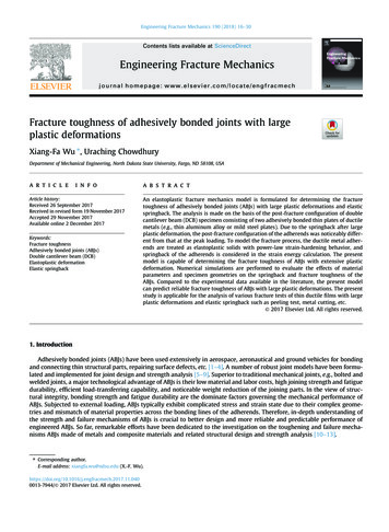
Stem Cells 1421Md.Ar/Ps.Cl.Ar (%)Cg.Ar/Ps.Cl.Ar (%)7706050403020100Days after fracture14⁎⁎⁎7101421Days after alinAmygdalinTRAPp-Smad1/5/8ControlOsteoclast gdalin(d)(e)Figure 3: Amygdalin accelerated formation of woven bone. (a) Histological sections of the bone fracture with Hematoxylin and Eosin (H&E)as well as Alcian blue staining. Black arrows represent the cartilage area; yellow arrows highlight the woven bone area. Scale bar 200 μm. (b)The cartilage area (blue) of callus tissues (%, Cg.Ar/http://Ps.Cl.Ar) was quantified at days 7, 10, 14, and 21 postfracture in the control andamygdalin-treated groups. (c) Newly formed woven bone in callus tissues (%, Md.Ar/http://Ps.Cl.Ar) was quantified at days 7, 10, 14, and21 postfracture in the control and amygdalin-treated groups. (d) Phospho-Smad2 and phospho-Smad1/5/8 protein expressions wereassessed by immunohistochemistry in control and amygdalin-treated groups at days 7 and 10, separately. Blue arrows: p-Smad2 positivecells. Scale bar 50 μm. Data are presented as mean standard deviation (SD). (e) Tartrate-resistant acid phosphatase (TRAP) stainingand quantification of osteoclast surface per bone surface (Oc. S./BS) of sections from control and amygdalin-treated groups at day 14 aftersurgery. Scale bar 100 μm. P 0:05, n 6.Laboratories, Burlingame, VT, USA). The blots were visualized using a LI-COR Odyssey scanner (LI-COR Biosciences,USA). The relative density of the p-Smad2/3 bands wasnormalized to their corresponding actin bands.2.10. Wound Scratch Test and Transwell Cell Migration.C3H10 T1/2 cells were seeded in six-well plates and culturedwith DMEM/F12 supplemented with 5% FBS, 1 105 units/Lpenicillin, and 100 g/L streptomycin. After reaching 80%-90% confluence, a scratch of 0.5 mm was made using asterile pipette tip and then washed with PBS to clear cellulardebris. After injury, the cells were cultured with low glucoseDMEM/F12 containing 5% (v/v) FBS with 0, 10 μM amygdalin, 10 ng/mL TGFβ1, 10 μM amygdalin plus 10 μMSB525334, 10 ng/mL TGFβ1 plus 10 μM SB525334, or10 μM SB525334. The migrations of C3H10 T1/2 cells intothe scratch were photographed using an inverted microscope(Olympus, Tokyo, Japan) at 24 hours post insult.
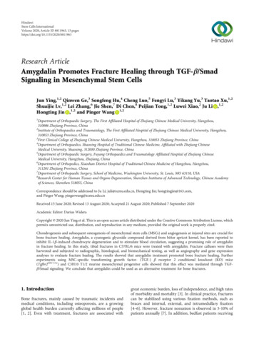
6Stem Cells International2.01.51.0⁎0.5071014Col2 expression(fold change)Sox9 expression(fold change)2.01.51.00.50217Days after fracture(a)1.00.57211014⁎2.0Runx2 expression(fold change)Col10 expression(fold change)⁎1.5014(b)⁎2.010Days after fracture1.51.00.5021⁎7Days after fracture101421Days after fractureControlAmygdalin(c)(d)⁎3.03.2⁎Vegf expression(fold change)OCN expression(fold change)4.02.01.007101421Days after fracture(e)⁎2.4⁎1.60.807101421Days after fracture(f)Figure 4: Amygdalin upregulated expression of chondrogenesis-, osteogenesis-, and angiogenesis-related genes in the callus. (a–f)Expressions of Sox9, Col2a1, Col10a1, Runx2, Ocn, and Vegf were measured by quantitative real-time polymerase chain reaction (PCR)between control and amygdalin groups at days 7, 10, 14, and 21 postfracture. Data are the relative expression versus housekeeping gene βactin and are presented as mean standard deviation (SD). P 0:05, n 6.Transwell assays were performed using 8 μm-pore Transwell polycarbonate membranes (Corning Inc., Corning, NewYork, USA). 1:0 104 cells were seeded with 200 μLDMEM/F12 containing 2% FBS in the upper chamber, andin the lower chamber, 500 μL DMEM/F12 containing 2%FBS with 0, 10 μM amygdalin, 10 ng/mL TGFβ1, 10 μMamygdalin plus 10 μM SB525334, 10 ng/mL TGFβ1 plus10 μM SB525334, or 10 μM SB525334. After incubating for48 h, the migratory cells were fixed with 4% paraformaldehyde for 15 min and stained with crystal violet. Threerandom images were taken in the fields.was injected with more pressure due to its viscosity and conserved overnight at 4 C. Muscles around the bone callus wereremoved to eliminate the influence of muscle blood vessels.μCT scanning was first used to locate the callus surroundingthe fracture. After decalcification of the harvested bone samples for 3 days, the samples were scanned again, using thesame volume of interest (VOI). The analysis area included400 slices by considering the fracture line as the midline.The CTAn Software (Skyscan, Version 1.16) was used tobuild three-dimensional (3D) models and to measure relativevessel volume.2.11. In Vivo Angiography. After injecting a mouse with ketamine (60 mg/kg), the right atrium was perforated, and 10 mLPBS/heparin sodium was injected through the apex of the leftventricle. Subsequently, 10 mL of 4% paraformaldehyde wasperfused using the same method. Finally, 10 mL MICROFIL2.12. Statistical Analysis. All data were obtained from 6individual mice in every experiment and 3 duplicated wellsof C3H10 T1/2 cells. Values are showed as the mean. Twoway ANOVA followed by the Tukey-Kramer posttest (multiple groups) and unpaired Student’s t-test (two groups) were
Stem Cells International7AmygdalinVessel volume trolAmygdalinCD31Negative(b)Figure 5: Amygdalin promoted angiogenesis in the newly formed callus around the fracture. (a) Three-dimensional images and quantificationof angiography around the fractures in control and amygdalin-treated mice 10 days after surgery. Quantification shows the vessel volume.Scale bar 1 mm. (b) CD31 protein expression was assessed by immunohistochemistry in control and amygdalin-treated groups (day 10).Blue arrows: CD31-positive cells. Scale bar 50 μm. Data are presented as mean standard deviation (SD). P 0:05, n 6.used for statistical analyses. The SPSS 17.0 software was usedto perform the statistical tests. P 0:05 was consideredsignificant.3. Results3.1. Amygdalin Facilitates Bone Fracture Healing in Mice. Toassess the effects of amygdalin on fracture healing, mice weretreated with PBS or amygdalin (0.5 mg/kg/day). X-ray resultsshowed that treatment with amygdalin promoted earlier formation of bone callus (Figure 1(b)). In the amygdalin group,a clear callus profile could be observed at day 10, and thecallus became more radiopaque at day 14 postfracture. Incontrast, the callus was not apparent in the control groupuntil day 14 postfracture.In addition, 3D reconstructions of μCT images showedthat obvious fracture gaps could be observed in the controlgroup at days 4, 7, 10, and 14, and newly formed bone hadnot fully bridged the gaps until day 21 (Figure 1(c), left). Incontrast, in the amygdalin-treated group, the fracture gapappeared indistinct at day 14, indicating a higher proportionof mineralized bone in the callus (Figure 1(c), right). μCTanalysis revealed that the bone volume (%, BV/TV) in theamygdalin group was significantly increased compared tothe control group at days 10 and 14 (Figure 1(d)), consistentwith the radiographic results.Furthermore, biomechanical testing showed that themodulus of elasticity and maximum loading increased significantly in tibia samples at days 10, 14, and 21 in theamygdalin-treated group compared to the PBS control group(P 0:05). These data demonstrated that bone mechanicalstrength has been improved by amygdalin treatment(Figure 2).Because radiographic evaluation cannot detect changes incartilage and soft tissues, Alcian blue/Hematoxylin/Orange G(ABH/OG) staining and histomorphometric analysis of thefractured bone were performed. The cartilage area (%,Cg.Ar/http://Ps.Cl.Ar) was significantly reduced by day 14postfracture, and the mineralized bone area was significantlyincreased (%, Md.Ar/http://Ps.Cl.Ar) at days 10-21 postfracture in amygdalin-treated mice compared to the control. Atday 21, the cartilage was no longer detectable in either group(Figures 3(a)–3(c)). In addition, phospho-Smad2 and phospho-Smad1/5/8 IHC was performed to observe activationof TGF-β/Smad and BMP2 signaling during fracture healing.As shown in Figure 3(d), amygdalin treatment could increasephospho-Smad2 expression at day 7 but have no obviouseffect on phospho- Smad1/5/8 expression at day 10. TRAPstaining was also performed in the fracture callus at day 14.No significant changes in osteoclast numbers were foundafter amygdalin treatment at day 14 (Figure 3(e)).We also measured the expression of marker genes relatedto bone fracture healing. The gene expression of early chondrogenesis, such as Sox9 and Col2a1, was similar inamygdalin-treated and control mice during fracture healing,except that Sox9 expression was reduced at day 21(Figures 4(a) and 4(b)). Levels of Col10a1, a marker of latehypertrophic cartilage, were upregulated at days 10 and 14in the amygdalin treatment group (Figure 4(c)); this supported the histologic results of decreased cartilage area atday 14. Osteogenic marker genes, runt-related transcriptionfactor 2 (Runx2), and osteocalcin (OCN), were also increased
8Stem Cells InternationalNegativep-Smad210 d14 d21 d5Radiographicunion Days after fracture(a)(b)10 d14 d21 d50403020100⁎710Bone volume (mm2)BV/TV 7d⁎1421Days after fractureCre-negative (amygdalin)Tgfbr2Gli1Cre (amygdalin)(c)54⁎⁎32107101421Days after fracture(d)Figure 6: Amygdalin promoted bone fracture healing in the Cre-negative group compared with the Tgfβr2Gli1Cre group. (a) Phospho-Smad2protein expression was assessed by immunohistochemistry in the Cre-negative and Tgfβr2Gli1Cre groups (day 7). Scale bar 50 μm. (b) X-rayresults of fractures in the amygdalin-treated Tgfβr2Gli1Cre group and Cre-negative mice. Red arrows indicate fracture lines. (c, d) μCT analysisof the bone volume and BV/TV (%) in callus tissues at days 7, 10, 14, and 21 postfracture in amygdalin-treated Tgfβr2Gli1Cre and Cre-negativemice. Scale bar 500 μm. Data are presented as mean standard deviation (SD). P 0:05, n 6.at days 10 and 14 in the amygdalin treatment group compared to the control group (Figures 4(d) and 4(e)). In addition, expression of Vegf at days 10 and 14 was increased inthe amygdalin-treated group compared to the controls, indicating that amygdalin may stimulate angiogenesis duringfracture healing (Figure 4(f)).Angiographic results demonstrated that amygdalin stimulated angiogenesis and increased the vessel volume in the cartilaginous bone around the fracture callus (P 0:05)(Figure 5(a)). Protein expression of the endothelial cell marker,CD31, was assessed by IHC at the callus area on day 10. Fracture callus samples from amygdalin-treated mice showedincreased CD31 expression (Figure 5(b)), consistent withupregulation of the angiogenesis-related marker gene Vegf.3.2. Amygdalin Accelerates Bone Fracture Healing throughTGF-β/Smad Signaling of MSCs In Vivo. TGF-β/Smad signaling plays fundamental roles in bone homeostasis [25],and amygdalin has been implicated in the regulation of thispathway. To determine whether TGF-β/Smad signaling inMSCs is directly required for amygdalin-induced fracturehealing, tibia fractures were performed on MSC-specificTgfβr2 conditional KO (Tgfβr2Gli1Cre) mice and Crenegative mice.To determine the Tgfβr2 knockout efficiency inTgfβr2Gli1Cre mice, IHC of p-Smad2 on fracture callus sections of Cre-negative mice and Tgfbr2Gli1Cre mice at day 10was performed. The protein expression of p-Smad2 was obviously reduced in Tgfbr2Gli1Cre mice, indicating inhibition ofTGFβ/Smad pathway signaling (Figure 6(a)). Following fractures, amygdalin was administrated to all mice. X-ray resultsshowed that fracture lines disappeared at days 14 and 21 inthe Cre-negative group but were still clearly visible untilday 21 in the Tgfβr2Gli1Cre group (Figure 6(b)). Furthermore,μCT results indicated that amygdalin treatment increasedbone volume and BV/TV around the fracture lines in Crenegative mice compared to the Tgfβr2Gli1Cre mice, especiallyat days 10 and 14 (Figures 6(c) and 6(d)).In histological analyses of fracture callus tissues, theTgfβr2Gli1Cre mice showed delayed fracture repair
Stem Cells (amygdalin)Cg.Ar/Ps.Cl.Ar (%)71050403020100⁎7101421Days after fracture14Cre-negative (amygdalin)Tgfbr2Gli1Cre dalin)Osteoclast e⁎TRAPMd.Ar/Ps.Cl.Ar 2Gli1Cre(amygdalin)Days after fractureCre-negative (amygdalin)Tgfbr2Gli1Cre (amygdalin)(c)(d)Figure 7: (a) Histological images of amygdalin-treated Tgfβr2and Cre-negative control mice stained with Alcian blue/Hematoxylinand Eosin (ABH). Black arrows designate chondrocytes; yellow arrows highlight woven bone. Scale bar 200 μm. (b, c) The cartilage areaof the callus (%, Cg.Ar/http://Ps.Cl.Ar) and newly formed woven bone (%, Md.Ar/http://Ps.Cl.Ar) were quantified at days 7, 10, 14, and21 in amygdalin-treated Tgfβr2Gli1Cre and Cre-negative control mice. (d) Tartrate-resistant acid phosphatase (TRAP) staining andquantification of osteoclast surface per bone surface (Oc. S./BS) of sections from amygdalin-treated Tgfβr2Gli1-Cre and Cre-negative mice(day 14). Scale bar 100 μm. Data are presented as mean standard deviation (SD). P 0:05, n 6.Gli1Cre(Figure 7(a)). The cartilage area (%, Cg.Ar/http://Ps.Cl.Ar) inTgfβr2Gli1Cre mice was significantly increased compared withcontrols at day 14 (Figure 7(b)). In addition, Tgfβr2Gli1Cremice showed a weaker ability for formation of woven boneat days 10, 14, and 21 postfracture (Figure 7(c)), indicatingits slower transformation from cartilage into woven bone.Even though the osteoclast number in Cre-negative m
Jun 15, 2020