Transcription
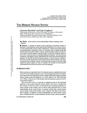
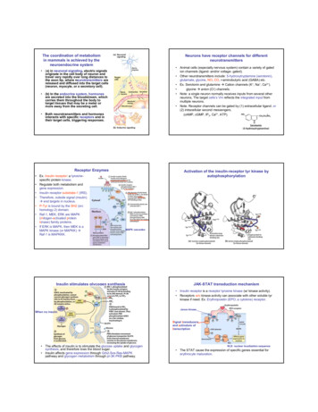
The coordination of metabolismin mammals is achieved by theneuroendocrine systemNeurons have receptor channels for differentneurotransmitters Animal cells (especially nervous system) contain a variety of gatedion channels (ligand- and/or voltage- gated). Other neurotransmitters include: 5-hydroxytryptamine (serotonin),glutamate, glycine, NO, CO, r-aminobutyric acid (GABA) etc. Ex. Serotonin and glutamine Æ Cation channels (K , Na , Ca2 ). glycine Æ anion (Cl-) channels. Note: a single neuron normally receives inputs from several otherneurons, The target cells’s Vm reflects the integrated input frommultiple neurons. Note: Receptor channels can be gated by (1) extracellular ligand. or(2) intracellular second messengers.(cAMP, cGMP, IP3, Ca2 , ATP). (a) in neuronal signaling, electric signalsoriginate in the cell body of neuron andtravel very rapidly over long distances tothe axon tip, where neurotransmitters arereleased and diffused into the target cells(neuron, myocyte, or a secretory cell). (b) In the endocrine system, hormonesare secreted into the bloodstream, whichcarries them throughout the body totarget tissues that may be a meter ormore away from the secreting cell. Both neurotransmitters and hormonesinteracts with specific receptors and intheir target cells, triggering responses.Receptor Enzymes Ex. Insulin receptor: a tyrosinespecific protein kinase. Regulate both metabolism andgene expression. Insulin receptor substrate-1 (IRS). Therefore, outside signal (insulin)Æ end targets in nucleus. P-Tyr is bound by the SH2 (srchomology 2) domain. Raf-1, MEK, ERK are MAPK(mitogen-activated proteinkinase) family proteins. If ERK is MAPK, then MEK is aMAPK kinase (or MAPKK) ÆRaf-1 is MAPKKK.Activation of the insulin-receptor tyr kinase byautophosphorylationMAPK cascadesInsulin stimulates glycogen synthesisJAK-STAT transduction mechanism Insulin receptor is a receptor tyrosine kinase (w/ kinase activity). Receptors w/o kinase activity can associate with other soluble tyrkinase if need. Ex. Erythropoietin (EPO; a cytokine) receptor.When no insulinJanus kinaseSignal transducersand activators oftranscription The effects of insulin is to stimulate the glucose uptake and glycogensynthesis, and therefore lows the blood sugar. Insulin affects gene expression through Grb2-Sos-Ras-MAPKpathway and glycogen metabolism through pI-3K-PKB pathway.NLS: nuclear localization sequence The STAT cause the expression of specific genes essential forerythrocyte maturation.1
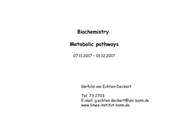
Receptor Guanylyl Cyclase (GC) generate the secondmessenger cGMPBy GCRenal excretion of Na (byion channel) ÆLoss water (osmoticpressure) Æreduce blood volumeIn smooth muscle cell,ANF Æ relaxation Æ lowblood pressurecGMP is degraded by cGMPphosphodiesterase (cGMP PDE)This serves as differentmessengers in differenttissueRegulate Cl- secretion in intestine ÆDecrease water absorption Æ (endotoxinby E. Coli.) diarrhea. The role of nitric oxide (NO) in smooth muscle relaxation in a blood vesselwall. Acetylcholine released by nerve terminals in the blood vessel wallactivates NO synthase in endothelial cells lining the blood vessel, causing theendothelial cells to produce NO. The NO diffuses out of the endothelial cellsand into the underlying smooth muscle cells, where it binds to and activatesguanylyl cyclase to produce cyclic GMP. The cyclic GMP triggers a responsethat causes the smooth muscle cells to relax, enhancing blood flow through theblood vessel .G protein-coupled receptors and second messengersThe cyclic GMP pathway in vasodilationPhosphodiesterase (PDE)Sildenafil (Viagra)PKG: cGMP-dependent protein kinaseor protein kinase G The third one (1st: gate ion channels; 2nd: receptor enzymes)consists: (1) a 7-TM membrane (serpentine) protein; (2) intracellularmessenger; (3) guanosine-nucleotide-binding protein (G protein). General mechanism: activated receptor activates G protein toexchange bound GDP for GTP. Æ activated G protein (dissociatedfrom receptor) alters the activity of nearby enzyme Æ secondmessenger Æ physiology effects. Human genome encodes more than 1,000 member of this typereceptors, which transduct the signals such as light, smells, tastes,and hormones. Ex. β-adrenergic receptor mediated by epinephrine (adrenaline). - released from adrenal gland. - regulates energy-yielding metabolism in muscle, liver, and adiposetissue.vasodilationEpinephrine and its synthetic analog Agonist: compound analogs that bind a receptor and mimic theeffects of its natural products. Ex. Epinephrine v.s. isoproterenol Antagonist: Compounds that bind a receptor without triggering theeffects. Ex. Epinephrine v.s. propranololG-protein-coupled receptors and second messengers G protein, stimulated by activated receptor, exchanges bound GDPfor GTP. Ex. β-adrenergic receptor7TM receptor or serpentine receptorNote: These are bindingaffinities to the β-adrenergicreceptor.Higher affinity does notmean higher effects.There are four types ofadrenergic receptors: α1, α2,β1, β2. Æ β type here.or self-inactivation2
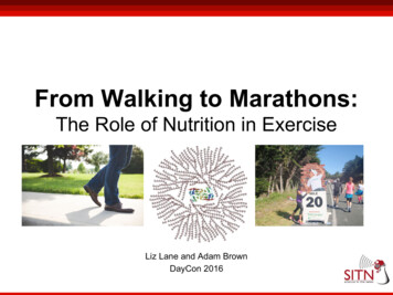
Self-inactivation of GsInteraction of Gsα with adenylyl cyclaseCatalytic subunit of ACDrug forskolinVery importantGTPGsαThe GTPase activity of G protein is generally very slow, so it allows Gprotein remains active in certain period. Once GTP is hydrolyzed backto GDP, G-protein goes back to inactive state.Epinephrine cascade Epinephrine Æ activate phosphorylase a(1 Æ 1000 molecules (underestimated)). PKA (cAMP-dependent protein kinase orprotein kinas A or PKA).Substrate binding site (catalytic subunit) of PKAIn the tetramer form(R2C2; inactive form),R domain occupied in PKAsubstrate binding site Æblock access to thesubstrateCatalytic subunit of PKASer residue inphosphorylase b that willbe phosphorylated byPKA.Once phosphorylated,phosphorylase b isactivated and then itactivates phosphylase a.Peptide substrate(i.e. phosphoylase b)Phosphylase a is theenzyme that breaksdown the glycogen.Two free PKA catalytic subunitIntracellular messenger cAMP is degraded by cyclicnucleotide phosphodiesterase The intracellular signal persists only as Epinephrine stimulationlong as the hormone receptor remainoccupied by epinephrine. Methyl xanthines inhibit thephosphodiesterase, increasing the life-lifeStimulateof cAMP and therefore potentiating agentsproduction ofglucosethat act by stimulate adenylyl cyclase.Methylxanthines(ex. Caffeine,theophylline)Cyclic nucleotidephosphodiesteraseNote: The ser residues are phosphorylated3
β-adrenergic receptor must be desensitized afterstimulationβARK is a member of Gprotein-coupled receptorkinases (GRKs). All of them phosphorylateserpentine receptors ontheir carboxyl-terminalcytosolic domain.βarr is a scaffold protein,which bring togetherseveral protein kinasesthat function in a cascade.Other signals also affect cAMP productionBARK: β-adrenergic receptor kinase(G Protein-coupled receptor kinase)Receptor of adipocytes Æ activate adenylylclcylase Æ cAMP Æ PKA Æ perilipin andtriacylglycerol lipase (to mobilization of fattyacid)Two second messengers are derived fromphosphatidylinositols A second class of serpentine receptors are coupled through Gprotein to a plasma membrane phospholipase C (PLC). PLC cleaves phosphatidylinositol 4,5-bisphosphate to DAG(diacylglycerol) and IP3 (inositol 3,4,5-triphosphate).For hormones, refer to table 12-5. IP3/receptor Æ open Ca2 channelon ER. Thus Ca2 acts as a secondmessenger. Tumor promoters: compounds acton PKC. ex. Phorbol ester- mimic DAG, but is notmetabolized rapidly.- interfere with normal regulation ofcell growth and cell division.Hormones trigger responsesCalcium is a second messenger in many signaltransduction Ca2 as a second messenger: exocytosis in neurons, contraction inmuscle, cytoskeletal rearrangement. Ca2 channels increase cytosolic Ca2 concentration Æ trigger acellular response. Ca2 concentration oscillate with a period of a few seconds probablyby phospholipid (IP3) or ion channels (Ca2 ).Calmodulin: a Ca2 binding protein that associates with avariety of other proteinsCaM: 17 KDa; acidic protein; when Ca2 is 1uM, the Ca2 binding inducesa conformational change.Enzymes regulated byCa2 /Calmodulin4 Ca2 ions4
Multivalent scaffold proteins and membrane raftsBinding modules of signaling molecules 10 % of human genome encodes signal proteins (receptors, Gproteins, proteins generating 2nd messenger, protein kinases;desensitization, ion channels). Q. How does one protein finds another in a signaling pathway? (1) protein modules binds phosphorylated Tyr, Ser, or Thr residuesin partner proteins. A. SH2 vs P-Tyr B. PTB (phosphotyrosine-binding domain) v.s p-Tyr (structure andsequence different). (2) with Proline-rich Æ SH3SH2 (3) PH to IP3Mechanism of autoinhibition of Src Tyr KinaseinactiveGSK3 is inactivated by PKBRemove the (p) groupIn this case, domainbinding partner isinternalInsulin-induced formation of supramolecular signalingcomplex 12 signaling molecules are involved.Note: phosphorylation is essential for this process.Note: dephosphrylation on any sites break the circuit.There are four types of general interactions: 1. SH2 or PDB to (p) 2.SH3 to proline-rich 3. PH to PIP3.Bacterial signaling Two component system: Receptor HisKinase and response regulatorTumbling is caused by an abruptreversal of the flagellar motor. Asecond reversal of the motorrestores smooth swimming, almostalways in a different direction.5
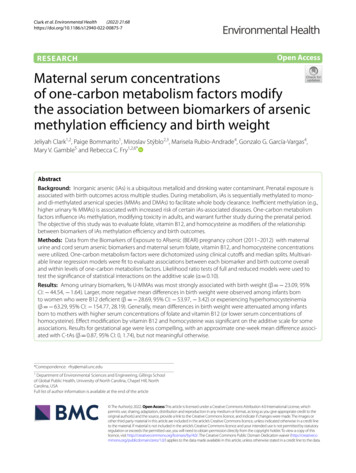
Stimuli produce response in plants Some plant mechanisms are similarto those in animals (kinases, scaffoldproteins, cNMP, ion pumps, ionchannels). Some are similar to two-componentsystem. Some are unique to plants (lightsensing mechanisms). Ex. Arabidopsis thaliana- 1,000 ser/thr kinases.- 60 MAPKs- 400 membrane-associatedreceptors.- 100 ion channels.etc.Note: PKG and PKA are absent inplants.Ethylene is a plant hormoneStructural Similarities between plant and animal signal Both plants and animals use compounds derived from aromaticamino acids as signals.Similarities between the signaling pathways thattrigger immune response in plants and animals Receptor-like kinase: triggered by bacterial pathogen. flg22: a peptide by breakdown of flagellin (C and D) The ethylene-mediated “triple response” that occurs whenthe growing shoot of a germinating seedling encounters an obstacleunderground. After such an encounter (D), the shoot thickens, and theprotective hook (at top) increases its curvature to protect the tip of theshoot.Sensory transduction in vision, olfaction, and gustation There are five major sensory systems: olfaction, taste (gustation),vision, hearing, and touch.Trigger the synthesisof proteins thatdefend against thebacterial infectionSensory transduction in vision, olfaction, andgustation The detection of lights, smells, andtastes. In animals, these are accomplished byspecialized sensory neurons. The light-sensing cells: 1. rod: sensitiveto light (high resolution and night version)but not to color. 2. cone: sensitive tocolor but not to light. The signals eventually pass fromganglion neurons through the optic nerveto the brain.innerouter6
Light-induced hyperpolarization of rod cells Outer segment: contains membrane-bound rhodopsin. Inner segment: contain nucleus and mitochondria (produces ATP). In the dark, membrane potential is around -45 mV (due to Na -K ATpase and ion-gate Na channel (now is open)). Light stimulates cGMP degradation Æ Vm - 75 mV Æ pass theelectric activity to the brain by interconnecting neuron.Light triggers conformational change n the receptorrhodopsin When a photon is absorbed by the retinalcomponent of rhodopsin Æ photochemicalchange Æ 11-cis-retinal changes to 11trans-retinal. This is a first stage of visual transduction. The excited rhodopsin interacts with atransducin (T) (GTP protein). In the dark, T/GDP Æ no signal With light, Tα/GTP Æ activate cGMP PDEÆ degrade cGMP to GMP. The cGMP-specific PDE is unique to thevisual cells of the retina. *** a photon changes the Vm of the cell.Molecular consequences of photon absorption by rhodopsin inthe red outer segmentHow we control the color vision Activation:Recovery [cGMP] Æ ion channels close Æ [Ca2 ] Æ GC activity Æ [cGMP] 1 photon Æ 500 transducins Æ 500 PDE Æ degrade 4,200cGMPs/sec; 1 photon Æ 1,000 channels to close Æ 1 mV change. The mechanism is similar to rod cells except using different pigment. There are three types of cone cells containing three different photoreactorproteins (opsins). Therefore, the visible spectrum for human is about 380 to 750 nm. Ex. Color blindness- results from different opsin mutations.- red- dichromates Æ loss of red photoreceptor.- mutations might also cause spectra shiftÆ abnormal color vision.ex. Red-anomalous trichromatesor green-anomalous trichormates.7
Birds have a total of six pigments: rhodopsin, four cone pigments, apinopsin Æ birds have hightly acute color perception.8
The third one (1st: gate ion channels; 2nd: receptor enzymes) consists: (1) a 7-TM membrane (serpentine) protein; (2) intracellular messenger; (3) guanosine-nucleotide-binding protein (G protein). General mechanism: activated receptor activates G protein to exchange bound GDP for GTP. Æactivated G protein (dissociated