Transcription
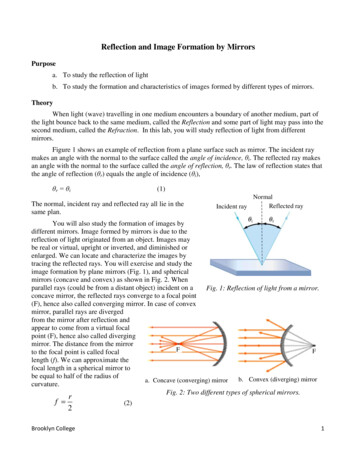
19 Jun 2e(2002/01/18)P1: IKH10.1146/annurev.neuro.27.070203.144230Annu. Rev. Neurosci. 2004. 27:169–92doi: 10.1146/annurev.neuro.27.070203.144230c 2004 by Annual Reviews. All rights reservedCopyright First published online as a Review in Advance on March 5, 2004THE MIRROR-NEURON SYSTEMGiacomo Rizzolatti1 and Laila Craighero2Annu. Rev. Neurosci. 2004.27:169-192. Downloaded from arjournals.annualreviews.orgby Princeton University Library on 03/26/08. For personal use only.1Dipartimento di Neuroscienze, Sezione di Fisiologia, via Volturno, 3, Università diParma, 43100, Parma, Italy; email: giacomo.rizzolatti@unipr.it;2Dipartimento SBTA, Sezione di Fisiologia Umana, via Fossato di Mortara, 17/19,Università di Ferrara, 44100 Ferrara, Italy; email: crh@unife.itKey Words mirror neurons, action understanding, imitation, language, motorcognition Abstract A category of stimuli of great importance for primates, humans inparticular, is that formed by actions done by other individuals. If we want to survive,we must understand the actions of others. Furthermore, without action understanding,social organization is impossible. In the case of humans, there is another faculty thatdepends on the observation of others’ actions: imitation learning. Unlike most species,we are able to learn by imitation, and this faculty is at the basis of human culture. Inthis review we present data on a neurophysiological mechanism—the mirror-neuronmechanism—that appears to play a fundamental role in both action understanding andimitation. We describe first the functional properties of mirror neurons in monkeys.We review next the characteristics of the mirror-neuron system in humans. We stress,in particular, those properties specific to the human mirror-neuron system that mightexplain the human capacity to learn by imitation. We conclude by discussing therelationship between the mirror-neuron system and language.INTRODUCTIONMirror neurons are a particular class of visuomotor neurons, originally discoveredin area F5 of the monkey premotor cortex, that discharge both when the monkeydoes a particular action and when it observes another individual (monkey or human)doing a similar action (Di Pellegrino et al. 1992, Gallese et al. 1996, Rizzolattiet al. 1996a). A lateral view of the monkey brain showing the location of area F5is presented in Figure 1.The aim of this review is to provide an updated account of the functionalproperties of the system formed by mirror neurons. The review is divided intofour sections. In the first section we present the basic functional properties ofmirror neurons in the monkey, and we discuss their functional roles in actionunderstanding. In the second section, we present evidence that a mirror-neuronsystem similar to that of the monkey exists in humans. The third section showsthat in humans, in addition to action understanding, the mirror-neuron systemplays a fundamental role in action imitation. The last section is more speculative.0147-006X/04/0721-0169 14.00169
19 Jun 200414:34170ARAR217-NE27-07.texRIZZOLATTI AR217-NE27-07.sgmLaTeX2e(2002/01/18)P1: IKHCRAIGHEROWe present there a theory of language evolution, and we discuss a series of datasupporting the notion of a strict link between language and the mirror-neuronsystem (Rizzolatti & Arbib 1998).THE MIRROR-NEURON SYSTEM IN MONKEYSAnnu. Rev. Neurosci. 2004.27:169-192. Downloaded from arjournals.annualreviews.orgby Princeton University Library on 03/26/08. For personal use only.F5 Mirror Neurons: Basic PropertiesThere are two classes of visuomotor neurons in monkey area F5: canonical neurons,which respond to the presentation of an object, and mirror neurons, which respondwhen the monkey sees object-directed action (Rizzolatti & Luppino 2001). In orderto be triggered by visual stimuli, mirror neurons require an interaction between abiological effector (hand or mouth) and an object. The sight of an object alone, ofan agent mimicking an action, or of an individual making intransitive (nonobjectdirected) gestures are all ineffective. The object significance for the monkey hasno obvious influence on the mirror-neuron response. Grasping a piece of food ora geometric solid produces responses of the same intensity.Mirror neurons show a large degree of generalization. Presenting widely different visual stimuli, but which all represent the same action, is equally effective. Forexample, the same grasping mirror neuron that responds to a human hand graspingan object responds also when the grasping hand is that of a monkey. Similarly, theresponse is typically not affected if the action is done near or far from the monkey,in spite of the fact that the size of the observed hand is obviously different in thetwo conditions.It is also of little importance for neuron activation if the observed action is eventually rewarded. The discharge is of the same intensity if the experimenter graspsthe food and gives it to the recorded monkey or to another monkey introduced inthe experimental room.An important functional aspect of mirror neurons is the relation between theirvisual and motor properties. Virtually all mirror neurons show congruence betweenthe visual actions they respond to and the motor responses they code. Accordingto the type of congruence they exhibit, mirror neurons have been subdivided into“strictly congruent” and “broadly congruent” neurons (Gallese et al. 1996).Mirror neurons in which the effective observed and effective executed actionscorrespond in terms of goal (e.g., grasping) and means for reaching the goal (e.g.,precision grip) have been classed as “strictly congruent.” They represent aboutone third of F5 mirror neurons. Mirror neurons that, in order to be triggered, donot require the observation of exactly the same action that they code motoricallyhave been classed as “broadly congruent.” They represent about two thirds of F5mirror neurons.F5 Mouth Mirror NeuronsThe early studies of mirror neurons concerned essentially the upper sector of F5where hand actions are mostly represented. Recently, a study was carried out on
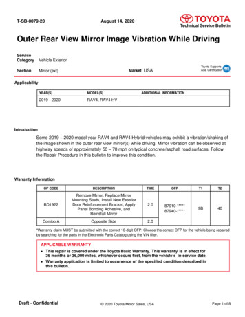
19 Jun 2e(2002/01/18)Annu. Rev. Neurosci. 2004.27:169-192. Downloaded from arjournals.annualreviews.orgby Princeton University Library on 03/26/08. For personal use only.MIRROR NEURONSP1: IKH171the properties of neurons located in the lateral part of F5 (Ferrari et al. 2003),where, in contrast, most neurons are related to mouth actions.The results showed that about 25% of studied neurons have mirror properties.According to the visual stimuli effective in triggering the neurons, two classes ofmouth mirror neurons were distinguished: ingestive and communicative mirrorneurons.Ingestive mirror neurons respond to the observation of actions related to ingestive functions, such as grasping food with the mouth, breaking it, or sucking.Neurons of this class form about 80% of the total amount of the recorded mouthmirror neurons. Virtually all ingestive mirror neurons show a good correspondencebetween the effective observed and the effective executed action. In about one thirdof them, the effective observed and executed actions are virtually identical (strictlycongruent neurons); in the remaining, the effective observed and executed actionsare similar or functionally related (broadly congruent neurons).More intriguing are the properties of the communicative mirror neurons. Themost effective observed action for them is a communicative gesture such as lipsmacking, for example. However, from a motor point of view they behave as theingestive mirror neurons, strongly discharging when the monkey actively performsan ingestive action.This discrepancy between the effective visual input (communicative) and theeffective active action (ingestive) is rather puzzling. Yet, there is evidence suggesting that communicative gestures, or at least some of them, derived from ingestiveactions in evolution (MacNeilage 1998, Van Hoof 1967). From this perspectiveone may argue that the communicative mouth mirror neurons found in F5 reflecta process of corticalization of communicative functions not yet freed from theiroriginal ingestive basis.The Mirror-Neuron CircuitNeurons responding to the observation of actions done by others are present notonly in area F5. A region in which neurons with these properties have been described is the cortex of the superior temporal sulcus (STS; Figure 1) (Perrett et al.1989, 1990; Jellema et al. 2000; see Jellema et al. 2002). Movements effective ineliciting neuron responses in this region are walking, turning the head, bendingthe torso, and moving the arms. A small set of STS neurons discharge also duringthe observation of goal-directed hand movements (Perrett et al. 1990).If one compares the functional properties of STS and F5 neurons, two pointsemerge. First, STS appears to code a much larger number of movements than F5.This may be ascribed, however, to the fact that STS output reaches, albeit indirectly(see below), the whole ventral premotor region and not only F5. Second, STSneurons do not appear to be endowed with motor properties.Another cortical area where there are neurons that respond to the observation ofactions done by other individuals is area 7b or PF of Von Economo (1929) (Fogassiet al. 1998, Gallese et al. 2002). This area (see Figure 1) forms the rostral part of the
19 Jun 200414:34Annu. Rev. Neurosci. 2004.27:169-192. Downloaded from arjournals.annualreviews.orgby Princeton University Library on 03/26/08. For personal use only.172ARAR217-NE27-07.texRIZZOLATTI AR217-NE27-07.sgmLaTeX2e(2002/01/18)P1: IKHCRAIGHEROinferior parietal lobule. It receives input from STS and sends an important outputto the ventral premotor cortex including area F5.PF neurons are functionally heterogeneous. Most of them (about 90%) respondto sensory stimuli, but about 50% of them also have motor properties dischargingwhen the monkey performs specific movements or actions (Fogassi et al. 1998,Gallese et al. 2002, Hyvarinen 1982).PF neurons responding to sensory stimuli have been subdivided into “somatosensory neurons” (33%), “visual neurons” (11%), and “bimodal (somatosensory and visual) neurons” (56%). About 40% of the visually responsive neuronsrespond specifically to action observation and of them about two thirds have mirrorproperties (Gallese et al. 2002).In conclusion, the cortical mirror neuron circuit is formed by two main regions:the rostral part of the inferior parietal lobule and the ventral premotor cortex. STSis strictly related to it but, lacking motor properties, cannot be considered partof it.Function of the Mirror Neuron in the Monkey: ActionUnderstandingTwo main hypotheses have been advanced on what might be the functional roleof mirror neurons. The first is that mirror-neuron activity mediates imitation (seeJeannerod 1994); the second is that mirror neurons are at the basis of actionunderstanding (see Rizzolatti et al. 2001).Both these hypotheses are most likely correct. However, two points should bespecified. First, although we are fully convinced (for evidence see next section) thatthe mirror neuron mechanism is a mechanism of great evolutionary importancethrough which primates understand actions done by their conspecifics, we cannotclaim that this is the only mechanism through which actions done by others maybe understood (see Rizzolatti et al. 2001). Second, as is shown below, the mirrorneuron system is the system at the basis of imitation in humans. Although laymenare often convinced that imitation is a very primitive cognitive function, they arewrong. There is vast agreement among ethologists that imitation, the capacity tolearn to do an action from seeing it done (Thorndyke 1898), is present amongprimates, only in humans, and (probably) in apes (see Byrne 1995, Galef 1988,Tomasello & Call 1997, Visalberghi & Fragaszy 2001, Whiten & Ham 1992).Therefore, the primary function of mirror neurons cannot be action imitation.How do mirror neurons mediate understanding of actions done by others? Theproposed mechanism is rather simple. Each time an individual sees an actiondone by another individual, neurons that represent that action are activated in theobserver’s premotor cortex. This automatically induced, motor representation ofthe observed action corresponds to that which is spontaneously generated duringactive action and whose outcome is known to the acting individual. Thus, themirror system transforms visual information into knowledge (see Rizzolatti et al.2001).
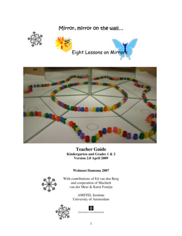
19 Jun 2e(2002/01/18)MIRROR NEURONSP1: IKH173Annu. Rev. Neurosci. 2004.27:169-192. Downloaded from arjournals.annualreviews.orgby Princeton University Library on 03/26/08. For personal use only.Evidence in Favor of the Mirror Mechanismin Action UnderstandingAt first glance, the simplest, and most direct, way to prove that the mirror-neuronsystem underlies action understanding is to destroy it and examine the lesion effecton the monkey’s capacity to recognize actions made by other monkeys. In practice,this is not so. First, the mirror-neuron system is bilateral and includes, as shownabove, large portions of the parietal and premotor cortex. Second, there are othermechanisms that may mediate action recognition (see Rizzolatti et al. 2001). Third,vast lesions as those required to destroy the mirror neuron system may producemore general cognitive deficits that would render difficult the interpretation of theresults.An alternative way to test the hypothesis that mirror neurons play a role in actionunderstanding is to assess the activity of mirror neurons in conditions in whichthe monkey understands the meaning of the occurring action but has no access tothe visual features that activate mirror neurons. If mirror neurons mediate actionunderstanding, their activity should reflect the meaning of the observed action, notits visual features.Prompted by these considerations, two series of experiments were carried out.The first tested whether F5 mirror neurons are able to recognize actions from theirsound (Kohler et al. 2002), the second whether the mental representation of anaction triggers their activity (Umiltà et al. 2001).Kohler et al. (2002) recorded F5 mirror neuron activity while the monkey wasobserving a noisy action (e.g., ripping a piece of paper) or was presented with thesame noise without seeing it. The results showed that about 15% of mirror neuronsresponsive to presentation of actions accompanied by sounds also responded to thepresentation of the sound alone. The response to action sounds did not depend onunspecific factors such as arousal or emotional content of the stimuli. Neurons responding specifically to action sounds were dubbed “audio-visual” mirror neurons.Neurons were also tested in an experimental design in which two noisy actionswere randomly presented in vision-and-sound, sound-only, vision-only, and motorconditions. In the motor condition, the monkeys performed the object-directedaction that they observed or heard in the sensory conditions. Out of 33 studiedneurons, 29 showed auditory selectivity for one of the two hand actions. Theselectivity in visual and auditory modality was the same and matched the preferredmotor action.The rationale of the experiment by Umiltà et al. (2001) was the following. Ifmirror neurons are involved in action understanding, they should discharge also inconditions in which monkey does not see the occurring action but has sufficientclues to create a mental representation of what the experimenter does. The neuronswere tested in two basic conditions. In one, the monkey was shown a fully visibleaction directed toward an object (“full vision” condition). In the other, the monkeysaw the same action but with its final, critical part hidden (“hidden” condition).Before each trial, the experimenter placed a piece of food behind the screen so
19 Jun 200414:34Annu. Rev. Neurosci. 2004.27:169-192. Downloaded from arjournals.annualreviews.orgby Princeton University Library on 03/26/08. For personal use only.174ARAR217-NE27-07.texRIZZOLATTI AR217-NE27-07.sgmLaTeX2e(2002/01/18)P1: IKHCRAIGHEROthat the monkey knew there was an object there. Only those mirror neurons werestudied that discharged to the observation of the final part of a grasping movementand/or to holding.Figure 2 shows the main result of the experiment. The neuron illustrated in thefigure responded to the observation of grasping and holding (A, full vision). Theneuron discharged also when the stimulus-triggering features (a hand approachingthe stimulus and subsequently holding it) were hidden from monkey’s vision (B,hidden condition). As is the case for most mirror neurons, the observation of amimed action did not activate the neuron (C, full vision, and D, hidden condition).Note that from a physical point of view B and D are identical. It was therefore theunderstanding of the meaning of the observed actions that determined the dischargein the hidden condition.More than half of the tested neurons discharged in the hidden condition. Outof them, about half did not show any difference in the response strength betweenthe hidden- and full-vision conditions. The other half responded more strongly inthe full-vision condition. One neuron showed a more pronounced response in thehidden condition than in full vision.In conclusion, both the experiments showed that the activity of mirror neuronscorrelates with action understanding. The visual features of the observed actionsare fundamental to trigger mirror neurons only insomuch as they allow the understanding of the observed actions. If action comprehension is possible on anotherbasis (e.g., action sound), mirror neurons signal the action, even in the absence ofvisual stimuli.THE MIRROR-NEURON SYSTEM IN HUMANSThere are no studies in which single neurons were recorded from the putativemirror-neuron areas in humans. Thus, direct evidence for the existence of mirrorneurons in humans is lacking. There is, however, a rich amount of data proving,indirectly, that a mirror-neuron system does exist in humans. Evidence of thiscomes from neurophysiological and brain-imaging experiments.Neurophysiological EvidenceNeurophysiological experiments demonstrate that when individuals observe anaction done by another individual their motor cortex becomes active, in the absenceof any overt motor activity. A first evidence in this sense was already provided inthe 1950s by Gastaut and his coworkers (Cohen-Seat et al. 1954, Gastaut & Bert1954). They observed that the desynchronization of an EEG rhythm recordedfrom central derivations (the so-called mu rhythm) occurs not only during activemovements of studied subjects, but also when the subjects observed actions doneby others.This observation was confirmed by Cochin et al. (1998, 1999) and by Altschuleret al. (1997, 2000) using EEG recordings, and by Hari et al. (1998) using
19 Jun 2e(2002/01/18)Annu. Rev. Neurosci. 2004.27:169-192. Downloaded from arjournals.annualreviews.orgby Princeton University Library on 03/26/08. For personal use only.MIRROR NEURONSP1: IKH175magnetoencephalographic (MEG) technique. This last study showed that the desynchronization during action observation includes rhythms originating from the cortex inside the central sulcus (Hari & Salmelin 1997, Salmelin & Hari 1994).More direct evidence that the motor system in humans has mirror propertieswas provided by transcranial magnetic stimulation (TMS) studies. TMS is a noninvasive technique for electrical stimulation of the nervous system. When TMSis applied to the motor cortex, at appropriate stimulation intensity, motor-evokedpotentials (MEPs) can be recorded from contralateral extremity muscles. The amplitude of these potentials is modulated by the behavioral context. The modulation of MEPs’ amplitude can be used to assess the central effects of variousexperimental conditions. This approach has been used to study the mirror neuronsystem.Fadiga et al. (1995) recorded MEPs, elicited by stimulation of the left motorcortex, from the right hand and arm muscles in volunteers required to observe anexperimenter grasping objects (transitive hand actions) or performing meaninglessarm gestures (intransitive arm movements). Detection of the dimming of a smallspot of light and presentation of 3-D objects were used as control conditions.The results showed that the observation of both transitive and intransitive actionsdetermined an increase of the recorded MEPs with respect to the control conditions.The increase concerned selectively those muscles that the participants use forproducing the observed movements.Facilitation of the MEPs during movement observation may result from a facilitation of the primary motor cortex owing to mirror activity of the premotorareas, to a direct facilitatory input to the spinal cord originating from the sameareas, or from both. Support for the cortical hypothesis (see also below, BrainImaging Experiments) came from a study by Strafella & Paus (2000). By using adouble-pulse TMS technique, they demonstrated that the duration of intracorticalrecurrent inhibition, occurring during action observation, closely corresponds tothat occurring during action execution.Does the observation of actions done by others influence the spinal cord excitability? Baldissera et al. (2001) investigated this issue by measuring the sizeof the H-reflex evoked in the flexor and extensor muscles of normal volunteersduring the observation of hand opening and closure done by another individual.The results showed that the size of H-reflex recorded from the flexors increasedduring the observation of hand opening, while it was depressed during the observation of hand closing. The converse was found in the extensors. Thus, whilethe cortical excitability varies in accordance with the seen movements, the spinalcord excitability changes in the opposite direction. These findings indicate that,in the spinal cord, there is an inhibitory mechanism that prevents the execution ofan observed action, thus leaving the cortical motor system free to “react” to thataction without the risk of overt movement generation.In a study of the effect of hand orientation on cortical excitability, Maedaet al. (2002) confirmed (see Fadiga et al. 1995) the important finding that, inhumans, intransitive movements, and not only goal-directed actions, determine
19 Jun 200414:34Annu. Rev. Neurosci. 2004.27:169-192. Downloaded from arjournals.annualreviews.orgby Princeton University Library on 03/26/08. For personal use only.176ARAR217-NE27-07.texRIZZOLATTI AR217-NE27-07.sgmLaTeX2e(2002/01/18)P1: IKHCRAIGHEROmotor resonance. Another important property of the human mirror-neuron system,demonstrated with TMS technique, is that the time course of cortical facilitationduring action observation follows that of movement execution. Gangitano et al.(2001) recorded MEPs from the hand muscles of normal volunteers while theywere observing grasping movements made by another individual. The MEPs wererecorded at different intervals following the movement onset. The results showedthat the motor cortical excitability faithfully followed the grasping movementphases of the observed action.In conclusion, TMS studies indicate that a mirror-neuron system (a motor resonance system) exists in humans and that it possesses important properties notobserved in monkeys. First, intransitive meaningless movements produce mirrorneuron system activation in humans (Fadiga et al. 1995, Maeda et al. 2002, Patuzzoet al. 2003), whereas they do not activate mirror neurons in monkeys. Second, thetemporal characteristics of cortical excitability, during action observation, suggestthat human mirror-neuron systems code also for the movements forming an actionand not only for action as monkey mirror-neuron systems do. These properties ofthe human mirror-neuron system should play an important role in determining thehumans’ capacity to imitate others’ action.Brain Imaging Studies: The Anatomy of the Mirror SystemA large number of studies showed that the observation of actions done by othersactivates in humans a complex network formed by occipital, temporal, and parietalvisual areas, and two cortical regions whose function is fundamentally or predominantly motor (e.g., Buccino et al. 2001; Decety et al. 2002; Grafton et al. 1996;Grèzes et al. 1998; Grèzes et al. 2001; Grèzes et al. 2003; Iacoboni et al. 1999,2001; Koski et al. 2002, 2003; Manthey et al. 2003; Nishitani & Hari 2000, 2002;Perani et al. 2001; Rizzolatti et al. 1996b). These two last regions are the rostralpart of the inferior parietal lobule and the lower part of the precentral gyrus plusthe posterior part of the inferior frontal gyrus (IFG). These regions form the coreof the human mirror-neuron system.Which are the cytoarchitectonic areas that form these regions? Interpretationof the brain-imaging activations in cytoarchitectonic terms is always risky. Yet, inthe case of the inferior parietal region, it is very plausible that the mirror activationcorresponds to areas PF and PFG, where neurons with mirror properties are foundin the monkeys (see above).More complex is the situation for the frontal regions. A first issue concerns thelocation of the border between the two main sectors of the premotor cortex: theventral premotor cortex (PMv) and the dorsal premotor cortex (PMd). In nonhumanprimates the two sectors differ anatomically (Petrides & Pandya 1984, TannéGariepy et al. 2002) and functionally (see Rizzolatti et al. 1998). Of them, PMvonly has (direct or indirect) anatomical connections with the areas where there isvisual coding of action made by others (PF/PFG and indirectly STS) and, thus,where there is the necessary information for the formation of mirror neurons(Rizzolatti & Matelli 2003).
19 Jun 2e(2002/01/18)Annu. Rev. Neurosci. 2004.27:169-192. Downloaded from arjournals.annualreviews.orgby Princeton University Library on 03/26/08. For personal use only.MIRROR NEURONSP1: IKH177On the basis of embryological considerations, the border between human PMdand PMv should be located, approximately, at Z level 50 in Talairach coordinates(Rizzolatti & Arbib 1998, Rizzolatti et al. 2002). This location derives from theview that the superior frontal sulcus (plus the superior precentral sulcus) representsthe human homologue of the superior branch of the monkey arcuate sulcus. Becausethe border of monkey PMv and PMd corresponds approximately to the caudalcontinuation of this branch, the analogous border should, in humans, lie slightlyventral to the superior frontal sulcus.The location of human frontal eye field (FEF) supports this hypothesis (Corbetta1998, Kimming et al. 2001, Paus 1996, Petit et al.1996). In monkeys, FEF lies in theanterior bank of the arcuate sulcus, bordering posteriorly the sector of PMv wherearm and head movements are represented (area F4). If one accepts the location ofthe border between PMv and PMd suggested above, FEF is located in a similarposition in the two species. In both of them, the location is just anterior to theupper part of PMv and the lowest part of PMd.The other issue concerns IFG areas. There is a deeply rooted prejudice that theseareas are radically different from those of the precentral gyrus and that they areexclusively related to speech (e.g., Grèzes & Decety 2001). This is not so. Alreadyat the beginning of the last century, Campbell (1905) noted clear anatomical similarities between the areas of posterior IFG and those of the precentral gyrus. Thisauthor classed both the pars opercularis and the pars triangularis of IFG togetherwith the precentral areas and referred to them collectively as the “intermediate precentral” cortex. Modern comparative studies indicate that the pars opercularis ofIFG (basically corresponding to area 44) is the human homologue of area F5 (VonBonin & Bailey 1947, Petrides & Pandya 1997). Furthermore, from a functionalperspective, clear evidence has been accumulating in recent years that human area44, in addition to speech representation, contains (as does monkey area F5) a motor representation of hand movements (Binkofski et al. 1999, Ehrsson et al. 2000,Gerardin et al. 2000, Iacoboni et al. 1999, Krams et al. 1998). Taken together, thesedata strongly suggest that human PMv is the homologue of monkey area F4, andhuman area 44 is the homologue of monkey area F5. The descending branch of theinferior precentral sulcus (homologue to the monkey inferior precentral dimple)should form the approximate border between the two areas (for individual variations of location and extension area 44, see Amunts et al. 1999 and Tomaiuoloet al. 1999).If the homology just described is correct, one should expect that the observationof neck and proximal arm movements would activate predominantly PMv, whereashand and mouth movements would activate area 44. Buccino et al. (2001) addressedthis issue in an fMRI experiment. Normal volunteers were presented with videoclips showing actions performed with the mouth, hand/arm, and foot/leg. Bothtransitive (actions directed toward an object) and intransitive actions were shown.Action observation was contrasted with the observation of a static face, hand, andfoot (frozen pictures of the video clips), respectively.Observation of object-related mouth movements determined activation of thelower part of the precentral gyrus and of the pars opercularis of the inferior frontal
19 Jun 200414:34Annu. Rev. Neurosci. 2004.27:169-192. Downloaded from arjournals.annualreviews.orgby Princeton University Library on 03/26/08. For personal use only.178ARAR217-NE27-07.texRIZZOLATTI AR217-NE27-07.sgmLaTeX2e(2002/01/18)P1: IKHCRAIGHEROgyrus (IFG), bilaterally. In addition, two activation foci were found in the parietallobe. One was loca
Mirror neurons in which the effective observed and effective executed actions correspond in terms of goal (e.g., grasping) and means for reaching the goal (e.g., precision grip) have been classed as "strictly congruent." They represent about one third of F5 mirror neurons. Mirror neurons that, in order to be triggered, do