Transcription
ARTICLEReceived 12 Jan 2014 Accepted 19 Sep 2014 Published 18 Nov 2014DOI: 10.1038/ncomms6342OPENEvidence for henipavirus spillover into humanpopulations in AfricaOlivier Pernet1, Bradley S. Schneider2,*, Shannon M. Beaty1,3,*, Matthew LeBreton2, Tatyana E. Yun4,Arnold Park1,3, Trevor T. Zachariah5, Thomas A. Bowden6, Peta Hitchens7, Christina M. Ramirez8,Peter Daszak9, Jonna Mazet7, Alexander N. Freiberg4, Nathan D. Wolfe2 & Benhur Lee1,3Zoonotic transmission of lethal henipaviruses (HNVs) from their natural fruit bat reservoirs tohumans has only been reported in Australia and South/Southeast Asia. However, a recentstudy discovered numerous HNV clades in African bat samples. To determine the potential forHNV spillover events among humans in Africa, here we examine well-curated sets of bat(Eidolon helvum, n ¼ 44) and human (n ¼ 497) serum samples from Cameroon for Nipah virus(NiV) cross-neutralizing antibodies (NiV-X-Nabs). Using a vesicular stomatitis virus (VSV)based pseudoparticle seroneutralization assay, we detect NiV-X-Nabs in 48% and 3–4% ofthe bat and human samples, respectively. Seropositive human samples are found almostexclusively in individuals who reported butchering bats for bushmeat. Seropositive humansera also neutralize Hendra virus and Gh-M74a (an African HNV) pseudoparticles, as well aslive NiV. Butchering bat meat and living in areas undergoing deforestation are the mostsignificant risk factors associated with seropositivity. Evidence for HNV spillover eventswarrants increased surveillance efforts.1 Department of Microbiology, Immunology and Molecular Genetics, David Geffen School of Medicine at UCLA, Los Angeles, California 90095, USA. 2 GlobalViral/Metabiota Laboratory Sciences, San Francisco, California 90104, USA. 3 Department of Microbiology, Icahn School of Medicine at Mount Sinai, NewYork, New York 10029, USA. 4 Department of Pathology, University of Texas Medical Branch, Galveston, Texas 77555, USA. 5 Brevard Zoo Veterinary Services,Brevard Zoo, Melbourne, 32940 Florida, USA. 6 Division of Structural Biology, Wellcome Trust Centre for Human Genetics, University of Oxford, Oxford,OX3 7BN UK. 7 Department of Medicine and Epidemiology, School of Veterinary Medicine, UC Davis, Davis, California 95616, USA. 8 Department ofBiostatistics, School of Public Health, UCLA, Los Angeles, California 90095, USA. 9 EcoHealth Alliance, New York, New York 10001, USA. * These authorscontributed equally to this work. Correspondence and requests for materials should be addressed to B.L. (email: Benhur.Lee@mssm.edu).NATURE COMMUNICATIONS 5:5342 DOI: 10.1038/ncomms6342 www.nature.com/naturecommunications& 2014 Macmillan Publishers Limited. All rights reserved.1
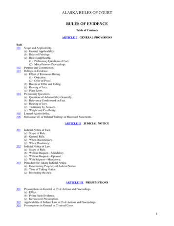
ARTICLENNATURE COMMUNICATIONS DOI: 10.1038/ncomms6342ipah (NiV) and Hendra viruses (HeV) are highlypathogenic paramyxoviruses of the henipavirus (HNV)genus that cause acute encephalitis and respiratory illness.Their mortality rate in humans can be greater than 90% (refs 1,2)and they are the only paramyxoviruses that are classified asbiosafety level 4 (BSL-4) pathogens. Until recently, theHenipavirus genus contained only two species—HeV and NiVviruses—which are phylogenetically closely related and exhibitserological cross-reactivity3. Fruit bats within the suborderMegachiroptera, particularly those of the genus Pteropus, havebeen identified as the natural reservoir for HNVs4–7. Thegeographical distribution of these reservoir bats partiallycoincides with the distribution of HNV outbreaks and spilloverevents around the Indian Ocean, reaching from Australia (HeV)to Southeast Asia and the Indian subcontinent (NiV). Ecologicalstudies8 have revealed several characteristics common to allregions of HNV outbreaks: (i) they are the natural habitat ofPteropid bats (Pteropus spp.), (ii) bat habitats in the region havebeen dramatically altered by the introduction of domestic plantand animal species and concomitant deforestation of the naturallandscape9 and (iii) humans or domestic animals have directcontact with bats in the area. Although these characteristics canbe observed in other locations around the world, to date, HNVoutbreaks and spillovers into human populations have only beenrecognized in Australia and South Asia.The geographic distribution of Pteropus and other Pteropodids(Old World fruit bats) extends well beyond areas withdocumented HeV and NiV outbreaks. In 2007, a survey ofPteropodid species in Madagascar10 reported that 2.3% and19.2% of serum samples from Pteropus rufus and Eidolondupreanum, respectively, tested positive for cross-reactive antiHNV antibodies. Malagasy fruit bats share ecological niches,either roosting in the same caves or feeding in the same fruittrees. As Eidolon species are extremely mobile (they can fly up to2,500 km per year11,12) and are present all around sub-SaharanAfrica, Iehlé et al. raised the possibility of lateral transfer of HNVfrom or to other Eidolon species on mainland Africa andhypothesized a much wider distribution of HNV10. Indeed, antiHNV antibodies were soon found in E. helvum (the commonstraw-coloured African fruit bat) from Ghana13 on the west coastof Africa, and more recently on Annobón island14 in the Gulf ofGuinea. Furthermore, HNV-like RNA sequences have beenidentified in faecal droppings of urban roosting bats inGhana15, and more ominously, in fruit bat bushmeat in theRepublic of Congo16.Recently, sequence analysis of a larger sample set collectedfrom western and southern Africa revealed a surprising diversityof paramyxoviruses in African bats, including 19 new species ofHNV-like viruses distinct from the Nipah and Hendra virusesfound in Southeast Asia and Australia17. However, only onealmost complete African HNV-like genome sequence (Gh-M74aclone) has been published to date, and the corresponding viralisolate has not been reported. This sequence was derived from abat specimen originating in Ghana. We will refer to this putativeHNV-like virus as the Ghana virus (GhV), and GhV-F andGhV-G when referring to its fusion (F) and attachment (G)envelope glycoproteins, respectively. In contrast to the 80–90%sequence identity shared between the F and G envelopeglycoproteins of NiV and HeV, GhV-F and GhV-G share onlyabout 70 and 40% sequence homology and even lower sequenceidentity (56 and 26%) with their respective NiV and HeVcounterparts. Given this poor overall sequence conservation, it isunclear whether humoral responses elicited against the F/Gproteins from African clades of HNV-like viruses would crossreact with F/G from NiV or HeV. This sequence divergencehighlights the limitations faced by current seroprevalence studies2that rely mostly on ELISA- or Luminex-based assays usingrecombinant NiV-G or HeV-G proteins as the targetantigen10,13,14,18.ELISA-based screening assays, although efficient, can yieldhigh false positive and false negative rates compared withfunctional seroneutralization (SN) assays19. Thus, wheneverpossible, ELISA/Luminex-positive samples are confirmed by aSN assay. Although SN assays are considered a gold standard forseroprevalence studies19–21, follow-up confirmation with livevirus SN assays is limited by the amount of sample available, andthe requirement to work with live HNV in a high-containmentfacility (BSL-4). Consequently, in many prior studies only ELISA/Luminex-positive samples, and often only a small subset such asthose with the highest binding activity, were confirmed with abiological or surrogate SN assay (reviewed in LF Wang et al.21; forexample, AJ Peel et al.22). The latter is based on serum antibodycompetition of soluble receptor (sEphrinB2-Fc) binding torecombinant NiV-G or HeV-G conjugated to Luminex beads18.Although these procedures can guard against false positives, theydo not address the loss of potential false negatives10,13,14.Given the recent reports that a diversity of HNV-like virusesare present and may be widely distributed in the bat reservoirhost population across Africa17,22, in the present study, we soughtto evaluate the seroprevalence of HNV-like infections in both thebat and proximate human populations in Cameroon, and toassess risk factors that might be associated with any putativezoonotic transmission of African HNV-like viruses. To avoid thespecificity and sensitivity issues associated with ELISA-basedassays, as well as the impracticalities of using a live virus SN assayin BSL-4 as a screening test, we developed a VSV-based HNVenvelope pseudotype particle (VSV-HNVpp) infectious SNassay20, which can be used at BSL-2 conditions as a primaryscreen for anti-NiV cross-neutralizing antibodies (anti-NiV-XNabs). Wang and Daniels raised the possibility that the highsensitivity and specificity of the pseudotyped particle platformmay allow for the combination of screening and confirmatorytests in a single assay21. Thus, we screened serum samples fromhunted bats (E. helvum) in an urban area in Yaoundé, Cameroon,and from almost 500 humans living in various villages across thesouth of Cameroon. The specificity, breath and potency of antiNiV-X-Nabs were confirmed using numerous specificity controlsunique to our infectious SN assay, including isogenic virusespseudotyped with irrelevant (VSV-G) or related HNV envelopes(HeV and GhV), and follow-up confirmation with a recombinantreplication-competent reporter NiV specifically engineered forhigh-sensitivity detection of anti-NiV-X-Nabs. Remarkably, theseropositive human samples were found almost exclusively inindividuals who reported butchering bats for bushmeat. Thegeographical and temporal clustering of these seropositive casesprovides evidence for recent HNV-like spillover events into thehuman population in this part of Africa.ResultsNiV cross-neutralizing activity in bat serum samples. Despitethe overall low sequence homology between GhV-G and NiV-G/HeV-G, mapping of the GhV-G sequence onto the crystalstructure of NiV-G complexed with ephrinB2 (ref. 23) indicatedthat the vast majority of the sequence conservation was located atthe receptor-binding interface, suggesting that GhV-G may alsouse ephrinB2 (and likely ephrinB3) as receptors for cell entry(Fig. 1). The clustering of conserved sequences around thereceptor-binding site raises the possibility of biologically significant anti-NiV cross-neutralizing antibodies (anti-NiV-XNabs) in African bats exposed to African clades of HNV-likeviruses despite their overall low sequence identity with NiV.NATURE COMMUNICATIONS 5:5342 DOI: 10.1038/ncomms6342 www.nature.com/naturecommunications& 2014 Macmillan Publishers Limited. All rights reserved.
ARTICLENATURE COMMUNICATIONS DOI: 10.1038/ncomms6342To determine the prevalence of anti-NiV-X-Nabs in that geographically proximate part of Western Africa, we screened fruitbat (E. helvum) serum samples from Cameroon, collected andPhe120Trp125ConservedSimilarFigure 1 Mapping sequence conservation of GH-M74a onto the surfaceof NiV-G. NiV-G surface is coloured according to residue conservation withGH-M74a: red, conserved; maroon, similar; white, no sequenceconservation. EphrinB2 residues 107–127 are represented as sticks.Although NiV-G shares relatively low sequence conservation with GhV-G(approximately 25%), it shares greater sequence conservation (45%) inresidues that make up the receptor-binding site (calculation performed withPISA EBI server).abccurated by Global Viral/Metabiota. To conserve the use of suchsera, we further optimized a previously validated VSV-based(VSV-DG-rLuc) NiV envelope pseudotyped particle (NiVpp) SNassay for high-specificity screening, and established appropriatecontrol sera as described in Methods (Supplementary Fig. 1).In our screen of bat serum samples (Fig. 2), B48% (21/44)were classified as being positive for anti-NiV cross-neutralizingactivity (Fig. 2d; Supplementary Fig. 2a) when compared againstthe fetal calf serum (FCS)-negative control group (Fig. 2a) asdetermined by the Dunnett’s test for multiple comparisonsagainst a single control group. The specificity of our NiVpp SNassay is underscored by the lack of inhibition of the relevantserum samples against vesicular stomatitis virus-based pseudoparticles (VSVpp). Furthermore, ‘normal bat sera’ (NBS) fromcaptive-bred bats in the United States did not show significantneutralization activity against NiVpp or VSVpp (Fig. 2b).Anti-NiV X-Nabs in human serum samples from Cameroon.The relatively high prevalence of anti-NiV-X-Nabs in the batpopulations surrounding Yaoundé in southern Cameroonprompted us to examine archival human sera collected from thisregion of Africa for the presence of similar anti-NiV-X-Nabs,which might indicate potential spillover event(s). Thus, weanalysed almost 500 blood samples collected from healthy adultsby Global Viral/Metabiota in southern Cameroon betweenFebruary 2001 and January 2003 in 13 different locations. Allsamples were collected in rural areas, but represent differenthabitats (savanna, gallery forest and lowland forest) supportingwild game populations that provided a source for the bushmeattrade (Supplementary Table 1).The careful curation of samples allowed us to segregate the serainto various dichotomous groups such as those who reportedcontacts with bats and those that did not (Table 1). Sera wereanalysed using the same SN assay and criteria as described for thebat serum analysis except that normal human sera (NHS) fromblood donors were used as complementary negative controls todMax75%MedianNormalized % infection25%100Min50VSVppNiVppDunnett testnegative10Dunnett testpositive5FCSNBSNegative controlsAnti-NiVCameroon bat samplesPositive controlFigure 2 Prevalence of anti-NiV cross-neutralizing antibodies in bat sera from Cameroon. Box-and-whisker plots showing the infection (normalizedto the negative control fetal calf serum, FCS) of Vero cells by VSVpp (isogenic control, striped pattern box) and NiVpp (white box) in the presence of100 diluted sera from different groups: (a) FCS and (b) normal bat sera (NBS), negative control sera; (c) rabbit anti-NiV, positive control; (d) bat serafrom Cameroon (n ¼ 44). Seropositive (n ¼ 21, median bar in red) and seronegative (n ¼ 23, median bar in yellow) bat sera in d were segregated basedon the Dunnett’s test for significance using the FCS control group. The boxes represent the first and the third quartiles, and the solid horizontal lineswithin the box represent the median values. The whiskers represent the lowest and highest value. Each sample was tested in quadruplicate. The datafor each serum sample are shown in Supplementary Fig. 2a.NATURE COMMUNICATIONS 5:5342 DOI: 10.1038/ncomms6342 www.nature.com/naturecommunications& 2014 Macmillan Publishers Limited. All rights reserved.3
ARTICLENATURE COMMUNICATIONS DOI: 10.1038/ncomms6342Table 1 Risk factor analysis.Risk factorContact with batsButchering batsHunting Seronegative (n)220260Seropositive (n)70Seroprevalence (%)3.10P value*0.0045Odds ratiow (95% CI)17.72P –84.48)CI, confidence interval.*Two-tailed Fisher’s Exact test.w0.5 added to all cells where there is a zero cell count, see statistical methods.Bold entries indicate significant statistical differences.500abcedMax75%MedianNormalized % infection25%MinVSVpp100NiVpp50Dunnett testnegativeDunnett testpositive105FCSNHSNegative controlsAnti-NiVPositive controlBat-exposedhuman samplesNon-exposedhuman samplesFigure 3 Seroneutralization activity of NiV pseudoparticle infection by human sera collected from Cameroon villagers with documented differentialexposure to bats. Box-and-whisker plots showing the normalized % infection of Vero cells by VSVpp (isogenic control, striped pattern box) and NiVpp(white box) in the presence of sera diluted 1:100 from the indicated sample groups (Panels 1–5). (a) All infections were normalized to the infectivityobserved for NiVpp in the presence of the fetal calf serum (FCS) negative control (set at 100%); (b) normal human sera (NHS) from Los Angeles blooddonors served as additional negative controls; (c) hyperimmune rabbit anti-NiV sera (positive control); (d,e) human sera from the bat-exposed (n ¼ 227) ornon-exposed (n ¼ 260) cohort of Cameroon villagers, respectively. For the bat-exposed group, the Dunnett’s test could stratify the NiVpp SN results intoseropositive (n ¼ 7, median bar in red) and seronegative (n ¼ 220, median bar in yellow) subsets. In contrast, in the non-bat-exposed group, the Dunnett’stest could not identify any serum sample as being significantly different from the negative control group (FCS; e), asterisk indicates no seropositivesamples). Boxes encompass the first and the third quartiles, and the solid horizontal lines within the boxes represent the median values. The whiskersrepresent the lowest and highest values in each sample group. The data for individual serum samples (n ¼ 487), each tested in quadruplicates, are shown inSupplementary Fig. 2b and c.FCS (Fig. 3). Using the Dunnett’s test as a stringent measure ofsignificance, 7 out of 227 samples (B3%) in the bat-exposedgroup were considered to have significant anti-NiVpp crossneutralizing activity when compared against the FCS group(Fig. 3d, Table 1 and Supplementary Fig. 2c and f). On the otherhand, none of the 260 samples in the non-bat exposed group werestatistically different from the FCS control group (Fig. 3e, Table 1and Supplementary Fig. 2d and f). Seropositive samples exhibiteddifferent neutralizing potencies. One seropositive sample gavean IC50 titre ofB1:1,000, whereas the other only achievedB40% inhibition at 1:50 (the lowest serum dilution tested;4Supplementary Fig. 2g). As expected, our hyperimmune anti-NiVcontrol serum exhibited the highest IC50 titre of B1:15,000.Interestingly, representative seronegative samples showed a slightenhancing effect as serum concentrations were increased,suggesting that we may even have underestimated the numberof true positives.Specificity of anti-NiV X-Nabs in human serum samples.To further confirm the specificity of the human sera positive foranti-NiV-X-Nabs, and to determine the potential breadth ofNATURE COMMUNICATIONS 5:5342 DOI: 10.1038/ncomms6342 www.nature.com/naturecommunications& 2014 Macmillan Publishers Limited. All rights reserved.
ARTICLENATURE COMMUNICATIONS DOI: 10.1038/ncomms6342*Infection in RLU 106Normalized % infectionP 0.00110050141062FCSNHS Anti-NiV Neg1NegativecontrolsSeVHeVNiVFAn CStiNiPo Vs1Pos2FAn CStiNiPo VsPo 1s2FAn CStiNiVPos1Pos2FAn CStiNiVPosPo 1s20GhVFigure 4 Characterization of seropositive human sera for antihenipavirus cross-neutralizing antibodies. Two seropositive humansamples (Pos1 and Pos2) from the bat-exposed group were tested forseroneutralization activity against different HNV pseudoparticles: Hendravirus (HeV, yellow), Nipah virus (NiV, orange) and Ghana virus (GhV, grey).Infection of Vero cells was performed in the presence of the indicated seradiluted 1:100 as in Fig. 3. As a specificity control for the virus, we used arecombinant GFP-expressing Sendai virus GFP (SeV, blue). Data arepresented as normalized % infection (mean s.d. from three independentreplicates) as in Fig. 3. FCS, fetal calf serum; anti-NiV, hyperimmune rabbitanti-NiV serum. Significant differences (asterisks) of inhibition wasobserved between the NiVpp and GhVpp for the anti-NiV serum (two-tailedStudent’s t-test, Po0.001).cross-reactivity of these antibodies, we chose two seropositivesamples and tested them in our SN assay against three HNVpseudoparticles: NiVpp, HeVpp and GhVpp, the latter bearingF and G from the Gh-M74a clone reported by Drexler et al.17,and an enhanced green fluorescent protein (eGFP)-expressingrecombinant Sendai virus (rSeV-eGFP). SeV is a murineparamyxovirus (Respirovirus genus) not normally found inhumans that enter cells via a pH-independent pathway usingsialic acid-based receptors, and thus, should not be enhanced orinhibited by the seropositive human samples identified in Fig. 3.Indeed, neither the seropositive human Cameroon sera northe hyperimmune rabbit anti-NiV serum inhibited the pHindependent entry of rSeV-eGFP (Fig. 4, blue bars). In contrast,seropositive human Cameroon samples inhibited NiVpp, HeVppand GhVpp to varying degrees (Fig. 4, red, orange and grey bars,respectively). Interestingly, our hyperimmune anti-NiV serashowed good to moderate (50–80%) inhibition of NiVpp andHeVpp infection but only minimal inhibition (B10–15%) ofGhVpp infection. The implications of these results will bediscussed.As a final confirmation regarding the specificity of the antiNiV-X-Nabs that we detected in the seropositive human samples,we repeated the SN assay with live NiV under BSL-4 conditions.In order to conserve the use of these archival sera, which wereonly available in small amounts, we generated and rescued arecombinant NiV expressing secreted Gaussia Luciferase (rNiVGLuc; see Methods and Supplementary Fig. 3a). In vitro growthkinetics of rNiV-GLuc was comparable to the parental NiVMalaysian strain and pathogenicity was demonstrated in thehamster model where bioluminescence in ex vivo harvestedorgans from the moribund animal correlated well with viral titresmeasured by traditional plaque assays (Supplementary Fig. 3b).Furthermore, when we infected Vero cells with rNiV-GLuc acrossa broad range of multiplicity of infection (MOI) (0.01–3), GLucactivity in cell culture supernatant at 24 h.p.i. was Cameroon seraFigure 5 Seropositive human samples neutralize live recombinant NiVinfection. Infection of Vero cells by live recombinant NiV expressingsecreted Gaussia luciferase (rNiV-GLuc, MOI ¼ 1) in the presence of 1:100dilution of the indicated sera: FCS (fetal calf serum, negative control), NHS(normal human serum, negative control), rabbit anti-NiV (positive control),two seronegative samples (picked randomly among the 480 seronegativesamples) and four seropositive samples. 20 ml (out of 150 ml) of infectedcell culture supernatant was collected and analysed for Gaussia luciferaseactivity at 24 h.p.i. Infectivity data are presented as mean relative light units(RLUs) s.d. from three independent replicates. Significant differences(asterisk) were observed between seropositive and seronegative sera(Po0.05; one-tailed Student’s t-test followed by the Holm step-downprocedure for multiple comparisons).and positively correlated with viral titres determined bytraditional plaque assays (Supplementary Fig. 4A, r2 ¼ 0.93,P ¼ 0.008). Thus, rNiV-GLuc allowed us a highly sensitive anddynamic method to monitor NiV infection (or inhibition thereof)in cell culture by sampling infected cell culture supernatant forGaussia luciferase activity. We then chose four seropositive andtwo seronegative human samples from our Cameroon cohort, andperformed our SN assay with rNiV-GLuc along with the otherappropriate negative and positive controls.Figure 5 shows that only the seropositive samples significantlyinhibited rNiV-GLuc infection, albeit with varying degrees ofpotency (15–65% reduction in GLuc activity). A relatively highinoculum was used, and raw luciferase activity values (4107relative light units (RLUs)) are presented to demonstrate therobustness of our assay. Supplementary Fig. 4B and C shows howinhibition of GLuc activity correlated with decreases in viral titresacross a range of MOI, and also indicates that we used a highMOI inoculum (Z3). Seronegative samples showed no significantinhibition of rNiV-GLuc infection, and the GLuc activity detectedwas not different from the FCS-negative or NHS-negativecontrols. As expected, the hyperimmune rabbit anti-NiV seruminhibited rNiV-GLuc infection by close to 90%.Risk factor analysis. The questionnaire filled out by the participants before blood sample collection covered their contacts withsome animals known to be HNV hosts, some of their ‘at risk’activities and the location of the village with its associatedenvironmental features. We analysed their answers to uncoverany risk factors that might be associated with seropositivity.Table 1 shows that all of the seropositive samples came from thegroup that reported contact with bats in one form or another withthose exposed to bats being 17 times more likely to be HNVseropositive (odds ratio ¼ 17.72, P ¼ 0.0021, two-tailed Fisher’sExact test with zero-cell correction24,25). The highly statisticallysignificant difference between seroprevalence rates in thebat-exposed (7/227, 3%) versus non-exposed groups (0/260;P ¼ 0.0045, Fisher’s Exact test) supports the hypothesis thatcontact with bats increases one’s risk of being infected by anantigenically related African HNV-like virus.NATURE COMMUNICATIONS 5:5342 DOI: 10.1038/ncomms6342 www.nature.com/naturecommunications& 2014 Macmillan Publishers Limited. All rights reserved.5
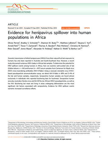
ARTICLENATURE COMMUNICATIONS DOI: 10.1038/ncomms634250 kmLow / no forest coverForest1MBNJHAArea IDn 55MNMAn 3PositivesHAn 5NDn 82Non-exposedn 16n 16YISAn 37DoualaNGSOn 374LEn 16MVn 5n 181MOn 3BCDParticipantsin the area3n 101YaoundéAExposed5EFG n 20HFigure 6 Map of collection sites in Southern Cameroon. For each location, the proportion of participants with self-reported contacts with bats(exposed) or not (non-exposed) is indicated, respectively, by the brown or yellow segment of the accompanying pie-chart. The superimposed redsegments in some pie-charts represent the seropositive samples identified in Fig. 4 (also Supplementary Fig. 2b). More detailed information on thesamples collected from each region is presented in Supplementary Table 1. The number of participants from each location is shown below thecorresponding pie chart. Green- or beige-shaded areas represent regions with high or low/no forest cover, respectively (see ‘Geographical data’section in Methods for further details).We next tried to determine whether a particular type of batexposure was more significantly associated with seropositivity.The detailed questionnaire allowed us to segregate the testedserum samples into other dichotomous groups such as those whobutchered bats (or not), those who hunted bats (or not) and thosewho remembered bites/scratches from bats (or not). Intriguingly,hunting bats alone was not a sufficient risk factor; however, thosebutchering bats were 29 times more likely to be seropositive thanthose not having contact with bats (7/164 (4%) versus 0/316,respectively; P ¼ 0.0002; Table 1). Although there was nostatistically significant association with gender, it is interestingto note that with the exception of one 85-year-old case there wereno seropositives above the age of 45 years (Supplementary Fig. 5).Finally, we examined environmental and geographic parameters. Figure 6 shows a map of the indicated areas of our samplecollection sites, and Supplementary Table 1 lists the mapcoordinates of the indicated areas shown in Fig. 6, for example,ND is in grid C3. The majority of the seropositive participantswere from the ND area (4/7), whereas the others were from MB(C1), MN (A2) and HA (F2) areas. Ground reporting fromGlobal Viral/Metabiota staff at the time of collection revealed thatseropositive samples came almost invariably from low forestcover areas (Fig. 6). Indeed, Table 1 shows that seroprevalence forHNV-like viruses were significantly higher in samples originatingfrom these four areas compared with those that did not (3.2%versus 0.3%, respectively, P ¼ 0.0136). In effect, those living inareas of putative deforestation were ten times more likely to beHNV-seropositive than those who were not (odds ratio ¼ 10.1,P ¼ 0.0088). In addition to the seven ‘true’ seropositive samplesclassified by the Dunnett’s test, there were three borderlinepositive samples (B50% inhibition) in the bat-exposed group(Supplementary Fig. 2e, blue) that also came from areasassociated with deforestation: ND, MN and HA (located in C3,A2 and F3, respectively, in Fig. 6 map). Within the ND area, 3 ofthe 4 seropositive samples were from the same village, and only12 persons from this village with known bat contacts volunteeredfor this study, which would have resulted in a 25% seroprevalencerate among the bat-exposed group if this village were consideredalone.6DiscussionIn this study, we provide multiple lines of evidence that suggestHNV-like spillover events from its natural bat reservoir into thehuman population in southern Cameroon. Using our optimizedpseudotyped virus SN assay as a primary screen for almost 500human serum samples, we confidently identified at least sevenHNV-seropositive samples based on stringent statistical criteria,internal specificity controls for both the virus and the sera, andfollow-up confirmation with live recombinant NiV (rNiV-GLuc)or SeV, a paramyxovirus from a different genus. Together, theseprecautions enabled the risk factor analysis that revealed highlysignificant associations of seropositivity with the behavioural andenvironmental parameters that are known to facilitate zoonoticemergence26–29.By combining behavioural, geographic and serological data, wecan provide more conclusive results than that which would beavailable from inclusion of one set of data alone. For example,that seropositivity was exclusively and most significantlyassociated with intimate bat exposure (P ¼ 0.0006), such as theslaughtering of bats for bushmeat (Table 1), is more informativethan results from a SN assay with perfect sensitivity andspecificity unlinked to an exposure risk. As for environmentalfactors, all seropositive samples come from villages located nearopen savannah lands or areas of deforestation as documented byour local field staff. In toto, we provide a strong body of evidenceindicating spillover of HNV-like viruses into the humanpopulation in Africa.In recent years, several groups have detected HNV-likesequences in African wildlife (bat) and
1Department of Microbiology, Immunology and Molecular Genetics, David Geffen School of Medicine at UCLA, Los Angeles, California 90095, USA. 2Global Viral/Metabiota Laboratory Sciences, San Francisco, California 90104, USA. 3 Department of Microbiology, Icahn School of Medicine at Mount Sinai, New