Transcription
Orphanet Journal of Rare DiseasesBioMed CentralOpen AccessReviewFamilial adenomatous polyposisElizabeth Half*1, Dani Bercovich2,3 and Paul Rozen4,5Address: 1Familial Cancer Clinic, Gastroenterology Dept, Meir Hospital, Kfar Saba, Israel, 2Human Molecular Genetics & Pharmacogenetics, Migal- Galilee Bio-Technology Center, Kiryat-Shmona, 11016, Israel, 3Tel-Hai Academic College, Israel, 4Sestopali Fund for Gastrointestinal CancerPrevention, Dept of Gastroenterology, Tel Aviv Medical Center, 6 Weizmann St, Tel Aviv, 64239, Israel and 5Tel Aviv University Medical School,IsraelEmail: Elizabeth Half* - eohalf@yahoo.com; Dani Bercovich - danib@migal.org.il; Paul Rozen - prozen@012.net.il* Corresponding authorPublished: 12 October 2009Orphanet Journal of Rare Diseases 2009, 4:22doi:10.1186/1750-1172-4-22Received: 18 February 2009Accepted: 12 October 2009This article is available from: http://www.ojrd.com/content/4/1/22 2009 Half et al; licensee BioMed Central Ltd.This is an Open Access article distributed under the terms of the Creative Commons Attribution License (http://creativecommons.org/licenses/by/2.0),which permits unrestricted use, distribution, and reproduction in any medium, provided the original work is properly cited.AbstractFamilial adenomatous polyposis (FAP) is characterized by the development of many tens to thousands of adenomas in the rectumand colon during the second decade of life. FAP has an incidence at birth of about 1/8,300, it manifests equally in both sexes,and accounts for less than 1% of colorectal cancer (CRC) cases. In the European Union, prevalence has been estimated at 1/11,300-37,600. Most patients are asymptomatic for years until the adenomas are large and numerous, and cause rectal bleedingor even anemia, or cancer develops. Generally, cancers start to develop a decade after the appearance of the polyps. Nonspecificsymptoms may include constipation or diarrhea, abdominal pain, palpable abdominal masses and weight loss. FAP may presentwith some extraintestinal manifestations such as osteomas, dental abnormalities (unerupted teeth, congenital absence of one ormore teeth, supernumerary teeth, dentigerous cysts and odontomas), congenital hypertrophy of the retinal pigment epithelium(CHRPE), desmoid tumors, and extracolonic cancers (thyroid, liver, bile ducts and central nervous system). A less aggressivevariant of FAP, attenuated FAP (AFAP), is characterized by fewer colorectal adenomatous polyps (usually 10 to 100), later ageof adenoma appearance and a lower cancer risk. Some lesions (skull and mandible osteomas, dental abnormalities, and fibromason the scalp, shoulders, arms and back) are indicative of the Gardner variant of FAP. Classic FAP is inherited in an autosomaldominant manner and results from a germline mutation in the adenomatous polyposis (APC) gene. Most patients ( 70%) have afamily history of colorectal polyps and cancer. In a subset of individuals, a MUTYH mutation causes a recessively inheritedpolyposis condition, MUTYH-associated polyposis (MAP), which is characterized by a slightly increased risk of developing CRCand polyps/adenomas in both the upper and lower gastrointestinal tract. Diagnosis is based on a suggestive family history, clinicalfindings, and large bowel endoscopy or full colonoscopy. Whenever possible, the clinical diagnosis should be confirmed bygenetic testing. When the APC mutation in the family has been identified, genetic testing of all first-degree relatives should beperformed. Presymptomatic and prenatal (amniocentesis and chorionic villous sampling), and even preimplantation genetictesting is possible. Referral to a geneticist or genetic counselor is mandatory. Differential diagnoses include other disorderscausing multiple polyps (such as Peutz-Jeghers syndrome, familial juvenile polyps or hyperplastic polyposis, hereditary mixedpolyposis syndromes, and Lynch syndrome). Cancer prevention and maintaining a good quality of life are the main goals ofmanagement and regular and systematic follow-up and supportive care should be offered to all patients. By the late teens orearly twenties, colorectal cancer prophylactic surgery is advocated. The recommended alternatives are total proctocolectomyand ileoanal pouch or ileorectal anastomosis for AFAP. Duodenal cancer and desmoids are the two main causes of mortalityafter total colectomy, they need to be identified early and treated. Upper endoscopy is necessary for surveillance to reduce therisk of ampullary and duodenal cancer. Patients with progressive tumors and unresectable disease may respond or stabilize witha combination of cytotoxic chemotherapy and surgery (when possible to perform). Adjunctive therapy with celecoxib has beenapproved by the US Food and Drug Administration and the European Medicines Agency in patients with FAP. Individuals withFAP carry a 100% risk of CRC; however, this risk is reduced significantly when patients enter a screening-treatment program.Page 1 of 23(page number not for citation purposes)
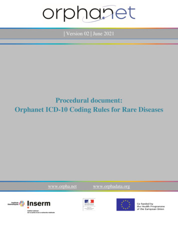
Orphanet Journal of Rare Diseases 2009, 4:22http://www.ojrd.com/content/4/1/22Disease namesFamilial adenomatous polyposis (FAP)SynonymsFamilial polyposis coliVariants or FAP-associated conditionsAttenuated FAP is a milder form of FAPGardner's syndrome is a clinical variant of FAP where theextra-colonic features are prominentTurcot syndrome refers to FAP and having a medulloblastoma brain tumorDefinitionFAP is an autosomal dominant disease that is classicallycharacterized by the development of hundreds to thousands of adenomas in the rectum and colon during thesecond decade of life. Almost all patients will developcolorectal cancer (CRC) if they are not identified andtreated at an early stage. However, today it is unusual forpatients to present with CRC as the majority of patientsare diagnosed before cancer develops.FigureRelativetheincidence1and approximateof colorectalcontributionscancerof familial causes toRelative and approximate contributions of familialcauses to the incidence of colorectal cancer. FAP Familial adenomatous polyposis, JP - Familial juvenile polyposis, PJ - Peutz-Jeghers syndrome, HNPCC - Hereditary nonpolyposis colorectal cancer (Lynch Syndrome). Note thevery small contribution of FAP to the etiology of cancer.Clinical descriptionAttenuated FAP is a milder form that is characterized byfewer adenomas, a later age of adenoma development andcancer diagnosis.EpidemiologyWorldwide, CRC is a major cause of cancer associatedmorbidity and mortality. Its incidence varies considerablyamong different populations with the highest incidencereported from Western and industrialized countries.Worldwide, about 85% of CRCs are considered to be sporadic, while approximately 15% are familial with FAPaccounting for less than 1% (Fig. 1). However, FAP is oneof the best known and understood genetic diseases.In many countries there are local FAP registries; howeverit is difficult to obtain accurate nation-wide data. In theUK, Reed and Neel presented a detailed genetic study in1955 and calculated the incidence of FAP at birth to be1:8,300 [1]. In 1975, Alm presented an incidence rate of1:7,645 in Sweden [2]. These estimates were based onclinical criteria before the availability of mutation analysisand recognition of all the clinical variants and differentialdiagnoses. In 2009, the European Medicines Agency(EMEA) estimated that FAP affected approximately 3-10/100,000 people in the European Union which is equivalent to 11,300 - 37,600 individuals [3]. Clinically, FAPmanifests equally in both sexes by the late teens and in thetwenties age group.Symptoms are uncommon in the child and adolescentuntil the adenomas are large and numerous so as to causerectal bleeding or even anemia. Other non-specific complaints such as change in bowel habits, constipation, ordiarrhea, abdominal pains or palpable abdominal massesor weight loss in young patients can lead to recto-sigmoidexamination and identification of polyps suggestive ofFAP. FAP can present with extraintestinal manifestationssuch as osteomas, dental abnormalities (unerupted teeth,congenital absence of one or more teeth, supernumeraryteeth, dentigerous cysts, and odontomas), congenitalhypertrophy of the retinal pigment epithelium (CHRPE),desmoid tumors, or extracolonic cancers (thyroid, liver,bile ducts, central nervous system), (see below). Somelesions (skull and mandible osteomas, dental abnormalities, fibromas on the scalp, shoulders, arms, and back) areindicative of the Gardner variant of FAP. Today the condition should rarely present as a colonic or even as an extracolonic malignancy.Colonic manifestationsClassic FAP is characterized by the presence of hundredsto thousands of colorectal adenomas of different sizes.Today this is rarely seen in countries with well developedpublic health services. In the majority of patients polypsbegin to develop during childhood, mostly in the distalcolon (rectosigmoid) as small intramucosal nodules (Fig.2A). By the time of adolescence, the polyps are usuallyidentified throughout the colon and, thereafter, increasein size and numbers (Fig. 2B). About half of FAP patientsPage 2 of 23(page number not for citation purposes)
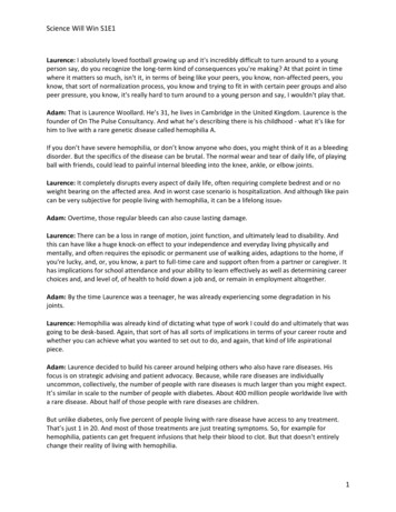
Orphanet Journal of Rare Diseases 2009, 4:22http://www.ojrd.com/content/4/1/22yps, progress to cancer within 10 years [10]. While rare inthe general population, the risk of duodenal or periampullary cancer is increased several hundred fold in FAPpatients [7]. Duodenal polyposis usually progresses in anorderly fashion through increasing Spigelman stage [7],but cancer can present in patients under surveillance withlower Spigelman stages being identified in their penultimate examinations [10-12]. Pancreatitis can be the resultof ampullary adenoma or as a presentation of el2showsB theshowsFAPendoscopicadenomasthe endoscopicappearanceappearanceof earlyofFAPestabadePanel A shows the endoscopic appearance of earlyFAP adenomas; panel B shows the endoscopicappearance of established, multiple, FAP adenomas.develop adenomas by 15 years of age and 95% by age 35years [4].Generally, cancers start to develop a decade after theappearance of the polyps. So, if the colon is left intact, themajority of patients with FAP eventually develop CRC bythe ages 40-50 years. However, it should be emphasizedthat, although uncommon, CRC can develop in childrenor in older adults.Small bowel adenomas. It is well known that individualswith FAP carry a risk of small bowel polyps and even cancer although at a much lower rate than duodenal andampulary neoplasms. The exact incidence of small bowelpolyps is unknown and is generally dependent on theexamining methodology used for evaluation. Bertoni G etal., in 1993, studied 16 patients with FAP by push enteroscopy and detected jejunal polyps in 50% of subjects[13]. Later, with the use of double balloon enteroscopy orcapsule endoscopy, the rate of jejunal and ileal polyps wasestimated to be between 30-75% [14-16]. The rate ofsmall bowel malignancy is much lower than ampullary orduodenal cancer. However, the treating physician shouldbe aware of this possibility and be ready to implementsurveillance, which will be discussed.In the stomach, fundic gland polyps (FGP) develop in 90%of patients with FAP. They are of special interest since, incontrast to the benign nature of sporadic FGP, 40% ofthese lesions in individuals with FAP have been shown tohave adenomatous features, but rarely do progress to cancer [5]. FGPs in FAP patients are pathogenetically distinctfrom sporadic FGPs. Somatic, second-hit APC gene alterations, which precede morphological dysplasia in manyFAP-associated FGPs, indicate that FGPs arising in the setting of FAP are neoplastic lesions [6].Extra-intestinal manifestationsExtraintestinal manifestations of FAP, which are rarelymalignant, include: cutaneous lesions such as fibromas, lipomas, sebaceous and epidermoid cysts, and nasopharyngealangiofibromas (Fig 4A). Osteomas can be palpated or seen,they can be identified as occult radio-opaque jaw lesions,and dental abnormalities which can be disfiguring (Fig.4B). Gardner described the presence of these extraintestinal lesions and this phenotypic variant is named after himas "Gardner syndrome" [17]. Congenital hypertrophy of theretinal pigment epithelium (CHRPE) is a patch(s) of discoloration in the ocular fundus but is not specific for FAP(Fig. 4C). Almost all patients with CHRPE do not havesymptoms and the lesions are found during examinationof the dilated eye. However, when multiple bilaterallesions occur it can be a sensitive phenotypic marker andcalls for screening. Low-grade adenocarcinoma has beendescribed in these lesions [18,19].Adenomatous polyps in the duodenum (mainly in the 2nd and3rd parts) and periampullary region. In one series they developed in approximately 90% of individuals with FAP, 1020 years after diagnosis of colorectal polyps [7]. The lifetime risk of duodenal adenomas has been reported toreach 100% [8,9]. Spigelman's classification of duodenalpolyps is a scale, based on polyp number, size, histology,and severity of dysplasia (Table 1). It is estimated thatabout 5% of duodenal, and specifically periampulary pol-Individuals with FAP may develop soft-tissue tumors(desmoid tumors) in the mesentery, abdominal wall orareas of scars. These tumors are considered benign, but byprogressive enlargement and consequently pressure ongastrointestinal or urinary tracts, local nervous or vascularsystem, can be life threatening. Desmoids can be both acause of severe morbidity as well as mortality. Over a lifetime, desmoid tumors occur in approximately 8% of menand 13% of women with FAP [20].Other gastrointestinal manifestationsIndividuals with FAP can also develop a variety of extracolonic gastrointestinal manifestations (3A).Page 3 of 23(page number not for citation purposes)
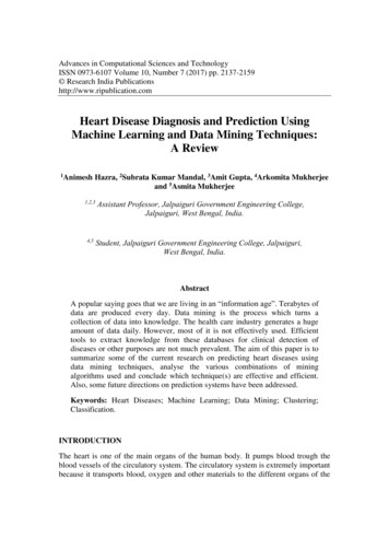
Orphanet Journal of Rare Diseases 2009, eltheAmericanA3illustratesGastroenterologythe extracolonicAssociationupper gastrointestinalInstitute's Clinicalpolyp TeachingmanifestationsUnit 11,of FAPcopyright)(Used with permission and adapted fromPanel A illustrates the extracolonic upper gastrointestinal polyp manifestations of FAP (Used with permissionand adapted from the American Gastroenterology Association Institute's Clinical Teaching Unit 11, copyright). Fig. B is a schematic illustration of a "J-pouch", formed from the distal small intestine and attached to the anal sphincter.The shorter limb of the "J" acting as a fecal reservoir; Fig. C is an x-ray demonstration of this pouch (Figures provided by Dr. A.Deutsch, Petach Tikva and reproduced, with permission of the publishers, from reference [70]: Rozen P, Levin B, Young GP:Who are at risk for familial colorectal cancer and how can they be managed? In Colorectal Cancer in Clinical Practice: Prevention,Early Detection and Management Edited by: Rozen P, Young GP, Levin B, Spann SJ. London, Ed 2, Taylor and Francis 2002:55-66).Table 1: Spigelman classification for duodenal polyposis in FAPPolyp numberPolyp size (mm)HistologyDysplasiaStage 1Stage 2Stage 31-41-4TubularLow grade5-205-10TubulovillousLow grade 20 10VillousHigh gradeStage 0, 0 points; stage I, 1-4 points; stage II, 5-6 points; stage III, 7-8points; stage IV, 9-12 points. Points are accumulated for polyps'number, size, histology and severity of dysplasia. Stage I (1-4 points)indicates mild disease, whereas stage III-IV (7-12 points) impliessevere duodenal polyposis. Derived from reference [7].Other extracolonic malignanciesOther extra-colonic malignancies that are associated withFAP include pancreatic mucinous adenocarcinomas, liver(hepatoblastoma), and brain tumors. In 1959, Turcot andcolleagues described two teenaged siblings with adenomatous polyps of the colorectum in whom malignanttumors of the central nervous system developed [21].Later it was noticed that "Turcot syndrome" is heterogeneous, encompassing at least two subtypes. The first is characterized by a germline mutation in one of the DNAmismatch repair genes, such as hPSM2 or hMLH1 andpresents with the occurrence of glioblastoma in patientswith Lynch syndrome (hereditary non-polyposis coloncancer, HNPCC). A second Turcot subtype is characterizedby medulloblastoma, or rarely glioblastoma, in the settingPage 4 of 23(page number not for citation purposes)
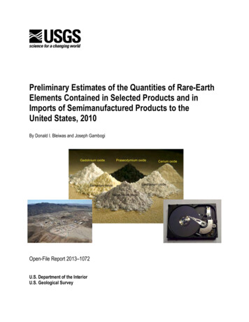
Orphanet Journal of Rare Diseases 2009, nelterologyA alClinicalmanifestationsTeaching Unitof11,FAPcopyright)(Used with permission and adapted from the American GastroenPanel A illustrates the extraintestinal manifestations of FAP (Used with permission and adapted from theAmerican Gastroenterology Association Institute's Clinical Teaching Unit 11, copyright). Panel B is a panoramicjaw X-ray showing mandibular areas of osteoslerosis that can be found in some FAP patients but is not diagnostic of FAP. PanelC shows retinal pigmentation (CHRPE) that can be found in some FAP patients but is not diagnostic of FAP.of FAP and APC germline mutations [22]. Thyroid cancer ina FAP patient was first described in 1949 by Crail. Youngwomen (less than 35 years of age) are at particular highrisk of developing thyroid cancer, at a rate of approximately 160 times that of the general population [23,24].Attenuated familial adenomatous polyposis (AFAP)This is a less aggressive variant of FAP that is characterizedby fewer colorectal adenomatous polyps (usually 10 to100), later age of adenoma appearance (mean age ofpolyp diagnosis is 44 years) and cancer (mean age 56years). Clinically, it can be confusing as often there ismainly proximal colonic involvement with polyps, andinfrequent rectal involvement. Thus, it can be misdiagnosed as occurring in a patient with sporadic adenomas(Figs. 5A-C) however, it can also occur in families withmembers having full clinical features of FAP.Although these individuals have a smaller polyp burdenrelative to classic FAP, they still have an increased risk ofcancer that, in general, occurs approximately 10-15 yearslater. As in FAP the most prominent extracolonic findingsare upper gastrointestinal polyps specifically, duodenaland gastric adenomas and fundic gland polyps. Gastricand breast adenocarcinomas, as well as hepatoblastoma,have also been documented in AFAP. Other extracolonicmanifestations of FAP are rare [25].EtiopathogenesisFAP is a genetic disorder resulting from a mutation in theadenomatous polyposis gene (APC) gene.Most FAP patients have a family history of colorectal polyps and cancer, however, 25-30% of them are "de novo",without clinical or genetic evidence of FAP in familyPage 5 of 23(page number not for citation purposes)
Orphanet Journal of Rare Diseases 2009, reproducedthe ,early2006;inkFFAP5:227-335,Macrae,contrastand matouspermissionin identifyingnotepolyposis:ofadenomas;Springerthe f clinPanel A shows the endoscopic appearance of early FAP and the difficulty in identifying adenomas; Panel Bshows the endoscopic appearance after spraying with dilute Indian ink contrast chromoendoscopy; note theeasier identification of small polyps (Figures reproduced from reference [85]: P Rozen, F Macrae, Familial adenomatous polyposis: The practical applications of clinical and molecular screening. Familial Cancer, 2006;5:227-335, with kind permission of Springer Science and Business Media). Panel C shows the occasional adenomaseen in attenuated FAP and the difficulty in making the endoscopic diagnosis of FAP. Panel D demonstrates the usefulness oftaking random biopsies in attenuated FAP and finding an intramucosal microadenoma, consistent with the diagnosis of FAP.Note the appearance of dark stained and elongated dysplastic nuclei in a distorted crypt that does not protrude above the surrounding normal surface epithelium (Figures reproduced, with permission of the publishers, from reference [70]: Rozen P,Levin B, Young GP: Who are at risk for familial colorectal cancer and how can they be managed? In Colorectal Cancer in ClinicalPractice: Prevention, Early Detection and Management Edited by: Rozen P, Young GP, Levin B, Spann SJ. London, Ed 2, Taylor andFrancis 2002:55-66.members [26,27]. It is now recognized that this can bepartially explained by being the result of germline mosaicism [28]. Classic FAP is inherited as an autosomal dominant trait and results from a germline APC mutation;AFAP is mostly caused by specific APC mutations. A subsetof individuals with clinical features of FAP will insteadcarry a mutation in the MUTYH gene (to be discussedlater).Normal APC structure and functionsThe APC gene spans a region of 108,353bp (NC 000005).The mRNA is 10,719 bp long (NCBI# MN 000038) andhas 16 exons. The mRNA codes for a protein of 2,843amino acids long with a molecular weight of 310 kDa(NCBI# NP 000029). Most of the amino acids are in thelast exon (exon-16 that is 8,689 bp long with 6,574 bpcoding sequences). Only exons 2-15 are coding exons andhave 653 amino acids and exon 16 has 2,190.The APC gene in briefAPC is a tumor suppressor gene located on the long armof chromosome 5 in band q21 (5q21). The coding regionis divided into 15 exons and encodes a large protein (309kilo-Daltons) [29]. The APC protein has multipledomains that mediate oligomerization as well as bindingto a variety of intracellular proteins, which have an important role in cell adhesion, signal transduction and transcriptional activation.APC is a classical tumor suppressor protein that plays acentral role in Wnt signaling, in part by regulating the degradation of -catenin. Wnt signals influence the stabilityof a protein complex containing -catenin, conductin andGSK3 (glycogen synthase kinase 3). In the absence of Wntor the presence of wild-type APC protein, -catenin isdegraded. In the presence of Wnt, or the absence of APC(as occurs in many colon cancers), -catenin target genesPage 6 of 23(page number not for citation purposes)
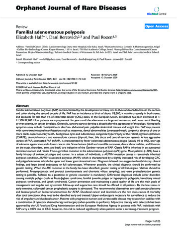
Orphanet Journal of Rare Diseases 2009, 4:22including c-myc are expressed. Myc expression, in turn,leads to the expression of the polyamine ornithine decarboxylase (ODC) which is a proto-oncogene (Fig. 6). TheAPC gene product indirectly regulates transcription of anumber of critical cell-proliferation genes, through itsinteraction with the transcription factor -catenin. APCbinding to -catenin leads to ubiquitin-mediated -catenin destruction; loss of APC function increases transcription of -catenin targets. Homozygous APC truncationhas been shown to affect chromosome attachment in cultured cells. Roles for APC in cell migration have beendemonstrated in-vitro and in mouse models.Disturbed APC structure and functions in briefMore than 300 different types of mutations are recognizedtoday as the cause of FAP. Most of these mutations (insertions, deletions, nonsense mutations, etc.), result in atruncated protein. The most common mutation, occurring in about 10% of FAP patients, is a deletion mutationin codon 1309, the next most common, occurring in 5%of the patients, is a deletion at codon 1061.APC disturbed structure and function in FAP in humans and animalmodelsLoss of normal APC function is known to be an early eventin both familial and sporadic colon cancer pathogenesis,wntCONDUCTINAPCGSK3 ȕ CATENIN WNT/ -APC- WNT/ APCProteasomal degradationNUCLEOUSȕ CATENINTCF-4Target genes e.g c-MYCMYC target mactivity)betweenin signaling -cateninin ship between Wnt signaling and the APCtumor-suppressor gene in activating the -catenin toenter the nucleus (no APC activity) for promotinggenes expression and polyamine metabolism (ODC)in the colonic epithelium; or (with APC activity) forthe proteasomal degradation in the colonic ring at the pre-adenoma stage [30]. Generally, coloncancers show either chromosomal instability (CIN),which correlates with loss of APC function, or microsatellite instability, which correlates with loss of mismatchrepair function, but not both. The genetically manipulated mouse model provides an excellent in-vivo system ofhuman diseases and an opportunity to test therapies. Themouse model of FAP contains a point mutation in theAPC gene; it develops numerous adenomas and was thefirst model used to study the involvement of the APC genein intestinal tumorigenesis. The model has providedexamples of modifying loci in mice and demonstratingthe principle of genetic modulation of disease severity[31].FAP is caused by a highly heterogeneous spectrum ofpoint mutations and this represents a problem for molecular genetic diagnosis; all the mutations are chain terminating. Mutations typically cluster in, or just distal, to thearmadillo repeat region and truncate near the middle ofthe protein [30,32] (Fig. 7). It is not known which ispathophysiologic - absence of the full-length protein orpresence of the truncated version; evidence exists for both.Mutational analysis of the APC gene indicates that themajority of germline mutations found in patients withFAP are nonsense mutations, leading to the formation ofa truncated protein. More than 60% of APC mutations arefound in the central region (between codons 1284 and1580) of the protein, which is called the mutation clusterregion (MCR) [33]. The MCR region coincides with aregion in the APC gene that is important for the down-regulation of -catenin, which suggests that this function isimportant for the pathogenesis of CRC. Subsequent studies demonstrated that APC and -catenin are importantparts of the Wnt signaling pathway (Fig. 6). The 5' codingregion of exon 15 includes a second mutation-clusterregion. In addition, there are several recurrently describedmutations, of which two, at codon positions 1309 and1061, account for as much as 30% of the germline APCmutations. The vast majority of mutations found in theAPC gene represent truncating mutations (small deletions, 46%; small insertions, 10%; nonsense mutations,28%). However, missense mutations (3%) and grossalterations (13%) have also been reported [34]. Recentdata suggest that gross alterations in the APC gene maycontribute more to this disease than previously reported;perhaps as many as 20% of FAP families may have a grossalteration.Genotype-phenotype correlations in briefSome correlation does exist between the sites of specificgenetic mutations and the clinical manifestations of thedisease, however, this correlation is not exact and differences do occur (to be discussed later) [35,36] (Fig. 7).Page 7 of 23(page number not for citation purposes)
Orphanet Journal of Rare Diseases 2009, 4:22Q769XR564XQ218XQ208X778del T FSl698XR499XR554XW157X 541X S713X1464del AG FSQ1338XQ1067XAPC 2842G1120ES1395CC1272X1244del G FSE1317I1307K1681580Classical FAP12501464severe modimerization domain15 Amino acids repeat20 Amino acids repeatArmadilo repeatSAMP repeatMicrotubule binding domainEB1 binding domainPDZ binding domaintypesproteinAPCFigure7 functional domains, several of the common mutations and their locations, in relation to the different FAP phenoAPC protein functional domains, several of the common mutations and their locations, in relation to the different FAP phenotypes.Mutations contributing to classical FAP occur betweenexon 5 and the 5' portion of exon 15, whereas those associated with AFAP tend to cluster in the extreme 5' portionof the gene and the 3' portion of exon 15 proximal tocodon 1517 or distal to codon 1900 [35,36]. Mutationsbetween codons 1250 - 1464 are associated with profusepolyposis. Mutations at specific positions can causeCHRPE [37]; it is almost always absent if the protein-truncating mutation in the APC gene occurs before exon 9, butis consistently present if it occurs after this exon. Patientswith a mutation between codons 1445 - 1578 do notexpress CHRPE, but can develop severe desmoid tumors[38].DiagnosisThe diagnosis of classic FAP is based on a suggestive family history and clinical findings. Whenever possible, theclinical diagnosis should be confirmed by genetic testing.Clinical diagnosisThe clinical diagnosis is dependent on the physician's suspicion and awareness. The patient may be completelyasymptomatic and obtaining a detailed family cancer history is essential for a correct diagnosis, since in most casessome grandparents, parents and siblings will be affected.Asking simple questions like "has anyone in your familyhad cancer? Which cancer? And at what age?" is importantinformation. Alternatively, rectal bleeding or abdominalcomplaints may develop depending on the stage of disease i.e., polyp burden or stage of cancer. For the astutephysician, identification of extra-colonic manifestationscan lead to performing endoscopic examination of thelarge bowel. For example, identifying a desmoid or a mandibular osteoma in an individual should lead to a workup for ruling-out FAP. This should initially be done bytaking a detailed extended family history and performinga sigmoidoscopy or a full colonoscopy depending on theage of the patient or whether we suspect FAP or AFAP.Page 8 of 23(page number not for citation purposes)
Orphanet Journal of Rare Diseases 2009, 4:22During childhood, only dimi
Review Open Access Familial adenomatous polyposis . Half*1, Dani Bercovich2,3 and Paul Rozen4,5 Address: 1Familial Cancer Clinic, Gastroenterology Dept, Meir Hospital, Kfar Saba, Israel, 2Human . Dept of Gastroenterology, Tel Aviv Medical Center, 6 Weizmann St, Tel Aviv, 64239, Israel and 5Tel Aviv University Medical School, Israel Email .