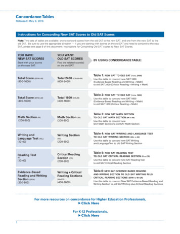
Transcription
DOI: 10.5152/eurjrheum.2019.19040Original ArticleJoint scores in hemophilic arthropathy in children:Developing country perspectivesAritra Guha1 , Akash Rai1 , Arnab Nandy1 , Tanushree Mondal2 , Narayan Pandit3 ,Sumantra Guha4 , Dipankar Gupta1, Rakesh Mondal1AbstractORCID IDs of the authors:A.G. 0000-0002-7798-4728;A.R. 0000-0002-6666-7663;A.N. 0000-0002-7075-1040;T.M. 0000-0002-2727-3354;N.P. 0000-0003-1466-3481;S.G. 0000-0002-9867-7065;R.M. 0000-0002-2241-3514.Cite this article as: Guha A, Rai A, NandyA, Mondal T, Pandit N, Guha S, et al.Joint scores in hemophilic arthropathyin children: Developing countryperspectives. Eur J Rheumatol 2020; 7(1):26-30.Pediatric Rheumatology Clinic,Department of Pediatric Medicine, NorthBengal Medical College, Darjeeling, India2Department of Community Medicine,Medical College, Kolkata, India3Department of Radiodiagnosis,Coochbehar Govt. Medical College,Coochbehar, India4IFMR graduate School of Business, KREAUniversity, Chennai, IndiaAddress for Correspondence:Rakesh Mondal; Pediatric RheumatologyClinic, Department of Pediatric Medicine,North Bengal Medical College, Darjeeling,IndiaE-mail: ivanrakesh2001@gmail.comSubmitted: February 26, 2019Accepted: September 9, 2019Available Online Date: December 16, 20191Copyright@Author(s) - Available online atwww.eurjrheumatol.org.Content of this journal is licensed under a CreativeCommons Attribution-NonCommercial 4.0International License.26Objective: Hemophilia is a common X-linked recessive coagulopathy causing recurrent bleeding intothe synovial joints and results in articular and periarticular abnormalities. To our knowledge, this is thefirst comprehensive study aimed at studying the clinico-radiological joint score evaluation in hemophilic arthropathy in children from a developing country and its possible impact on the quality of life.Methods: In this hospital-based, prospective, descriptive study, all children presenting to the pediatricrheumatology clinic were studied. The joint physical examination was scored using the HemophiliaJoint Health Score 2.1 (HJHS 2.1). The patients were then subjected to imaging of the most affectedjoint using ultrasonography (USG) and magnetic resonance imaging (MRI). Detailed USG and MRIradiological evaluation was recorded in the predesigned proforma using the Hemophilia Early Arthropathy Detection with Ultra Sound (HEAD-US) score and MRI DENVER score. The physical qualityof life as per Functional Independence Score in Hemophilia (FISH) was noted. The clinical, radiological,and functional scores were analyzed with an appropriate statistical measure.Results: The mean age at presentation was 7.4 years (interquartile range 4.9-10), with the knee beingthe most common joint involved. All of the USG score, MRI score, and FISH score have a significantcorrelation (p 0.05), with the HJHS 2.1 score with correlation coefficients of 0.7086, 0.8916, and0.8607, respectively. USG and MRI had a correlation coefficient of 0.7145 and 0.8326 with FISH,respectively.Conclusion: The degree of association between HJHS 2.1 score was found to be maximum with HEADUS score, whereas a negative correlation was seen evaluating FISH score with both HEAD-US and MRIDENVER scores. Use of these scores, specifically HEAD-US score, will result in consistent assessment ofhemophilic joints, optimizing the management of the destructive changes.Keywords: Hemophilia, arthropathy, degenerative arthropathy, quality of lifeIntroductionHemophilic arthropathy (HA), a crippling joint disease, leads to severe disability often necessitating joint replacement (1-3). Arthropathy continues to be the most common complication of hemophilia (4-6) despiteattempts to prevent bleeding by substitution therapy with the missing clotting factors (7, 8). Knee, ankle,elbow, shoulder, and hip are the joints usually affected in descending order of frequency (9). Early findingsinclude hemarthrosis and joint effusion, followed by articular and periarticular changes (including hemosiderin deposition and synovial hypertrophy). The later stages, as a result of hyperemia, exhibit osteopenia andepiphyseal overgrowth, destructive changes, such as loss of cartilage, along with early closure of physeal plate,bone erosions, and subchondral cyst formation (10, 11). Studies suggest that hemophilic arthropathy is a multifactorial event (12), including both inflammatory synovium-mediated and degenerative cartilage-mediatedcomponents (13). The early stages of HA have some similarities with arthritides, such as rheumatoid arthritis,whereas the later degenerative stages of HA have certain features in common with osteoarthritis (14). Mostof the arthritis commonly encountered among the pediatric population is inflammatory in nature. Thus, HApresents as one of the rare models of degenerative arthropathy in children. Being a resource-limited country,most of the children with HA in our set-ups are on “on-demand” clotting factor substitution therapy. Thus, thejoint damage in these patients in most cases tends to be quite extensive. However, the joint damage in HAand its impact on the quality of life is not well studied, and there is a definite paucity of data from developing countries. To the best of our knowledge, this is the first comprehensive study aimed at studying HA as amodel of degenerative arthropathy in children from developing countries. The aim of the present study wasto describe the clinico-radiological joint score evaluation of HA and its possible impact on the quality of life.
Eur J Rheumatol 2020; 7(1): 26-30MethodsStudy designThis was a hospital-based, prospective, descriptive study to assess all children presentingwith hemophiliac arthropathy over 1 year.Patient recruitmentInclusion criteria consisted of children aged1-12 years, diagnosed with hemophilia A or B,who presented to the pediatric rheumatologyclinic with permanent joint damage as a longterm consequence of repeated hemarthrosisand at the Department of Pediatrics, NorthBengal Medical College, Darjeeling, West Bengal, India with HA or history of HA from February 2018 to January 2019. Prior to assessments,parents of children with hemophilia were informed of the objectives of the study, and theysigned an informed consent document. Thestudy was approved by the Institutional EthicsCommittee.EvaluationsA detailed history was collected, and the jointphysical examination was scored using theHemophilia Joint Health Score 2.1 (HJHS 2.1).The patients were then subjected to imagingof the most affected joint using ultrasonography (USG) and magnetic resonance imaging(MRI). USG was performed using GE LOGIQ F8(8-10 MHz high frequency), and MRI was doneusing predominantly the T2-weighted sequence with a BRAVO Model 1.5 Tesla MRI (GEmake) under the guidance of a radiologist. TheUSG and MRI evaluation was recorded in thepredesigned proforma using the HemophiliaMain Points Haemophilic arthropathy is a multifactorial event having both inflammatorysynovium-mediated as well as degenerative cartilage-mediated components.This comprehensive study aimed atstudying hemophilic arthropathy as aproto-type of mechanical and degenerative arthropathy in children. Various radiological and functionalscores are available for the assessmentof the extent of joint disease in children. A quick and affordable technique, ultrasound imaging of joints emerges as efficient and reliable tool for early detectionof hemophilic arthropathy. Use of HEAD-US score will result in consistent assessment of hemophilic jointsand facilitate development of targetedtreatment to prevent the destructivechanges in the articular cartilage.Guha et al. Joint scores in hemophilic arthropathyEarly Arthropathy Detection with Ultra Sound(HEAD-US) score and MRI DENVER score. Thephysical quality of life as per Functional Independence Score in Hemophilia (FISH) was alsorecorded.Descriptive statistics were performed to calculate the mean, standard deviation, median, interquartile range (IQR), and percentage.Correlation coefficient has been computedbetween variables taking two at a time. Pearson’s correlation formula was applied. Simple linear regression model has been appliedas dependent and independent variables. Ap-value 0.05 was considered to be statisticallysignificant.The HJHS 2.1 is used for an objective observationof clinical changes in joints due to the development of HA. This scale consists of eight items perjoint (plus gait analyzed globally), and evaluatesthe following: (1) joint swelling, (2) duration ofswelling, (3) muscle atrophy, (4) strength, (5) crepitus on motion, (6) flexion loss, (7) extension loss,and (8) pain. The full range of score for the updated HJHS 2.1 is from 0 to 124 points (0-20 pointsfor each of the six evaluated joints, plus 4 pointsfor the overall assessment of gait).15The HEAD-US method estimates joint diseaseactivity by the presence of synovial hypertrophyand joint damage by looking at the cartilageand the bone and the presence of prominentosteophytes around the joint (16). The full rangeof score is from 0 to 8 points.The MRI Denver score is a progressive scorein which different stages of the pathology areclassified in relation to the most severe findingand assigned a score (maximum score of 10)(17).The FISH assesses seven daily activities of thepatients, and the patient is scored 1-4 for eachactivity based on his ability to perform the activity or lack of it. The total score ranges from 7to 28 (18).ResultsThe mean age at presentation is 7.4 years (IQR4.9-10). Of a total of 30 participants, 21 had hemophilia A and 9 had hemophilia B. The agesat presentation were 7.7 years (IQR 5.3-10.5)for hemophilia A and 6.7 years (IQR 3.8-9.5) forhemophilia B. However, as the two are similarin their clinical presentation and imaging findings (11), we did not study them as separateentities. Only three children were on weeklyprophylaxis, and the rest of the children wereon on-demand clotting factor replacement.Our study revealed the knee (50%) to be mostcommonly involved joint, followed by the elbow and ankle. The clinical characteristics ofthese patients are depicted in Table 1.Figure 1 shows the study flow diagram of theapproach followed for the evaluation of thesepatients with HA.The scores obtained from the HJHS 2.1, theHEAD-US score, the MRI DENVER score, and theFISH are compiled in Table 2.HEAD-US score was found to have a significantcorrelation with duration of disease, but MRIDenver score, FISH, and HJHS 2.1 were foundto be not statistically significant when equatedwith the same.We studied the HJHS 2.1 as a predictor ofHEAD-US score, MRI Denver score, and FISHscore using similar correlation coefficient andregression statistics. Here all three data werestatistically significant (p 0.05). HJHS 2.1 scorewhen compared with HEAD-US score (R20.50) (Figure 2), MRI Denver score, and FISHscore gave correlation coefficients of 0.7086, 0.8916, and 0.8607, respectively.Table 1. Characteristics of patients withhemophilic arthropathy (n 30).Variablen (%)Mean (IQR)TypeHemophilia A21 (70)-Hemophilia B9 (30)-Age at presentationHemophilia A7.7 years(IQR: 5.3-10.5)Hemophilia B6.7 years(IQR: 3.8-9.5)Duration of diseaseHemophilia A5.8 years(IQR: 3-8.8)Hemophilia B5.0 years(IQR: 2.5-7.5)Factor replacementOn demand27 (90)-Prophylaxis3 (10)-Joint involvedKnee15 (50)-Elbow9 (30)-Ankle6 (20)-27
Eur J Rheumatol 2020; 7(1): 26-30Guha et al. Joint scores in hemophilic arthropathyHEAD-US score versus FISH score had a correlation coefficient (r 0.714) suggesting a71.4% degree of association between FISH andHEAD-US score as depicted in Figure 3. In comparison, Figure 4 (R2 0.69) shows that MRI Denver score when equated with FISH score had acorrelation coefficient of ( ) 0.83.DiscussionBleeding frequency is the most important clinical endpoints used to assess the efficacy of hemophilia treatments (19). A dramatic reductionin the incidence of joint bleeds and arthropathy was shown in patients on prophylaxis byNilsson et al (20). However, our study conducted on episodic therapy compares the degreeof association between the joint health scoreand the HEAD-US score, MRI Denver score, andFISH. We found that the degree of associationbetween HJHS 2.1 score was at maximum withHEAD-US score, whereas a negative correlationcould be established while evaluating FISHscore with both HEAD-US and MRI DENVERscores. Thus, this highlights the importanceof USG in the assessment and monitoring ofthe “target” joint. It also makes USG the mostefficient tool for the early detection of arthropathy.Figure 1. Study flow diagram.Table 2. Joint scores compiled from patients (n 30).ScoreParameter studiedHJHS 2.1*HEAD-US**MRI ical 481-3.5Radiological814.532.162-7Functional assessment281318.934.4315.75-23*Hemophilia Joint Health Score 2.1.**Hemophilia Early Arthropathy Detection with Ultra Sound.#MRI DENVER score.¶Functional Independence Score for Hemophilia.In the recent Indonesian study by Sari et al.,they found that the joint outcome of childrenwith severe hemophilia A who were receivingepisodic treatment evaluated by HJHS andHEAD-US scoring shows more severe damagein older than in younger ones (21). Altisent etal. (22) utilized HJHS and HEAD-US scores inchildren with severe hemophilia treated withclotting factor prophylaxis and found thatHJHS and HEAD-US scores are concordant in91/124 (73.4%) joints. Thus, they inferred thatUSG with HEAD-US method detected a higher percentage of abnormalities than physicalevaluation. Our study in comparison showed a50% positive correlation between HJHS 2.1 andHEAD-US scores.In another Iranian cross-sectional study byTasbihi et al. on 25 patients with severe hemophilia, FISH, MRI, and sonography procedureswere performed. They found a statistically significant negative correlation between FISH andMRI Additive (A) scale (rs 0.537, p 0.006) (23).Our study also similarly showed a negative correlation between FISH and HEAD-US and MRIDENVER score.Figure 2. Scatter plot of HJHS 2.1 score with HEAD-US score.28Even objective joint assessment similar to theindexed study is useful for the evaluation ofpatients with hemophilic joint disease on earlyprophylaxis as reported by Fischer et al. (24).
Eur J Rheumatol 2020; 7(1): 26-30Guha et al. Joint scores in hemophilic arthropathyD.G., A.G., R.M.; Literature Search - T.M., A.G., R.M.; Writing Manuscript - A.G., R.M., T.M., A.n.; Critical Review- A.G., R.M., N.P., D.G., S.G.Conflict of Interest: The authors have no conflict ofinterest to declare.Financial Disclosure: The authors declared that thisstudy has received no financial support.References1.2.Figure 3. Scatter plot of HEAD-US score with FISH score.3.4.5.6.7.Figure 4. Scatter plot of MRI DENVER score with FISH score.To the best of our knowledge, this is the firstcomprehensive study that compiles all relevantscores across clinico-radiological evaluation ofHA as a model of mechanical and degenerativearthropathy in the pediatric population.Our study has limitations. This was a hospital-based study with a small sample size. Majority of the patients were on episodic factorreplacement as compared with prophylaxis, sowe have not analyzed separately the relationship of therapeutic practice in our study. Thelack of follow-up data for the re-evaluation ofthe patients hindered a better understandingof the disease process and its impact.Further multicentric community-based studyof joint scores in HA (both prophylaxis andepisodic) in larger population is warranted forthe in-depth assessment of joint damage andits impact.In conclusion, the degree of association between HJHS 2.1 score was found to be maxi-mum with HEAD-US score, whereas a negativecorrelation could be established while evaluating FISH score with both HEAD-US and MRIDENVER scores. USG emerges as the most efficient tool for the early detection of arthropathy, and the HEAD-US score can be used forthe consistent assessment of hemophilic jointsoptimizing the management of the destructive changes.Ethics Committee Approval: Ethics committee approval was received for this study from the Ethics Committee of North Bengal Medical College.8.9.10.11.12.Informed Consent: Written informed consent wasobtained from the patients who participated in thisstudy.Peer-review: Externally peer-reviewed.Author Contributions: Concept - A.G., A.R., A.N., T.M.,N.P., S.G., D.G., R.M.; Design - A.G., A.R., A.N., T.M., N.P.,S.G., D.G., R.M.; Supervision - A.G., A.R., A.N., T.M., N.P.,S.G., D.G., R.M.; Data Collection and/or Processing A.R., A.N., N.P.; Analysis and/or Interpretation - S.G.,13.14.Roosendaal, Rinsum V, Vianen, Berg VD, Lafeber, Bijlsma. Haemophilic arthropathy resembles degenerative rather than inflammatoryjoint disease. Histopathology 1999; 34: 144-53.[CrossRef]Arnold WD, Hilgartner MW. Haemophilic arthropathy. Current concepts of pathogenesisand management. J Bone Joint Surg 1977; 59;287-305. [CrossRef]Madhok R, York J, Sturrock RD. Haemophilic arthritis. Ann Rheum Dis 1991; 50; 588-91.[CrossRef]Aledort LM, Haschmeyer RH, Pettersson H. Alongitudinal study of orthopaedic outcomesfor severe factor-VIII-deficient haemophiliacs.The Orthopaedic Outcome Study Group. J IntMed 1994; 236; 391-9. [CrossRef]Petrini P, Lindvall N, Egberg N, Blomback M. Prophylaxis withfactor concentrates in preventinghaemophilic arthropathy. Am J Ped HematolOncol 1991; 13; 280-7. [CrossRef]Soreff J, Blomback M. Arthropathy in childrenwith severehemophilia A. Acta Paedriatr Scand1980; 69; 667-73. [CrossRef]Lusher JM. Considerations for recurrent andfuture management of haemophilia and itscomplications. Haemophilia 1995; 1; 2-10.[CrossRef]Nilsson IM, Bertorp E, Lofqvist T, Pettersson H.Twenty-five years’ experience of prophylactictreatment in severe haemophilia A and B. J IntMed 1992; 232; 25-32. [CrossRef]Resnick D. Diagnosis of Bone and Joint Disorders,4th edn. Philadelphia, PA: WB Saunders; 2002.Berquist TH. MR imaging of the musculoskeletalsystem, 5thedn. In: Elbow and Forearm. Philadelphia, PA: Lippincott Williams & Wilkins, 2006.p. 708-9.Jaganathan S, Gamanagatti S, Goyal A. Musculoskeletal Manifestations of Hemophilia: Imaging Features. Cur Prob Diagn Radiol 2011; 40:191-7. [CrossRef]Valentino LA, Hakobyan N, Rodriguez N, HootsWK. Pathogenesis of haemophilic synovitis:experimental studies on blood-induced jointdamage. Haemophilia 2007; 13 (Suppl 3): 10-3.[CrossRef]Roosendaal G, Lafeber FP. Pathogenesis of haemophilic arthropathy. Haemophilia. 2006; 12:117-21. [CrossRef]Haxaire C, Kalliolias G, Dicarlo E, Salmon J, Srivastava A, Blobel C. Blood-Induced Arthropathy in Hemophilia: Mechanisms and Heterogeneity. Semin Thromb Hemost 2015; 41: 832-7.[CrossRef]29
Eur J Rheumatol 2020; 7(1): 26-30Guha et al. Joint scores in hemophilic arthropathy15. Bladen M, Main E, Hubert N, Koutoumanou E,Liesner R, Khair K. Factors affecting the Haemophilia Joint Health Score in children with severehaemophilia. Haemophilia 2013; 19: 626-31.[CrossRef]16. Martinoli Carlo, Alberighi Ornella Della Casa, DiMinnoGiovanni, GrazianoErmelinda, MolinariAngelo Claudio,Pasta Gianluigi, et al. Development and definition of asimplified scanningprocedure and scoring method forHaemophiliaEarly Arthropathy Detection with Ultrasound(HEAD-US). J Thromb Haemost 2013; 109: 11709. [CrossRef]17. Abdelwahab M, Elsayed N. Radiological and clinical evaluation of hemophilic arthropathy in Egyptian patients. Acta Pediatr Esp 2010; 68: 71-8.18. Poonnoose PM, Manigandan C, Thomas R, Shyamkumar NK, Kavitha ML, Bhattacharji S, et al.30Functional Independence Score in Haemophilia: a new performance-based instrument tomeasure disability. Haemophilia 2005; 11: 598602. [CrossRef]19. Chai-Adisaksopha C, Hillis C, Thabane L, Iorio A.A systematic review of definitions and reporting of bleeding outcome measures in haemophilia. Haemophilia 2015; 21: 731-5. [CrossRef]20. Nilsson IM, Berntorp E, Löfqvist T, Pettersson H.Twenty-five years’ experience of prophylactictreatment in severe haemophilia A and B. J Intern Med 1992; 232: 25-32. [CrossRef]21. Sari TT, Chozie NA, Gatot D, Tulaar AB, DharmaR, Sukrisman L, et al. Clinical and ultrasoundjoint outcomes in severe hemophilia A childrenreceiving episodic treatment in IndonesianNational Hemophilia Treatment Center. Med JIndones 2017; 26: 47. [CrossRef]22. Altisent C, Martorell M, Crespo A, Casas L, Torrents C, Parra R. Early prophylaxis in childrenwith severe haemophilia A: clinical and ultrasound imaging outcomes. Haemophilia 2015;22: 218-24. [CrossRef]23. Tasbihi M, Pishdad P, Haghpanah S, ArdeshiriR, Tavoosi H, Karimi M. A comparison betweenMRI, sonography and Functional IndependenceScore in Haemophilia methods in diagnosis,evaluation and classification of arthropathy insevere haemophilia A and B. Blood Coagul Fibrinolysis 2016; 27: 131-5. [CrossRef]24. Fischer K, Nijdam A, Holmström M, Petrini P,Ljung R, Schouw YTVD, et al. Evaluating outcome of prophylaxis in haemophilia: objectiveand self-reported instruments should be combined. Haemophilia 2016; 22: e80-6. [CrossRef]
(MRI). USG was performed using GE LOGIQ F8 (8-10 MHz high frequency), and MRI was done using predominantly the T2-weighted se-quence with a BRAVO Model 1.5 Tesla MRI (GE make) under the guidance of a radiologist. The USG and MRI evaluation was recorded in the predesigned proforma using the Hemophilia Early Arthropathy Detection with Ultra Sound