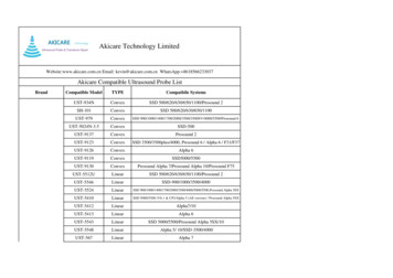
Transcription
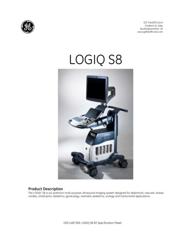
GE HealthcareChalfont St. GilesBuckinghamshire, UKwww.gehealthcare.comLOGIQ S8Product DescriptionThe LOGIQ* S8 is our premium multi-purpose ultrasound imaging system designed for abdominal, vascular, breast,cardiac, small parts, obstetrics, gynecology, neonatal, pediatrics, urology and transcranial applications.DOC1487369: LOGIQ S8 R2 Specification Sheet
General SpecificationsDimensions andWeight Height Maximum: 1750mm, 68.9in Minimum: 1150mm, 45.3in Width Keyboard: 500mm, 19.7 in Caster: 620mm, 24.4 in Depth Maximum: 880mm, 34.6 in Caster: 790mm, 31.1 in Weight 85 kg, 187.4 lbs Voltage: 100-120 Vac or 220-240Vac Frequency: 50/60 Hz Power consumption maximum of900VA with peripheralsConsole Design 19” high-resolution LCD LCD translation (independent ofconsole): 660 mm horizontal (end to end) 135 mm vertical (end to end) 90 swivel Fold-down and lock mechanism fortransportation Brightness and contrast adjustment Resolution: 1280 X 1024 Horizontal/Vertical viewing angle of /- 170 System OverviewApplicationsElectrical Power LCD Monitor4 Active probe ports and 1 parkingIntegrated HDD (at least 500 GB)Integrated DVD multi driveOn-board storage of thermalprinterIntegrated speakersLocking mechanism that providesrolling lock and caster swivel lockIntegrated cable managementFront and rear handlesEasily removable air filtersUser InterfaceOperator Keyboard Operating keyboard adjustable intwo dimensions: Height Rotation Backlit alphanumeric keyboard Ergonomic hard key layout Interactive back-lighting Integrated recording keys forremote control of up to 6peripheral or DICOM** devices Integrated gel warmerTouch Screen Wide 9” high-resolution, color,touch, LCD screen Interactive dynamic softwaremenu Brightness adjustment User-configurable layout AbdominalObstetricalGynecologicalBreastSmall partsVascular /PeripheralTranscranialPediatrics and neonatalMusculoskeletalUrologicalCardiacOperating Modes B-ModeM-ModeColor Flow Mode (CFM)TVI (Option)B-Flow*/B-Flow Color (Option)Extended Field of View (LOGIQView, Option)Power Doppler Imaging (PDI)PW DopplerCW Doppler (Option)Volume Modes (3D/4D) Static 3D Real Time 4D (option)Anatomical M-ModeCurved Anatomical M-ModeB Steer (Option)Coded Contrast Imaging (Option)Elastography (Option)Scanning Methods Electronic SectorElectronic ConvexElectronic LinearMechanical Volume SweepTransducer Types Sector Phased Array Convex ArrayDOC1487369: LOGIQ S8 R2 Specification SheetMicro convex ArrayLinear ArrayMatrix ArrayVolume Probes (4D) Convex Array Micro convex Array Split Crystal System StandardFeatures Advanced user interface with highresolution wide 9” LCD touch panel Automatic Optimization CrossXBeam* Speckle Reduction Imaging (SRI-HD) Fine Angle Steer Coded Harmonic Imaging Virtual Convex Patient information database Image Archive on integratedCD/DVD and hard drive 3D Raw Data Analysis Real-time automatic Doppler calcs OB Calcs Fetal Trending Multigestational Calcs Hip Dysplasia Calcs Gynecological Calcs Vascular Calcs Urological Calcs Renal Calcs Cardiac Calcs InSite*ExC capability On-board electronic documentationPeripheral Options Integrated options for: Digital BW thermal printer DVD video recorder Digital color thermal printer Digital A6 color thermal printer External USB printer connection DVI-D output available forcompatible devices Foot Switch, with programmablefunctionality Video ConverterDisplay Modes Live and Stored Display Format: Fullsize and split screen - both w/thumbnails. for still and Cine Review Image Format: 4x4 and"thumbnails". for still and Cine Simultaneous Capability B or CrossXBeam /PW B or CrossXBeam /CFM or PDIPage 2 of 14
B/M B/CrossXBeam Real-time Triplex Mode(B or CrossXBeam CFM orPDI/PW or CW(Option))Selectable alternating Modes B or CrossXBeam /PW B or CrossXBeam CFM(PDI)/PW(CW(Option)) B/CW (Option)Multi-image (split/quad screen) Live and/or frozen B or CrossXBeam B orCrossXBeam /CFM or PDI PW/M Independent Cine playbackTime line display Independent Dual B orCrossXBeam /PW Display CWDisplay Formats Top/ Bottom selectable format Side/Side selectable format Timeline onlyVirtual ConvexDisplay Annotation Patient Name: First, Last andMiddle Patient ID 2nd Patient ID Age, Sex and Birth Date Hospital Name Date format: 3 types selectableMM/DD/YY, DD/MM/YY, YY/MM/DD Time format: 2 types selectable:24 hours, 12 hours Gestational Age fromLMP/EDD/GA/BBT Probe Name Map names Probe Orientation Depth Scale Marker Lateral Scale Marker Focal Zone Markers Image Depth Zoom Depth B-Mode Gain Dynamic Range Imaging Frequency Frame Averaging Gray Map SRI-HD M-Mode Gain Dynamic Range Time Scale Doppler Mode Gain Angle Sample Volume Depth and Width Wall Filter Velocity and/or Frequency Scale Spectrum Inversion Time Scale PRF Doppler FrequencyColor Flow Mode Line Density Frame Averaging Packet Size Color Scale: 3 typesPower, Directional PDI, andSymmetrical Velocity Imaging Color Velocity Range and Baseline Color Threshold Marker Color Gain PDI Inversion Doppler FrequencyTGC CurveAcoustic Frame RateCine Gage, Image Number / FrameNumberBody Pattern: Multiple human andanimal typesApplication NameMeasurement ResultsOperator MessageDisplayed Acoustic Output TIS: Thermal Index Soft Tissue TIC: Thermal Index Cranial (Bone) TIB: Thermal Index Bone MI: Mechanical Index% of Maximum Power outputBiopsy Guide Line and ZoneHeart RateGeneral SystemParametersSystem Setup Pre-programmable Categories User Programmable PresetCapability Factory Default Preset Data Languages: English, French,German, Spanish, Italian,Portuguese, Russian, Greek,Swedish, Danish, Dutch, Finnish,Norwegian, Japanese (messageonly), Chinese (message only) OB Report Formats including TokyoUniv., Osaka Univ., USA, Europe, andASUM User Defined AnnotationsDOC1487369: LOGIQ S8 R2 Specification Sheet Body Patterns Customized Comment HomePositionUser Manual availableon board through Help(F1)User Manual and Service Manual areincluded on CD with each system. Aprinted manual is available uponrequest.Cine Memory/ImageMemory 776 MB of Cine Memory Selectable Cine Sequence for CineReview Prospective Cine Mark Measurements/ Calculations andAnnotations on Cine Playback Scrolling timeline memory Dual Image Cine Display Quad Image Cine Display Cine Gauge and Cine ImageNumber Display Cine Review Loop Cine Review Speed: 11 steps (11, 13,14, 17, 22, 25, 31, 48, 100, 200,400%)Image Storage On-board database of patientinformation from past exams Storage Formats: DICOM – compressed/uncompressed, single/ multiframe, with/ without Raw Data Export JPEG, JPEG2000, WMV(MPEG 4) and AVI formats Storage Devices: USB Memory Stick: 64MB to 4GB(for exporting individualimages/clips) CD-RW storage: 700MB DVD storage: -R (4.7GB) Hard Drive Image Storage: 112GB Compare previous exam imageswith current exam images Reload of archived data sets Network Storage support for Import,Export, DICOM Read, SaveAs,SaveAs Image, Report SaveAs,MPEGVuePage 3 of 14
Connectivity andDICOM Ethernet network connection DICOM 3.0 (Option) Verify Print Store Modality Worklist Storage Commitment Modality Performed ProcedureStep (MPPS) Media Exchange Off network / mobile storagequeue Query / Retrieve Public SR Template Structured Reporting –compatible with vascular andOB standard InSite ExC capabilityPhysiological InputPanel (Option) Physiological Input ECG, 2 lead Dual R-Trigger Pre-settable ECG R Delay Time Pre-settable ECG Position Adjustable ECG Gain Control Automatic Heart Rate DisplayReport Writer (Option) On-board reporting packageautomates report writing Formats various exam results intoa report suitable for printing orreviewing on a standard PC Exam result reports can includepatient info, exam info,measurements, calculations,images, comments and physiciandiagnosis Standard templates provided Customizable templatesScanning ParametersDigital Beamformer478,405 ChannelsFrame Rate: 2399Hz MaximumDisplayed Imaging Depth: 0 – 33cm Minimum Depth of Field: 0 – 2 cm(Zoom) (probe dependent) Maximum Depth of Field: 0 – 33cm (probe dependent) Continuous Dynamic Receive Focus/ Continuous Dynamic ReceiveAperture 274dB Dynamic Range System frequency range: 1-13MHz 256 shades of gray Adjustable Field of View (FOV) Image Reverse: Right/ Left Image Rotation of 0 , 180 Digital B-Mode Adjustable: Acoustic Power: 0 - 100%, 25steps Gain: 0 – 90 dB range, 1dB steps Dynamic Range: 36 – 96 dB, 16steps Frame Averaging: up to 8 steps Gray Scale Map: 10 maps Frequency: up to 6 steps Speed of Sound (probe,application dependent) Line Density: up to 5 steps Scanning Size (FOV or Angle depending on the probe, seeprobe specifications) B Colorization: 9 maps Reject: 6 steps Suppression: up to 6 steps SRI-HD: 0 – 5, 6 steps Edge Enhance: 7 stepsDigital M-Mode Adjustable: Acoustic Power: 0 - 100%, 25steps Gain: -20 – 20 dB (delta from B) Dynamic Range : 36 – 96 dB, 16steps Gray Scale Map: 10 maps Frequency: up to 6 steps Sweep Speed: 0–7, 8 steps M Colorization: 9 maps M Display Format: V-1/3B, V-1/2B,V-2/3B, H-1/2B, H-1/4B, TL Only Rejection: 6 stepsAnatomical M-Mode M-mode cursor adjustable at anyplane Can be activated from a Cine loopfrom a live or stored image M and A capability Available with Color Flow Mode Curved Anatomical M-ModeDOC1487369: LOGIQ S8 R2 Specification SheetDigital SpectralDoppler Mode Adjustable: Acoustic Power: 0-100%, 25 steps Gain: 0 – 85dB range, 1dB steps Gray Scale Map: 8 maps Transmit Frequency: Up to 5 steps Wall Filter: 18 settings, Min: 5 Hz,Max: 3,339Hz PW Colorization: 6 maps Velocity Scale Range: 6.9 –1032.8cm/s Sweep Speed: 0 – 7, 8 steps Sample Volume Length: 1 – 16mm,12 steps Angle Correction: -90 - 90 degrees,1 degree steps Steered Linear: 0 – 30 degrees Spectrum Inversion Trace Method Baseline Shift: 5 – 95%, 11 steps Doppler Auto Trace Compression: 0.5 – 2.4, 9 steps Trace Direction Trace SensitivityDigital Color FlowMode Adjustable: Acoustic Power: 0 - 100%, 25steps Color Maps, including velocityvariance maps: 15 maps Gain: 0 – 40dB, 81 steps Velocity Scale Range: 2 – 370cm/s Wall Filter: 0 – 3, 4 steps Packet Size: 5 – 24, 9 steps Line Density: 5 steps Spatial Filter: 6 steps Steering Angle: 0 – 20 degrees Baseline Shift: 0 – 100%, 11 steps Frame Average: 0 – 6, 7 steps Threshold: 0 – 100%, 11 steps Accumulation mode: 7 levels Sample Volume Control Flash SuppressionDigital Power DopplerImaging Adjustable: Acoustic Power: 0 - 100%, 25steps Color Map: 16 maps Gain: 0 – 40 dB, 81 steps Wall Filter: 0 – 3, 4 steps Packet Size: 5 – 24, 9 stepsPage 4 of 14
Line Density: 5 stepsSpatial Filter: 6 stepsSteering Angle: 0 – 20 degreesFrame Average: 0 – 6, 7 stepsThreshold: 0 – 100%, 11 stepsAccumulation mode:7 levelsSample Volume ControlFlash Suppression: 0 – 4, 5 stepsContinuous WaveDoppler (Option) Available on M5S-D, 3Sp-D, 6S-D,S4-10-D, 6Tc-RS, P2D, P6D Steerable CW mode includesAdjustable: Acoustic Power: 0 - 100%, 25steps Gain: 0 – 85dB range, 1dB steps Gray Scale Map: 8 maps Transmit Frequency: 1 or 2 steps Wall Filter: 18 settings, Min:5.5Hz,Max:5095Hz CW Colorization: 6 maps Velocity Scale Range: 20 – 1116cm/s (2000cm/s :P2D) Sweep Speed: 0 – 7, 8 steps Angle Correction: -90 - 90degrees, 1 degree steps Spectrum Inversion Trace Method Baseline Shift: 5 – 95%, 11 steps Doppler Auto Trace Compression: 0.5 – 2.4, 9 steps Trace Direction Trace SensitivityAutomaticOptimization Optimize B-Mode image to helpimprove contrast resolution. Selectable amount of contrastresolution enhancement (low,medium, high) Auto-Spectral Optimize – adjustsbaseline, invert, PRF (on live image),and angle correctionCoded HarmonicImaging Available on all 2D and 4D probesB-Flow (Option) Available on C1-5-D, C2-9-D, 9L-D,11L-D, ML6-15-D, M5S-D, S1-5-D,L8-18i-D, 10C-D Background: On/Off Sensitivity/PRI : 1 – 50, 17 steps Line Density: 5 stepsEdge Enhance: 7 stepsFrame Average: 0 – 7, 8 stepsGray Scale Map: 8 mapsTint Map :9mapsDynamic Range: 36 – 96 dB, 16stepsRejection: 6 stepsGain: 0 – 90 dB range, 1dB stepsB-Flow ColorAccumulation: 7 levelsB Steer (Option) Available on 9L-D, 11L-D, ML6-15-D,L8-18i-DCoded ContrastImaging (Option) Available on 3CRF-D, C1-5D, C2-9-D,C2-6b-D, IC5-9-D, 9L-D, 11L-D, ML615-D, M5S-D, L8-18i-D, RAB6-D 2 Contrast Timers Timed Updates: 0.05 – 10 seconds Accumulation mode, six levels Maximum Enhance Mode Flash Time Intensity Curve (TIC) Analysis Auto MI control The LOGIQ S8 is designed forcompatibility with commerciallyavailable ultrasound contrastagents. Because the availability ofthese agents is subject togovernment regulation andapproval, product features intendedfor use with these agents may notbe commercially marketed normade available before the contrastagent is cleared for use. Contrastrelated product features areenabled only on systems fordelivery to an authorized country orregion of use.LOGIQ View (Option) Extended Field of View Imaging Available on the following probes:9L-D, 11L-D, ML6-15-D, L8-18i-D,3CRF-D, C1-5D, C2-9-D, C2-6b-D,10C-D, IC5-9-D, S1-5-D, M5S-D,3Sp-D, 6S-D, S4-10-D, RAB6-D,RIC5-9-D, 6Tc-RS For use in B-Mode CrossXBeam is available on linearprobes Auto detection of scan direction Pre or post-process zoom RotationDOC1487369: LOGIQ S8 R2 Specification Sheet Auto fit on monitor Measurements in B-Mode Up to 60cm scan length3D Allows unlimited rotation andplanar translations 3D reconstruction from Cine sweepAdvanced 3D Acquisition of Color dataAutomatic rendering3D Landscape technology3D MovieReal Time 4D (Option) Acquisition Modes: Real Time 4D Static 3D Visualization Modes: 3D Rendering (diverse surfaceand intensity projection modes) Sectional Planes (3 Section planesperpendicular to each other) Volume Contrast Imaging-Static(option) Tomographic Ultrasound Imaging(option) Render Mode: Surface Texture, Surface Smooth,max-, min- and X-ray (averageintensity projection), mix mode oftwo render modes Curved 3 point Render start 3D Movie Scalpel: 3D Cut tool Display Format: Quad: A-/B-/C-Plane/3D Dual: A-Plane/3D Single: 3D or A- or B- or C-Plane Automated Volume Calculation VOCAL II (option) Betaview Auto SweepVolume Navigation(Option) Available on C1-5-D, C2-9-D, C2-6bD, 9L-D, ML6-15-D, 3CRF-D, IC5-9-D,S1-5-D, S4-10-D, M5S-D, L8-18i-D Sensor-based acquisition Position Markers Needle tip tracking Virtual tracking Tru3D feature includes: Display of data in: Main-, Parallel-,Angular-ModePage 5 of 14
Render Modes: Gray Surface,Texture, Min-, Max-, AverageIntensity Measurements: distance, angle,area, volume 3D MovieScan Assistant(Option)ElastographyQuantification (Option– not available in theUnited States) Relative quantification tool Available on ML6-15-D, 11L-D, 9L-D,IC5-9-D, C1-5-D, C2-9-D Factory Programs User –defined programs Steps include image annotations,mode transitions, basic imagingcontrols and measurementinitiationQuantitative FlowAnalysis (Option)Compare Assistant(Option) Myocardial Doppler Imaging withcolor overlay on tissue image Available on the sector probes Tissue color overlay can beremoved to show just the 2D image,still retaining the tissue velocityinformation Curved Anatomical M-mode: free(curved) drawing of M-modegenerated from the cursorindependent from the axial plane Q-Analysis: Multiple Time -Motiontrace display from selected pointsin the myocardium Allows side-by-side comparison ofprevious ultrasound and othermodality exams during livescanningPower Assistant(Option) Allows moving the system withouta complete system shutdown andboot-up power cycle.Breast ProductivityPackage (Option) Allows automatic contour andmeasurement of breast lesions Worksheet summary Feature Assessment BI RADS Assessment User editableThyroid ProductivityPackage (Option) Worksheet summary includesmeasurements and locations fornodule, parathyroid and lymphnode Feature Assessment User editableElastography (Option) Available on ML6-15-D, 11L-D, 9LD, C2-9-D, C1-5-D, IC5-9-D Available in Color and PowerDopplerTVI (Option)Stress Echo (Option) Advanced and flexible stress-echoexamination capabilities Provides exercise andpharmacological protocoltemplates 8 default templates Template editor for userconfiguration of existing templatesor creation of new templates Reference scan display duringacquisition for stress levelcomparison (dual screen) Baseline level/Previous levelselectable Raw data continuous capture Over 100 sec available Wall motion scoring (bulls-eye andsegmental) Smart stress: Automatically set upvarious scanning parameters (forinstance geometry, frequency, gain,etc.) according to same projectionon previous levelVirtual Convex Provides a convex field of viewDOC1487369: LOGIQ S8 R2 Specification Sheet Compatible with CrossXBeam Available on all linear and sectortransducers 9L-D, 11L-D, ML6-15-D,L8-18i-D, S1-5-D, M5S-D, 3Sp-D, 6SD, S4-10-D, 6Tc-RSSRI-HD Speckle Reduction Imaging Provides multiple levels of specklereduction Compatible with Side by SideDualView Display Compatible with ALL linear, convexand sector transducers Compatible w/ B-Mode, Color,Contrast Agent and 3D imagingCrossXBeam Provides 3,5,7, or 9 angles of spatialcompounding Live Side by Side DualView Display Compatible with: Color Mode PW SRI-HD Coded Harmonic Imaging Virtual Convex Available on 9L-D, 11L-D, ML6-15-D,L8-18i-D, 3CRF-D, C1-5D, C2-9-D,C2-6b-D, 10C-D, IC5-9-D, RAB6-D,RIC5-9-DControls AvailableWhile “Live” Write Zoom B/M/CrossXBeam-Mode Gain TGC Dynamic Range Acoustic Output Transmission Focus Position Transmission Focus Number Line Density Control Sweep Speed for M-Mode Number of Angles forCrossXBeam PW-Mode Gain Dynamic Range Acoustic Output Transmission Frequency PRF Wall Filter Spectral Averaging Sample Volume Gate Length Depth Velocity ScalePage 6 of 14
Time Resolution Color Flow Mode CFM Gain CFM Velocity Range Acoustic Output Wall Echo Filter Packet Size Frame Rate Control CFM Spatial Filter CFM Frame Averaging CFM Line Resolution Frequency / Velocity Base LineShiftControls Available on“Freeze” or Recall Automatic Optimization SRI-HD CrossXBeam – Display noncompounded and compoundedimage simultaneously in splitscreen 3D reconstruction from a storedCine loop B/M/CrossXBeam Mode Gray Map Optimization TGC Colorized B and M Frame Average (loops only) Dynamic Range Anatomical M Mode Max Read Zoom to 8x Base Line Shift Sweep Speed PW Mode Gray Map Post Gain Baseline shift Sweep Speed Invert Spectral wave form Compression Rejection Colorized Spectrum Display Format Doppler Audio Angle Correct Quick Angle Correct Auto Angle Correct Color Flow Overall Gain (loops and stills) Color Map Transparency Map Frame Averaging (loops only) Flash Suppression CFM Display Threshold Spectral Invert for Color/Doppler Anatomical M-Mode on cine loop 4D Gray Map, Colorize Post Gain Change display – single, dual,quad sectional or renderedMeasurements /CalculationsGeneral B-Mode Depth and DistanceCircumference (Ellipse / Trace)Area (Ellipse / Trace)Volume (Ellipsoid)% Stenosis (Area or Diameter)Angle between two linesGeneral M-Mode M-DepthDistanceTimeSlopeHeart RateGeneral DopplerMeasurements/Calculations Velocity Time A/B Ratio (Velocities / FrequencyRatio PS (Peak Systole) ED (End Diastole) PS/ED (PS/ED Ratio) ED/PS (ED/PS Ratio) AT (Acceleration Time) ACCEL (Acceleration) TAMAX (Time Averaged MaximumVelocity Volume Flow (TAMEAN and VesselArea) Heart Rate PI (Pulsatility Index) RI (Resistivity Index)Real-time DopplerAuto Measurements /Calculations PS (Peak Systole)ED (End Diastole)MD (Minimum Diastole)PI (Pulsatility Index)RI (Resistivity Index)AT (Acceleration Time)ACC (Acceleration)PS/ED (PS/ED Ratio)ED/PS (ED/PS Ratio)HR (Heart Rate)DOC1487369: LOGIQ S8 R2 Specification Sheet TAMAX (Time Averaged MaximumVelocity) PVAL (Peak Velocity Value) Volume Flow (TAMEAN and VesselArea)OB Measurements /Calculations Gestational Age by: GS (Gestational Sac) CRL (Crown Rump Length) FL (Femur Length) BPD (Biparietal Diameter) AC (Abdominal Circumference) HC (Head Circumference) APTD x TTD (Anterior/PosteriorTrunk Diameter by TransverseTrunk Diameter) FTA (Fetal Trunk Cross-sectionalArea) HL (Humerus Length) BD (Binocular Distance) FT (Foot Length) OFD (Occipital Frontal Diameter) TAD (Transverse AbdominalDiameter) TCD (Transverse CerebellumDiameter) THD (Thorax Transverse Diameter) TIB (Tibia Length) ULNA (Ulna Length) Estimated Fetal Weight (EFW) by: AC, BPD AC, BPD, FL AC, BPD, FL, HC AC, FL AC, FL, HC AC, HC BPD, APTD, TTD, FL BPD, APTD, TTD, SL Calculations and Ratios FL/BPD FL/AC FL/HC HC/AC CI (Cephalic Index) AFI (Amniotic Fluid Index) CTAR(Cardio-Thoracic Area Ratio) Measurements / Calculations by:ASUM, ASUM 2001, Berkowitz,Bertagnoli, Brenner, Campbell, CFEF,Chitty, Eik-Nes, Ericksen, Goldstein,Hadlock, Hansmann, Hellman, Hill,Hohler, Jeanty, JSUM, Kurtz,Mayden, Mercer, Merz, Moore,Nelson, Osaka University, Paris,Rempen, Robinson, Shepard,Shepard/Warsoff, Tokyo University,Tokyo/Shinozuka, YarkoniPage 7 of 14
Fetal Graphical TrendingGrowth PercentilesMulti-Gestational Calculations (4)Fetal Qualitative Description(Anatomical survey)Fetal Environmental Description(Biophysical profile)Programmable OB TablesOver 20 selectable OB CalcsExpanded WorksheetsOB Measure Assistant(Option) Allows automatic contour andmeasurement of BPD, HC, FL andAC User editableBreast MeasureAssistant (Option) Allows automatic contour andmeasurement of breast lesions User editableGYN Measurements/Calculations Right Ovary Length, Width, HeightLeft Ovary Length, Width, HeightUterus Length, Width, HeightCervix Length, TraceOvarian VolumeENDO (Endometrial thickness)Ovarian RIUterine RIFollicular measurementsSummary ReportsVascularMeasurements/Calculations SYS DCCA (Systolic Distal CommonCarotid Artery) DIAS DCCA (Diastolic DistalCommon Carotid Artery) SYS MCCA (Systolic Mid CommonCarotid Artery) DIAS MCCA (Diastolic Mid CommonCarotid Artery) SYS PCCA (Systolic ProximalCommon Carotid Artery) DIAS PCCA (Diastolic ProximalCommon Carotid Artery) SYS DICA (Systolic Distal InternalCarotid Artery) DIAS DICA (Systolic Distal InternalCarotid Artery) SYS MICA (Systolic Mid InternalCarotid Artery) DIAS MICA (Diastolic Mid InternalCarotid Artery) SYS PICA (Systolic Proximal InternalCarotid Artery) DIAS PICA (Diastolic ProximalInternal Carotid Artery) SYS DECA (Systolic Distal ExternalCarotid Artery) DIAS DECA (Diastolic Distal ExternalCarotid Artery) SYS PECA (Systolic Proximal ExternalCarotid Artery) DIAS PECA (Diastolic ProximalExternal Carotid Artery) VERT (Systolic Vertebral Velocity) SUBCLAV (Systolic SubclavianVelocity) Automatic IMT (Option) Summary ReportsAuto EF (Option) Allows semi-automaticmeasurement of the global EF(Ejection fraction) User editableUrological Calcs Bladder VolumeProstate VolumeLt/Rt Renal VolumeGeneric VolumePost-Void Bladder VolumeProbes (All Optional)3CRF-D Micro Convex Biopsy Probe Applications: Abdomen Biopsy Guide: Single-Angle,disposable with a reusable bracket(H40442LR), Multi-Angle with areusable bracket (H40452LP) Band Width: 1 - 6MHz Number of Elements: 128 Field of View (Max): 80 degree Physical Foot Print: 29 x 12 mm B-mode Frequency: 3, 3.5, 4MHz Harmonic Frequency: 3.6, 4, 4.4MHz PW Doppler Frequency: 2.1, 2.5,3.6MHz Color Doppler Frequency: 2.1, 2.5,3.6MHzC1-5-D Convex Probe Applications: Abdomen, OB,Gynecology, Urology, Vascular Biopsy Guide: Multi-Angle,disposable with a reusable bracket(H40432LE)DOC1487369: LOGIQ S8 R2 Specification SheetBand Width: 1 – 6MHzNumber of Elements: 192Field of View (Max): 70 degreePhysical Foot Print: 66 x 18mmB-mode Frequency: 2, 3, 4, 5MHzHarmonic Frequency: 3, 4, 5MHzPW Doppler Frequency: 1.8, 2.1, 2.5,3.6MHz Color Doppler Frequency: 1.8, 1.9,2.5, 3.1, 3.6MHz C2-9-D Convex Probe Applications: Abdomen, Pediatrics,OB, Gynecology, Neonatal,Vascular Biopsy Guide: Multi-Angle,disposable with a reusable bracket(H4913BA) Band Width: 2 – 9 MHz Number of Elements: 192 Field of View (Max): 65 degree Physical Foot Print: 52 x 9 mm B-mode Frequency: 2.5, 4.5, 6, 7.5,9MHz Harmonic Frequency: 5, 6, 7, 8,9MHz PW Doppler Frequency: 2.8, 3.1, 3.6,4.2, 5MHz Color Doppler Frequency: 2.8, 3.1,3.6, 4.2, 5MHzC2-6b-D Biopsy Convex Probe Applications: Abdomen, Intervention Biopsy Guide: Multi-Angle,disposable (H46332LY) Band Width: 2 – 6MHz Number of Elements: 128 Field of View (Max): 61degree Physical Foot Print: 65 (Normal) x13mm B-mode Frequency: 2, 3, 4, 5MHz Harmonic Frequency: 3.5, 4, 5MHz PW Doppler Frequency: 2.5, 3, 3.6,4.2MHz Color Doppler Frequency: 2.5, 3.1,3.6, 4.2MHz10C-D Micro convex Probe Applications: Neonatal, Pediatrics,Vascular Band Width: 4 – 12MHz Number of Elements: 128 Field of View (Max): 102degree Physical Foot Print: 28 x 13mm B-mode Frequency: 4, 6, 8, 10MHz Harmonic Frequency: 7, 8, 9, 10MHz PW Doppler Frequency: 4.2, 5, 6.3,8.3MHzPage 8 of 14
Color Doppler Frequency: 4.2, 5,6.3, 8.3MHzIC5-9-D Micro convex Probe Applications: OB, Gynecology,Urological Biopsy Guide: Single Angle,Disposable with a disposablebracket (E8385MJ) or reusablebracket (H40412LN) Band Width: 3 – 10MHz Number of Elements: 192 Field of View (Max): 145degree Physical Foot Print: 26 x 6mm B-mode Frequency: 4, 5, 6, 7, 8,9MHz Harmonic Frequency: 7, 8, 9MHz PW Doppler Frequency: 3.6, 4.2, 5,6.3MHz Color Doppler Frequency: 4.2, 6.3,8.3MHzS1-5-D Sector Probe Applications: Abdomen, OB,Gynecology, Vascular Biopsy Guide: Multi-angle,disposable with a reusablebracket (H4908SD) Band Width: 1 – 6 MHz Number of Elements: 128 Field of View (Max): 90degree Physical Foot Print: 28 x 14mm B-mode Frequency: 2, 3, 4MHz Harmonic Frequency: 2.5, 3, 4, 4.5,5MHz PW Doppler Frequency: 1.8, 2.1,2.5, 3.4MHz Color Doppler Frequency: 1.8, 1.9,2.1, 2.5MHzS4-10-D Sector Probe Applications: Pediatrics, Neonatal Band Width: 3 - 9MHz Number of Elements: 128 Field of View (Max): 90degree Physical Foot Print: 20 x 15mm B-mode Frequency: 4, 6, 8, 10MHz Harmonic Frequency: 5, 6, 7, 8.5,10MHz PW Doppler Frequency: 3.6, 4.2, 5,6.3MHz Color Doppler Frequency: 5, 6.3,8.3MHz CWD Doppler Frequency: 4.2,5MHzM5S-D Sector Probe Applications: Cardiac, Transcranial Band Width: 1 – 5MHz Number of Elements: 288Field of View (Max): 120degreePhysical Foot Print: 28 x 17mmB-mode Frequency: 2, 2.5, 3.5,4.5MHzHarmonic Frequency: 2.5, 3, 3.2, 3.3,3.5, 3.7, 4, 4.5, 4.6MHzPW Doppler Frequency: 1.8, 2.1, 2.5,3.1, 3.6MHzColor Doppler Frequency: 1.8, 2.1,2.5, 3.1, 4.2MHzCWD Doppler Frequency: 1.9,2.5MHz3Sp-D Sector Probe Applications: Cardiac, Transcranial Band Width: 1 – 6MHz Number of Elements: 64 Field of View (Max): 120degree Physical Foot Print: 15 x 14mm B-mode Frequency: 2, 3, 4, 5MHz Harmonic Frequency: 3, 3.2, 3.3, 3.5,3.7, 4, 4.5, 4.6MHz PW Doppler Frequency: 1.8, 2.1, 2.5,3.1, 4.2MHz Color Doppler Frequency: 1.6, 1.8,2.1, 2.5, 3.1, 4.2MHz CWD Doppler Frequency: 1.9,2.5MHz6S-D Sector Probe Applications: Cardiac, Pediatrics,Neonatal Band Width: 2 – 8 MHz Number of Elements: 128 Field of View (Max): 90degree Physical Foot Print: 20 x 15mm B-mode Frequency: 4, 5, 6.5, 8MHz Harmonic Frequency: 4.8, 5.4,6.2MHz PW Doppler Frequency: 2.8, 3.1, 3.6,5MHz Color Doppler Frequency: 2.8, 3.1,3.6, 5MHz CWD Doppler Frequency: 2.8, 3.6,5MHz9L-D Linear Probe Applications: Vascular, Small Parts,Pediatrics, Abdomen Biopsy Guide: Multi-angle,disposable with a reusable bracket(H4906BK) Band Width: 2 – 8MHz Number of Elements: 192 Field of View (Max): 44mm Physical Foot Print: 49 x 9mm B-mode Frequency: 5, 6, 7, 9MHz Harmonic Frequency: 8.5, 10MHzDOC1487369: LOGIQ S8 R2 Specification Sheet PW Doppler Frequency: 3.1, 3.6, 4.2,5MHz Color Doppler Frequency: 3.1, 3.6,4.2, 5, 6.3MHz11L-D Linear Probe Applications: Vascular, Small Parts,Pediatrics, Neonatal Biopsy Guide Available: Multi- Angle,disposable with a reusable bracket(H40432LC) Band Width: 3 – 11MHz Number of Elements: 192 Field of View (Max): 38mm Physical Foot Print: 44 x 7mm B-mode Frequency: 7, 8, 9, 11MHz Harmonic Frequency: 9, 11, 12MHz PW Doppler Frequency: 4.2, 5, 6.3,8.3MHz Color Doppler Frequency: 4.2, 5, 6.3,8.3MHzML6-15-D Matrix Array Linear Probe Applications: Small Parts, Vascular,Neonatal, Pediatrics Biopsy Guide Available: Multi-Angle,disposable with a reusable bracket(H40432LJ) Band Width: 4 – 15MHz Number of Elements: 1008 Field of View (Max): 50mm Physical Foot Print: 50 x 6mm B-mode Frequency: 9, 11, 13,15MHz Harmonic Frequency: 8, 9, 12,15MHz PW Doppler Frequency: 5, 6.3,8.3MHz Color Doppler Frequency: 5, 7.7, 10,12.5MHzL8-18i-D Linear Probe Applications: Small Parts, Vascular,Intraoperative Band Width: 4 – 15MHz Number of Elements: 168 Field of View (Max): 25mm Physical Foot Print: 35 x 10mm B-mode Frequency: 8, 9, 12, 15,18MHz Harmonic Frequency: 9, 15, 18MHz PW Doppler Frequency: 5, 6.3,8.3MHz Color Doppler Frequency: 5, 7.7, 10,12.5MHzRAB6-D Convex Volume Probe Applications: Abdomen, OB,Gynecology, PediatricsPage 9 of 14
Biopsy Guide: Multi Angle,disposable with a reusablebracket (H48681ML) Band Width: 2 – 8 MHz Number of Elements: 192 Field of View (Max): 80degree Physical Foot Print: 62 x 34mm B-mode Frequency: 2, 4, 6, 8MHz Harmonic Frequency: 5, 6.5, 8MHz PW Doppler Frequency: 2.5, 3, 3.6,4.2MHz Color Doppler Frequency: 2.5, 3,3.6, 4.2, 5MHzRIC5-9-D Convex Volume Probe Applications: OB, Gynecology,Urology Biopsy Guide: Single Angle,R
DOC1487369: LOGIQ S8 R2 Specification Sheet Page 2 of 14 General Specifications Dimensions and Weight Micro convex Array Height Maximum: 1750mm, 68.9in Minimum: 1150mm, 45.3in Width Keyboard: 500mm, 19.7 in Caster: 620mm, 24.4 in Depth Maximum: 880mm, 34.6 in