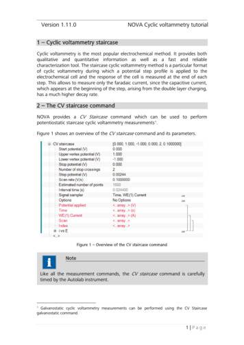
Transcription
This is an open access article published under an ACS AuthorChoice License, which permitscopying and redistribution of the article or any adaptations for non-commercial s Guide to Voltammetry: Acute and Chronic Electrodes forin Vivo Fast-Scan Cyclic VoltammetryNathan T. Rodeberg,† Stefan G. Sandberg,§, Justin A. Johnson,† Paul E. M. Phillips,*,§, and R. Mark Wightman*,†,‡†Department of Chemistry and ‡Neuroscience Center, University of North Carolina at Chapel Hill, Chapel Hill, North Carolina27599-3290, United States§Department of Psychiatry and Behavioral Sciences and Department of Pharmacology, University of Washington, Seattle,Washington 98195-6560, United StatesABSTRACT: Fast-scan cyclic voltammetry (FSCV) has beenused for over 20 years to study rapid neurotransmission inawake and behaving animals. These experiments were firstcarried out with carbon-fiber microelectrodes (CFMs) encasedin borosilicate glass, which can be inserted into the brainthrough micromanipulators and guide cannulas. More recently,chronically implantable CFMs constructed with small diameterfused-silica have been introduced. These electrodes can beaffixed in the brain with minimal tissue response, whichpermits longitudinal measurements of neurotransmission insingle recording locations during behavior. Both electrodedesigns have been used to make novel discoveries in the fieldsof neurobiology, behavioral neuroscience, and psychopharmacology. The purpose of this Review is to address importantconsiderations for the use of FSCV to study neurotransmitters in awake and behaving animals, with a focus on measurements ofstriatal dopamine. Common issues concerning experimental design, data collection, and calibration are addressed. Whennecessary, differences between the two methodologies (acute vs chronic recordings) are discussed. The topics raised in thisReview are particularly important as the field moves beyond dopamine toward new neurochemicals and brain regions.KEYWORDS: Fast-scan cyclic voltammetry, carbon-fiber microelectrodes, dopamine, principal component regression, chemometrics TECHNIQUES FOR MONITORING MOLECULES INNEUROSCIENCEThe monitoring of molecules in the brain has undergonesignificant advances in the past four decades. One of the earliesttechniques for measuring neurotransmitter release was push pull perfusion, a method that uses a cannula for sample collectionprior to downstream analysis.1,2 However, the direct interface ofthe perfusate with brain tissue raised concerns with samplecontamination and flow-induced damage to the surroundingenvironment. To address these issues, this procedure was lateradapted to incorporate a dialysis membrane, creating thetechnique known as microdialysis.3 6 Microdialysis restrictsflow to the probe, which minimizes brain damage and maintainssample purity. Equilibration of analytes across the membraneaccording to their concentration gradients results in concentration changes in the dialysate reflective of fluctuations in thebrain. Microdialysis is highly versatile, with its sensitivity,selectivity, and number of analytes that can be monitoredsimultaneously dependent on the detection method employed.Its main limitation is spatiotemporal resolution, as microdialysisprobes are typically at least 200 μm in diameter, and samples arehistorically collected approximately every 5 20 min to allowsufficient sample volume accumulation at low flow rates.5 8Recent improvements, largely due to reduction in the minimum 2017 American Chemical Societyvolume needed for sample analysis, have permitted microdialysismeasurements on a subminute time scale.9 12For the subset of brain molecules that are electroactive,particularly biogenic amines, electrochemical monitoring hasflourished as an alternative methodology.13,14 This approach wasfirst attempted in the Ralph Adams lab with a carbon pasteelectrode implanted in the striatum of an anesthetized rat.15 Slowpotential sweeps between 0.2 to 0.6 V vs Ag/AgCl revealedpeaks in current corresponding to the oxidation and reduction ofelectroactive substances; however, the identity of the molecule(s) producing the signal was unclear, with the authorssuggesting it could arise from dopamine, norepinephrine, orascorbic acid. Indeed, early voltammetric measurements sufferedfrom poor chemical resolution between catecholamines andother easily oxidized species, often present in the brain at higherconcentrations.15 20 In response to these problems, criteria weredeveloped to ensure that intended analytes were indeed thesource of recorded signals, including electrochemical, anatomSpecial Issue: Monitoring Molecules in Neuroscience 2016Received: November 15, 2016Accepted: January 27, 2017Published: January 27, 2017221DOI: 10.1021/acschemneuro.6b00393ACS Chem. Neurosci. 2017, 8, 221 234
ReviewACS Chemical NeuroscienceFigure 1. Designs of (a) borosilicate glass (b) fused silica CFMs. (a) Carbon fibers are aspirated through borosilicate glass under vacuum. A seal iscreated by heating and pulling the capillary to a fine tip. The protruding fiber is then trimmed, typically between 75 and 125 μm. For optimalelectrochemical performance, epoxy resin is used to fill any leaks in the seal that occur during electrode fabrication. Left panel: Electron micrograph ofCFM. Reprinted with permission from ref 113. Copyright 2003 American Association for Clinical Chemistry. Middle panel: A rat with dual cannulas forlater acute implantation of a CFM and reference electrode. The rat is tethered to a swivel and commutator via fastening of the headstage to an implantedstimulating electrode. Right panel: Side view of cannula for acute implantation of electrodes (left) and a micromanipulator for precise driving of theCFM during in vivo recordings (right). (b) Carbon fibers are threaded through a small diameter fused silica capillary under isopropyl alcohol. Afterdrying, epoxy is placed on the fiber and wicked into the fused silica capillary to create a hemispherical seal (inset image). The protruding carbon fiber istrimmed between 150 and 200 μm long. Electrical connection is established between a silver pin and the fiber with silver epoxy, which is later insulatedwith clear epoxy. Reprinted with permission from ref 59. Copyright 2010 Nature Publishing Group.ical, pharmacological, and independent verification.21 23 A majoradvance to the field came with the development of fast-scancyclic voltammetry (FSCV), a technique that utilizes rapidpotential sweeps to oxidize and reduce analytes of interest.24 26This process produces cyclic voltammograms, which displaymeasured current as a function of the applied potential, that serveas “fingerprints” for compound identification, providing anadvantage over single potential techniques.26 28 This moderatechemical selectivity allows the use of chemometric methods toseparate, and subsequently quantitate, analytes with differentcurrent potential characteristics (see Chemometric DataAnalysis section below).28,29The development of carbon-fiber microelectrodes (CFMs)has aided the FSCV field in multiple ways.30 32 The small size,and thus reduced capacitance and time constant, of theseelectrodes permits rapid scan rates ( 100 V/s), which enablesmeasurements on a subsecond time scale. Additionally, thisrelatively small size compared to traditional probes increasesspatial resolution and permits localized measurements in discretebrain regions. Moreover, in contrast to tissue damage observednear microdialysis probes, minimal damage is seen surroundingfiber implantation sites.33,34 These probes are also easilymodified with a variety of surface coatings, which can improvechemical selectivity, electron transfer kinetics, and sensitivity.35 39 Lastly, carbon-based electrodes demonstrate strongbiocompatibility, and are more resistant to biofouling than metalelectrodes. These advantages make FSCV with CFMs anattractive measurement technique for rapid neurotransmitterdynamics.glass-encased CFMs lowered into the brain using head-mountedmicrodrives.42 44 Later, behavioral evoked dopamine wasdetected by this approach,45 and these types of recordingsbecame routine, primarily due to improved sensitivity obtainedby increasing the anodic limit of the waveform46,47 to maintainoxygen-containing moieties on the electrode surface whichenhance adsorption of positively charged analytes (such asdopamine).27 FSCV has been adapted for multimodal recordingswith simultaneous extracellular electrophysiological recordings48 51 and iontophoresis51 54 at the same probe.The most recent generation of FSCV use in behaving animalshas been to adapt CFMs for chronic implantation, permittinglongitudinal measurements over an extended time scale in thesame animal. This is not a novel direction for electrochemicalmonitoring, as earlier methodologies had adopted such anapproach.55 57 These papers, which utilize amperometry, arenotable because they clearly show the need for more chemicalspecificity in the measurements due to difficulty assigning thesource of the signal. The standard fabrication of CFMs for FSCVusing a glass-encased design had limited success when chronicallyimplanted.58 However, the chronic CFMs used today employ abasic design where a carbon fiber is sealed in a small diameterfused-silica tube.59 Similar to results at acutely implantedCFMs33 and other miniaturized devices,60 these electrodeswere demonstrated to avoid the progressive immune responseand cell death that can impair measurements at larger probes.34With the improved sensitivity27 and low-noise61 of modernapproaches to using FSCV in vivo, recordings in striatal regionspermit detection of dopamine elicited by task-related events suchas the delivery of primary rewards,62,63 including pharmacological rewards,29,64 67 or reward-associated stimuli.51,62,68 70 Inaddition, spontaneous dopamine “transients” (i.e., briefelevations in extracellular dopamine concentration above theambient level, produced by release events) can be observed.These do not appear to be time locked to overt stimuli71 73 butare dependent upon activity in the ventral tegmental area,74 andhave been suggested to be a contributor to ambient extracellulardopamine levels in the nucleus accumbens.75 While the functionof these dopamine transients has not been fully characterized, DEVELOPMENT OF FSCV FOR FREELY BEHAVINGANIMALSMeasurements using FSCV with CFMs were originallyconducted in anesthetized animals.25,26,40,41 However, thesestudies could not reveal direct information about neurotransmission during behavior. The first FSCV measurements infreely moving animals detected dopamine release in terminalregions, evoked by electrical stimulation of afferent axonalpathways in rats. These experiments used acutely implanted222DOI: 10.1021/acschemneuro.6b00393ACS Chem. Neurosci. 2017, 8, 221 234
ReviewACS Chemical Neurosciencetheir activity can be altered by behavioral context73 as well aspharmacological agents including drugs of abuse.64,71The objective of this paper is to discuss the nuances of usingFSCV in behaving animals, based primarily on experience onmeasuring striatal dopamine. We will attempt to discuss thepotential pitfalls that can make the use of FSCV or relatedapproaches challenging, and then summarize how these caveatsdifferentially affect alternative approaches, with a particular focuson the use of acute or chronic electrodes.Not all electrodes are created equal. For glass electrodes, thestructural integrity of the pulled seal is the most importantdeterminant of electrode performance. Large cracks or gaps inthe seal will lead to exposed fiber and/or increased fragility,which will impede electrochemical measurements. Theseproblems can be alleviated by the use of epoxy to reinforce theseal (see above). For fused-silica encased CFMs, it is importantthat no epoxy remains on the fiber itself and that the seal forms aconvex, rather than concave, seal (see Figure 1b for illustration).Electrochemical characteristics of either electrode design can betested pre-experiment, either in vitro (i.e., in buffer) or in vivo(i.e., during surgical implantation of chronic CFMs beforecementation, or after lowering acute CFMs), to observe noiselevels and ensure electrical connectivity.Experimental Design for FSCV Recordings in FreelyMoving Animals. Measurements in freely moving animals areconducted using head-mounted amplifiers (“headstages”), whichconnect to the CFM and reference electrodes and transduce theexperimental current into voltages for downstream datacollection and analysis.82,83 These headstages are anchored tothe animal’s heads either directly via an electrical connector or ata separate point, such as the pedestal for the stimulating electrodeassembly (Figure 1a, middle panel). The headstage is alsoconnected to a swivel and commutator that permits movementwithin the behavioral chamber. Depending on the type ofelectrode used, cannulae may be affixed to the skull for laterimplantation of fresh CFM or reference electrodes (Figure 1a,middle panel) or fused-silica CFMs can be cemented directly tothe skull.The electrode design used influences both the type ofexperimental questions that can be answered and the overallsuccess rate of recordings. The fragility of borosilicate glasselectrodes can lead to a lower yield of successful experimentswith respect to fused-silica implantations. Moreover, tissuedamage from repeated insertion limits the number of withinsubject recordings.42,83 Once successfully lowered, however,these electrodes tend to be stable over the course of individualmeasurement periods. Thus, these electrodes are best served forstudies in which experimentally relevant manipulations occurduring single recording sessions.Conversely, the flexibility of fused-silica electrodes permits ahigher success rate for implantation compared to glasselectrodes. For chronic recordings, fused-silica electrodes areaffixed to the skull with dental cement and left unused for at least1 month to allow the immune response to these probes todissipate.59 Following this waiting period, it is possible toconduct many recordings at each electrode, increasing the datayield of chronic electrodes over acute electrodes. Longitudinalmeasurements permit the monitoring of dopamine overextended behavioral training and treatments. This is particularlyrelevant for models of disease states in which conditions developslowly over time.84 These electrodes are routinely used forperiods up to 4 months of recording. Naturally, there is someattrition of usable electrodes over that time. The majority of thisattrition pertains to physical failures (e.g., separation of surgicalimplant from subject’s head, loss of electrical continuity) and ismuch more infrequently due to altered electrochemical properties of the electrode (see Figure 3a, Supplementary Table 1 ofClark et al.59).In Vivo Electrode Positioning. For different applications,recording electrodes can either be fixed in the brain, or can behoused in a microdrive that allows their position to be adjusted(Figure 1a, right panel). The former is amenable to multiple EXPERIMENTAL CONSIDERATIONSElectrode Materials and Design. The most commonconstruction of recording electrodes for FSCV in behavinganimals uses carbon fibers housed in glass or fused-silicacapillaries. These carbon fibers host surface moieties, such ascarbonyl, hydroxyl, or more complex groups,76,77 which can alterthe electrochemical properties of the carbon fiber by changing itssurface charge and steric properties. The constellation offunctional groups on the carbon surface can be tuned withelectrochemical,78 thermal,79 or chemical80 pretreatment, andwill determine the selectivity of the adsorption of molecules tothe surface, including fouling agents and electrochemicalanalytes. A popular approach using FSCV at CFMs is to usethe applied waveform on each FSCV scan to electrochemicallycondition the electrode, essentially “pretreating” the electrodesurface each time a measurement is made.47 Specifically,increasing the anodic limit of the waveform above 1.0 V versusAg/AgCl substantially enhances the sensitivity to dopamine byincreasing dopamine-adsorbing oxide groups on the carbonsurface and slowly etching the fiber surface to mitigate the effectsof irreversible fouling.27,47,77 This approach has the advantageover traditional pretreatment strategies in that equilibrium ismaintained throughout the experiment, providing stablesensitivity.Construction of CFMs involves housing a carbon fiber in acapillary insulator with an exposed length at one end (the sensor)and an electrical connection at the other. For glass-basedelectrodes, a single carbon fiber is aspirated into a borosilicateglass capillary (600 1000 μm outer diameter, 400 500 μminner diameter) (Figure 1a, left panel). The capillary is thenpulled on a commercial glass-electrode puller (either vertical orhorizontal) to produce a tapered seal onto the carbon fiber.Sometimes the glass seal is then deliberately broken and resealedusing epoxy (Epon 828 with 14% m-phenylenediamine byweight). This approach increases the robustness (i.e. preventsunintentionally exposed carbon fiber from providing a lowresistance path for current) and reduces the shunt capacitance ofthe electrode taper by providing a thicker insulating layerbetween the carbon fiber and the extracellular fluid. For fusedsilica-based electrodes, a single carbon fiber is loaded into apolyimide-coated fused-silica tube (90 μm outer diameter, 20 μminner diameter, 8 12 mm length) submerged in isopropylalcohol (Figure 1b). With the carbon fiber protruding, one end ofthe tube is sealed with epoxy (Devcon 20845). For eitherelectrode type, the carbon fiber protruding from the seal is thentrimmed to the desired length, and an electrical connection ismade at the other end. Lengths of the trimmed carbon fiberusually range from 50 to 200 μm, where longer exposed fibers aremore sensitive, but have lower spatial resolution. Typically, glassbased electrodes have been used for acute implantation, whilefused-silica-based electrodes are favored for chronic implantationdue to both their durability, and biocompatibility arising fromtheir narrow diameter and polyimide coating.81223DOI: 10.1021/acschemneuro.6b00393ACS Chem. Neurosci. 2017, 8, 221 234
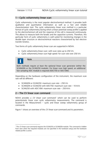
ReviewACS Chemical Neuroscienceelectrodes in the same animal,59 whereas the latter permitssystematic mapping of heterogeneity of electrically or naturallyevoked dopamine release along the dorsal-ventral axis.85Microdrives also allow selection of a recording site within thisheterogeneity by identifying “hot spots” (i.e., areas with a highdensity of release sites).73,85 Placement of fixed electrodes doesnot typically use this type of feedback-based selection. Therefore,positioning of these electrodes is more akin to random samplingof the tissue, and so signals converge upon the populationaverage rather than local maxima. Consequently, signalsmeasured with fixed electrodes tend to be smaller than thosefrom drivable electrodes due to unbiased selection of recordingsites (Figure 2).86,87 Dopamine signals in regions without releaseand so spontaneous transients have seldom been reported.Nonetheless, on the rare occasion when they were quantified,they were comparable in detected frequency as those measuredwith drivable electrodes.72 Figure 3 demonstrates examples ofpharmacologically and behaviorally evoked dopamine transients,as well as spontaneous transients, measured at chronicallyimplanted CFMs.Acute electrodes have the advantage of being drivable.However, in addition to the concerns with electrode fragilityduring repeated use mentioned above, electrode insertionimposes restraint stress on the animals. This could impactbehavioral assays that study stress under controlled conditions.89Figure 2. Comparison of concentrations measured at acute and chronicCFMs without optimization for dopamine release sites. In a study withacute CFMs (Owesson-White et al., ref 87), electrode placement wasoptimized for extracellular electrophysiological signals rather thandopamine release, resulting in recording locations without (top) andwith (middle) phasic dopamine release. The concentration profilecorresponds well with values from chronically implanted CFMs thatwere not optimized for recording location (bottom), indicating thelower concentrations measured with chronic CFM may be a result ofrecording site selection. Reprinted with permissions from ref 86.Copyright 2012 PNAS.Figure 3. Dopamine transients at chronically implanted CFMs. (a)Pharmacologically induced dopamine transients at a chronic CFM inresponse to i.v. cocaine infusion (red bar, 1.5 s duration). Backgroundsubtraction is denoted by the white dashed line. (b) Measurements at achronic CFM during a behavioral session of Pavlovian conditioning.Spontaneous dopamine transients are observed preceding cue onset(white asterisks). Moreover, both cue onset (left red dotted line) andreward delivery immediately following cue offset (right red dotted line)evoked phasic dopamine release. Background subtraction is denoted bythe white dashed line. Dopamine traces were extracted with PCR using astandard training set. Both measurements were made in the nucleusaccumbens core.or uptake sites rely on diffusion from nearby terminals, and thesesites exhibit both slower rises and decays compared to “hotspots”.88 As a result, electrodes not deliberately targeted atregions of high terminal density would be expected to haveslower signals due to heterogeneity of release sites.85 For similarreasons, one would intuit that fixed electrodes should detectfewer spontaneous transients. However, most studies usingchronic electrodes focus on the analysis of task-related events,224DOI: 10.1021/acschemneuro.6b00393ACS Chem. Neurosci. 2017, 8, 221 234
ReviewACS Chemical Neuroscienceequilibrium can vary between electrodes, however, it is morereliable to assess electrode stability via the background CV, whichshould remain relatively consistent in shape and amplitudefollowing cycling. With either approach, additional cycling at thedata-collection repetition rate (usually 10 Hz) for at least 10 minis required to re-establish equilibrium at this waveformapplication frequency. Nonetheless, even with extensive cyclingof the electrode before the experiment, some drift may stillpersist.Another type of background-current drift can be caused byetching of the carbon fiber during voltage scans. Etching drivesevolution of the electrode surface and thereby affects both thefaradaic and nonfaradaic currents. The extent that an appliedwaveform will produce etching of carbon fiber is dependent on itsduration at higher potentials, specifically the period in which theapplied potential remains greater than 1.0 V versus Ag/AgCl.97With the waveforms typically used in FSCV for in vivo dopaminedetection, the excursion above 1.0 V is relatively short (1.5 ms/scan) and so, any etching that takes place is incremental overmillions of scans ( 0.002 Å/scan).97 Therefore, drift attributableto this process occurs at a much lower rate than that fromchanges in surface chemistry. Thus, two main sources ofbackground drift are augmented using voltage waveforms thatthat have an anodic limit in excess in 1.0 V. This drift is a trade-offwith the increase in sensitivity afforded by these waveforms.27The structural quality of the electrode and its connection tothe headstage can also impact the stability of the signal. Forexample, if the seal between the carbon fiber and the insulatingcapillary is compromised then fluid can leak into the capillaryincreasing the background size (i.e., producing drift). Thelikelihood of this problem occurring can be reduced using epoxyto make, or reinforce, the seal. The integrity of electricalconnections between the electrode and headstage are alsoimportant, especially with regard to movement artifacts. Thesetypes of problems are largely eliminated with practice inelectrode fabrication, combined with robust quality controlprior to implantation.These instabilities in the signal can interfere with reliable signalanalysis. While the reduction of noise can lessen this issue (e.g.,with good electrode quality control), background drift poses aparticular problem. Background drift, by definition, is anaccumulative process where the level of interference in ananalytical signal increases from the baseline (subtraction) period,limiting the effective window of analysis. Heien and colleaguessuggested, as a guideline, that with standard parameters for FSCVin behaving animals, chemometric data analysis (see below)remains reliable for CVs taken up to 90 s from the baseline.29This window is sufficient for the routine use of peri-eventhistograms to test changes in analyte concentration time lockedto a stimulus or action. However, as discussed below, the exactsize of a reliable analysis window will be dependent upon thequality of the data, and will be assessed as part of the data-analysisprocess. To attempt to remove the influence of drift, therebyincreasing the analysis window, one strategy has been toincorporate CVs representing the drift in to the training setsused for analysis.98,99When considering different types of electrode with respect tosignal stability, a number of factors come into play. Glass-basedelectrodes are more fragile than fused-silica-based electrodes andare therefore more susceptible to noise from compromised sealsor other structural damage. Fixed electrodes have a low profilewith connectors cemented in place, reducing movement artifactsand overall noise due to the absence of pendulum effects from aBecause chronic electrodes do not require repeated insertion,they do not share these issues. Although chronic electrodes arenot drivable in regular use, chronically implanted electrode arrayshave been used that permit independent movement of electrodeswithin the array.90Reference Electrodes. Experiments using FSCV in freelymoving animals generally use chronically implanted Ag/AgClreference electrodes.83 An issue with this approach is that halfcell reaction is not maintained over time, producing a shift in thereference potential and polarizing the reference electrode, mostlikely due to dechlorination.91 Further, fouling of the referenceelectrode would be expected upon insertion. This status isevident from an altered shape of the background current.92 Whilethe shift in reference potential can be compensated for bypositive offsets to the applied potential, some nonlinearity maybe introduced by the polarization if voltage error persists.93 Useof a polymer coating on the Ag/AgCl surface has been shown todelay dechlorination.91,94 Alternatively, reference electrodes canbe implanted on the day of recordings through a guidecannula.46,95,96Signal Stability. During each voltage scan with FSCV, acyclic voltammogram (CV) is generated that contains faradaic(redox) current from electroactive neurochemicals. In addition,there are other sources of current, primarily from the electrodeitself, which produces both faradaic current from redox processesat its surface moieties, and nonfaradaic current due to itsresistive-capacitive properties. The “background” current fromthe electrode is quantitatively much greater than the currentproduced by physiological levels of neurochemicals. For thisreason, background subtraction is used with FSCV to measurechanges in analyte concentration from a baseline reference point:CVs obtained during the baseline period are averaged andsubtracted from each of the subsequent CVs in the time series.This approach allows the detection of bidirectional changes inthe concentrations of electroactive neurochemicals from thebaseline. However, any changes in the other components of theCV following the baseline period will necessarily also contributeto background-subtracted CVs. The electrode backgroundcurrent described above is quite stable from scan to scan, butbecause it dominates the CV, even very small changes in theelectrode’s chemical or physical properties following the baselineperiod can contaminate background-subtracted CVs in the formof “drift”.The first type of drift we will discuss is that relating to thechemical properties of the electrode surface. This type of drift ismost prevalent when applying waveforms to the electrode thathave anodic limits that exceed 1.0 V versus Ag/AgCl. Applicationof these waveforms in aqueous solutions such as the interstitialfluid in the brain, changes the surface chemistry of carbon fibersby introducing surface oxide groups,77 increasing the faradaiccurrent in the CV. Until this “activation” process reachesequilibrium, there will be progressive increase in the overallcurrent in the background CV, as well as a net negative potentialshift in the background peak.To get to equilibrium more expediently, waveforms can beapplied (“cycled”) at a higher repetition rate than that used fordata collection (typically 60 Hz). The required time to reachequilibrium differs across electrodes and implantations. Inpractice, acutely implanted electrodes are cycled for 15 30min at 60 Hz before use. Chronically implanted electrodes aretypically cycled more extensively, as much as 2 h on the first use,followed by shorter durations (30 60 min) for each subsequentrecording. As the necessary amount of cycling to reach225DOI: 10.1021/acschemneuro.6b00393ACS Chem. Neurosci. 2017, 8, 221 234
ReviewACS Chemical Neurosciencemicrodrive, or movement of wires relative to the electrode andheadstage. However, these electrodes cannot be easily replacedwith a fresh electrode in the event of a failure. Drift relating to thesurface chemistry of the electrode is dependent on the type ofcarbon fiber used and the waveform applied. These aspects arenot systematically different between acute and chronic electrodesand so neither application appears to be more susceptible to thistype of background drift.By the same rationale, the rate of background drift due toetching should not differ between acute and chronic electrodes.However, because the cumul
ABSTRACT: Fast-scan cyclic voltammetry (FSCV) has been used for over 20 years to study rapid neurotransmission in awake and behaving animals. These experiments were first carried out with carbon-fiber microelectrodes (CFMs) encased in borosilicate glass, which can be inserted into the brain through micromanipulators and guide cannulas. More .