Transcription
Khatri et al. Stem Cell Research & Therapy (2018) 9:17DOI 10.1186/s13287-018-0774-8RESEARCHOpen AccessMesenchymal stem cell-derivedextracellular vesicles attenuate influenzavirus-induced acute lung injury in a pigmodelMahesh Khatri1*, Levi Arthur Richardson1 and Tea Meulia2AbstractBackground: Mesenchymal stem (stromal) cells (MSCs) mediate their immunoregulatory and tissue repair functionsby secreting paracrine factors, including extracellular vesicles (EVs). In several animal models of human diseases,MSC-EVs mimic the beneficial effects of MSCs. Influenza viruses cause annual outbreaks of acute respiratory illnessresulting in significant mortality and morbidity. Influenza viruses constantly evolve, thus generating drug-resistantstrains and rendering current vaccines less effective against the newly generated strains. Therefore, new therapiesthat can control virus replication and the inflammatory response of the host are needed. The objective of this studywas to examine if MSC-EV treatment can attenuate influenza virus-induced acute lung injury in a preclinical model.Methods: We isolated EVs from swine bone marrow-derived MSCs. Morphology of MSC-EVs was determined byelectron microscopy and expression of mesenchymal markers was examined by flow cytometry. Next, we examinedthe anti-influenza activity of MSC-EVs in vitro in lung epithelial cells and anti-viral and immunomodulatoryproperties in vivo in a pig model of influenza virus.Results: MSC-EVs were isolated from MSC-conditioned medium by ultracentrifugation. MSC-EVs were round-shapedand, similarly to MSCs, expressed mesenchymal markers and lacked the expression of swine leukocyte antigens I and II.Incubation of PKH-26-labeled EVs with lung epithelial cells revealed that MSC-EVs incorporated into the epithelial cells.Next, we examined the anti-influenza and anti-inflammatory properties of MSC-EVs. MSC-EVs inhibited thehemagglutination activity of avian, swine, and human influenza viruses at concentrations of 1.25–5 μg/ml.MSC-EVs inhibited influenza virus replication and virus-induced apoptosis in lung epithelial cells. The antiinfluenza activity of MSC-EVs was due to transfer of RNAs from EVs to epithelial cells since pre-incubation ofMSC-EVs with RNase enzyme abrogated the anti-influenza activity of MSC-EVs. In a pig model of influenza virus,intratracheal administration of MSC-EVs 12 h after influenza virus infection significantly reduced virus shedding in thenasal swabs, influenza virus replication in the lungs, and virus-induced production of proinflammatory cytokines in thelungs of influenza-infected pigs. The histopathological findings revealed that MSC-EVs alleviated influenza virus-inducedlung lesions in pigs.(Continued on next page)* Correspondence: khatri.14@osu.edu1Food Animal Health Research Program, Ohio Agricultural Research andDevelopment Center, The Ohio State University, 1680 Madison Avenue,Wooster, OH 44691, USAFull list of author information is available at the end of the article The Author(s). 2018 Open Access This article is distributed under the terms of the Creative Commons Attribution 4.0International License (http://creativecommons.org/licenses/by/4.0/), which permits unrestricted use, distribution, andreproduction in any medium, provided you give appropriate credit to the original author(s) and the source, provide a link tothe Creative Commons license, and indicate if changes were made. The Creative Commons Public Domain Dedication o/1.0/) applies to the data made available in this article, unless otherwise stated.
Khatri et al. Stem Cell Research & Therapy (2018) 9:17Page 2 of 13(Continued from previous page)Conclusions: Our data demonstrated in a relevant preclinical large animal model of influenza virus that MSC-EVspossessed anti-influenza and anti-inflammatory properties and that EVs may be used as cell-free therapy for influenzain humans.Keywords: Mesenchymal stem cells, Extracellular vesicles, Influenza, Acute lung injury, Stem cell therapy, Large animalmodelBackgroundInfluenza A viruses (IAV) cause an acute respiratorydisease in humans and animals. Annual outbreaks andoccasional pandemics of influenza result in millions ofdeaths, suffering, and economic losses. In the US alone,since 2010 influenza viruses have caused 140,000–710,000hospitalizations resulting in 12,000–56,000 deaths annually .The elderly, infants, and people with underlying conditions are at high risk of influenza-associated mortality. Inaddition to seasonal and pandemic viruses, highly pathogenic avian influenza (HPAI) H5N1 virus has been repeatedly transmitted directly from avian species to humans. Inhumans, H5N1 virus is associated with severe diseaseresulting in multi-organ failure and high mortality rates[1, 2]. As of 30 October 2017, HPAI H5N1 viruses havecaused 860 human infections resulting in 454 deaths since2003 (http://www.who.int/influenza/human animal interface/2017 10 30 tableH5N1.pdf?ua 1) Severe cases of influenza cause significant mortality due to their ability toinduce cytokine-mediated immune lung pathology with features of moderate to severe acute respiratory distress syndrome (ARDS) [3].Influenza virus infections are generally controlled byannual vaccination. However, these vaccines providelimited protection against new reassortants which aregenetically different from the vaccine virus. In the eventof a pandemic, generation of a new vaccine containingcirculating viruses takes approximately 6 months. Moreover, influenza viruses continually undergo mutationsresulting in the generation of new viral strains that canbecome resistant to currently available antiviral drugs.Thus, alternative therapies capable of inhibiting influenza virus replication and attenuating the inflammatoryresponse of the host are needed.Mesenchymal stem (stromal) cells (MSCs) are multipotent cells that were first identified in bone marrow (BM) as plastic adherent fibroblast-like cells.MSCs possess multilineage differentiation, and immunomodulatory and tissue repair properties [4]. Due tothese properties MSCs are attractive as cellular therapy for inflammatory and autoimmune diseases, andregenerative medicine. Several studies of ARDS inanimal models have shown beneficial effects of MSCadministration, and clinical trials have shown thefeasibility of MSC administration in patients withARDS [5–7]. Therapeutic studies are underway. Similarly to ARDS, severe influenza virus infections inhumans and animal models show acute inflammatoryresponse and lung damage [8]. As MSCs suppressinflammation and have tissue repair and regenerativeability, ARDS and influenza are appropriate targetsfor MSC therapy. However, MSC therapy in micemodels of influenza show inconsistent results [9–12].Also, we and others have shown that influenza virusinfects MSCs and that infection may alter theimmunoregulatory and differentiation properties ofMSCs [13–15].Several studies indicated that the beneficial actionsof MSCs are due to release of paracrine factors sinceonly a few transplanted MSCs engraft at the site ofinjury [16]. Recently, extracellular vesicles (EVs) thatinclude exosomes (Exo) which are released frommultivesicular bodies and microvesicles (MVs) thatare shed from the cell surface, and apoptotic bodieswere identified in MSC secretions [17]. In this paperEVs, Exo, and MVs will be collectively referred to asEVs. MSC-derived EVs have similar expression ofsurface molecules and contain MSC-specific proteins,mRNAs, microRNAs (miRNAs), organelles, and lipids[18–20]. In diseased tissue, MSC-EVs interact withinjured cells and transfer proteins, mRNA, and bioactive lipids from MSCs to injured cells resulting intissue repair [21, 22]. In rodent models, these EVswere as therapeutically efficacious as MSCs in E. coliendotoxin-induced acute lung injury (ALI) and E.coli-induced severe pneumonia in mice [20, 23].Due to their close similarity in anatomy, physiology,and immunology to humans, pigs are used as a largeanimal preclinical model for several human diseases,including respiratory diseases and regenerative medicine [24, 25]. In addition, pigs are naturally infectedwith influenza virus as the respiratory epithelium ofpigs expresses receptors utilized by avian and mammalian influenza viruses [26]. Influenza virus pathogenesis and clinical signs in pigs are also similar tothose observed in humans. Thus, pigs are a suitablelarge animal model to study human influenza viruspathogenesis and to test the efficacy of therapeuticsincluding MSCs or their derivatives for influenza.
Khatri et al. Stem Cell Research & Therapy (2018) 9:17MethodsIsolation of MSCs and MSC-derived EVsMSCs from femur bones of 2- to 6-week-old commercialpigs were isolated as described previously [27, 28].Briefly, the tip of each bone was removed and themarrow was harvested by inserting a syringe needle intoone end of the bone and flushing with Dulbecco’smodified Eagle’s medium (DMEM; Gibco). The BM cellswere filtered through a 70-μm nylon mesh filter (BD,Falcon, USA) and mononuclear cells were obtained bydensity gradient centrifugation over Ficoll-Hypaque.Cells (1–5 105/cm2) were plated in 75-cm2 cell cultureflasks in DMEM containing 10% fetal bovine serum(FBS; Gibco) and 1% antibiotic solution (Gibco) (CDMEM). Cultures were incubated at 37 C in a humidified atmosphere containing 95% air and 5% CO2. Thenonadherent cells were removed after 72 h of cultureand cells were passaged when they were 90% confluentby treating them with 0.25% trypsin containing 0.02%EDTA. MSCs passaged between three and five timeswere used for the generation of MSC-EVs for in vitroand in vivo experiments. For the isolation of MSC-EVs,MSCs were cultured in C-DMEM in T225 flasks andMSC-EVs were isolated as previously described [23, 29,30]. Briefly, cells when 80% confluent were washed withserum-free DMEM and cultured in DMEM containing0.5% bovine serum albumin. After 48 h, conditionedmedium (CM) from MSC cultures was collected andcentrifuged at 3000 rpm for 20 min to remove the cellular debris. Next, CM was ultracentrifuged at 25,000 rpmfor 70 min at 4 C. The EV pellet was washed withDMEM by ultracentrifugation at 25,000 rpm for 70 minat 4 C. The EV pellet was then suspended in phosphatebuffered saline (PBS; 10 μl PBS/million MSCs) andfurther dilutions were made in serum-free DMEM. Theprotein content of EVs was determined by microbicinchoninic acid protein assay kit (Thermofisher Scientific). MSC-EV RNA was isolated using the RNAeasykit (Qiagen) and RNA concentration was determined byNanoDrop (Thermofisher Scientific).Transmission electron microscopy (TEM)EVs (10 μl) were applied to a formvar/carbon-coatedgrid for 5 min; after blotting, the grid was stained with2% aqueous uranyl acetate for 1 min. After blotting, thegrids were air-dried and examined under TEM (HITACHI, H-7500, Japan).Flow cytometryMSCs and EVs were examined for surface expression ofmesenchymal markers and swine leukocyte antigen(SLA)-I and SLA-II, and EVs were also examined for EVmarkers (CD9, CD63, and CD81) by flow cytometry asdescribed previously [31]. EVs (30 μg in 50 μl PBS) werePage 3 of 13incubated with 10 μl of 4-μm diameter aldehyde/sulfatelatex beads for 15 min at room temperature followed bythe addition of 1 ml PBS and incubation was furthercontinued for 2 h with gentle shaking. The reaction wasstopped by incubation for 30 min in 100 mM glycine.MSCs were detached by treatment with 0.25% trypsinEDTA and single cell suspensions of MSC and EVcoated beads were stained with the following primaryantibodies: mouse anti-pig CD29, mouse anti-humanCD90 (BD Biosciences), mouse anti-pig CD44 and SLAI and SLA-II (VMRD). For the detection of EV markers,EV-coated beads were stained with the followingprimary antibodies cross-reactive with pig: mouse antihuman CD9 (GeneTex), mouse anti-human CD63 (BDBiosciences), and mouse anti-human CD81 (BD Biosciences) for 20 min at 4 C in dark. Cells and beads werewashed three times and incubated with secondaryantibodies conjugated with allophycocyanin or phycoerythrin for 30 min. Appropriate isotype and secondaryantibodies were used as controls for nonspecific binding.Cells and beads were acquired by C6 flow cytometer(BD Accuri Cytometers) and analyzed using CFlow plusSoftware (Accuri) as described previously [32].Incorporation of EVs in pig lung epithelial cellsWe examined whether MSC-EVs had the ability to entercells using pig lung epithelial cells (MK1-OSU; LECs).MK1-OSU is a spontaneously immortalized cell lineestablished in our laboratory. This cell line was derivedfrom the distal trachea and proximal lung tissue of a 5to 6-week-old pig. These cells express α2-3- and α2-6linked sialic acids (receptors for avian and mammalianinfluenza viruses, respectively), and support the replication of swine, avian, and human-origin influenza viruses(unpublished results). MSC-EVs were labeled usingPKH-26 Red Fluorescent Cell Linker Kit (Sigma-Aldrich)as per the manufacturer's instructions. LECs culturedovernight in a 24-well plate were incubated with labeledMSC-EVs for 24 h at 37 C. To confirm the internalization of EVs inside the cells, the cell cytoplasm wasstained using polyclonal rabbit anti-human β-tubulin asthe primary antibody (Thermofisher Scientific) andAlexa 488-conjugated goat anti-rabbit secondary antibody (Thermofisher Scientific). Cell nuclei were stainedwith 4′,6-diamidino-2-phenylindole (DAPI; Thermofisher Scientific Life Sciences). The cells were examinedunder a fluorescence microscope (Olympus, Japan). Forflow cytometry, a single cell suspension was obtained bytreating the cells with 0.25% trypsin-EDTA and washingwith PBS. Cells were acquired by C6 flow cytometer (BDAccuri Cytometers) and incorporation of PKH-26labeled EVs in cells was analyzed by CFlow plus Software (Accuri).
Khatri et al. Stem Cell Research & Therapy (2018) 9:17Hemagglutination inhibition assayThe effect of MSC-EVs on the ability of influenza virusto hemagglutinate red blood cells was examined byhemagglutination inhibition assay. Two-fold dilutions ofMSC-EVs (starting from 10 μg/ml) in 25 μl volume weremixed with 8 hemagglutination (HA) units of swine(swine/TX/98; H3N2 and Swine/MN/08; H1N1), avian(Gull/MD/1995; H9N5 and Chicken/NY/H7N2), andhuman (Human/CA/09; H1N1) influenza viruses andincubated for 20 min at 37 C followed by the additionof 50 μl 1% turkey red blood cell suspension. Plates wereincubated for 30 min at room temperature and theconcentration of MSC-EVs capable of inhibiting HAactivity of different influenza viruses was determined.Effect of MSC-EVs on influenza virus replication and apoptosis in LECsTo examine the effect of MSC-EVs on influenza virusreplication, two sets of experiments were conducted.In the first set, we incubated swine/MN/08; H1N1(SwIV) equivalent to MOI 1 with 10 μg/ml MSC-EVsfor 20 min at room temperature. LECs cultured overnight were then infected with SwIV alone or virusMSC-EV mixture for 1 h at 37 C. Cells were washedwith PBS and were incubated with or without MSCEVs. After 8 h, influenza virus nucleoprotein (NP)was detected using anti-NP monoclonal antibody inan immunofluorescence assay (IFA). Cells expressingNP protein were counted in at least five microscopicfields and virus titers in culture supernatants were determined by titration in MDCK cells [8]. At 24 hafter infection, apoptotic cells were detected byTUNEL assay using ApopTag Fluorescein ApoptosisDetection Kit (EMD Millipore). Apoptotic cells werecounted in five microscopic fields and data areexpressed as mean SD number of apoptotic cells/microscopic field.In the second set of experiment, LECs were infectedwith SwIV at an MOI 1 for 1 h at 37 C, followed bywashing with PBS, and LECs were incubated with orwithout MSC-EVs for 8 h. Influenza-infected cellswere detected by the expression of NP by IFA. Insome experiments, MSC-EVs were treated with 1 U/ml RNase for 1 h at 37 C, 1 U/ml RNase was thenadded to stop the reaction and MSC-EVs werewashed by ultracentrifugation [29].Immunofluorescence assayLECs were washed with PBS and fixed with 80%acetone for 10 min at –20 C. The expression ofinfluenza virus NP in infected cells was detectedusing mouse anti-NP antibody as a primary antibodyand Alexa 488-conjugated goat anti-mouse IgG as aPage 4 of 13secondary antibody. Nuclei were stained with DAPI(Thermofisher Scientific Life Sciences).AnimalsFive-week-old conventional large White-Duroc crossbred pigs were obtained from the OSU herd. Maintenance of pigs and all experimental procedures wereconducted in accordance with the guidelines of the Institutional Laboratory Animal Care and Use Committee,The Ohio State University (protocol #2014A00000040).Experimental design and sample collectionAt 8 weeks of age, nine pigs (mean weight 12.1 1.3 kg)were divided into three groups of three pigs each. Pigs ingroup 1 were inoculated intranasally with DMEM andthese pigs served as controls. Pigs in groups 2 and 3were similarly inoculated with SwIV (5 106 TCID50 perpig). Pigs had a hemagglutination inhibition (HI) antibody titer of 1:12 4 against SwIV before infection. Inour previous study, infection of 8-week-old commercialpigs with SwIV (5 106 TCID50 per pig) induced extensive lung lesions [33]. Twelve hours after SwIV infection,pigs in groups 2 and 3 were administered intratracheallywith DMEM and MSC-EVs (80 μg/kg body weight(BW)), respectively. In human clinical trials for ARDS[6], MSCs between 1 and 10 106 cells/kg BW wereused. In our study, EV dose per kg BW was calculatedby the protein content of EVs produced by 10 106MSCs cultured for 48 h. In a previous study [23] intratracheally administered EVs were effective in treating E.coli-induced severe pneumonia. Based on this observation, in this study EVs were administered by the intratracheal route. Pigs were monitored daily for clinical signsand nasal swabs were collected at 1 and 3 days postinfection (DPI) for virus titration in MDCK cells. At DPI3, pigs were euthanized and lungs were harvested. Lunglysate was prepared from the left lung for virus quantification and cytokine analysis, and the entire right lungwas fixed in 10% buffered formalin for microscopicexamination. The time-point DPI 3 was selected basedon our earlier publication showing higher virus replication and proinflammatory cytokine production at DPI 3compared with DPI 6 [8]. Paraffin-embedded sections oflung tissues were stained with hematoxylin and eosinand examined with the help of a trained pathologist whowas not aware of the experimental design. The slideswere examined for bronchiolar epithelial changes, peribronchiolar inflammation, and interstitial pneumonia,and lesions were scored from 0–3 as described previously [34, 35].Detection of SwIV in nasal swabs and lungsA 10% (w/v) lung homogenate was prepared from leftapical lobe lung tissue and virus titer was determined by
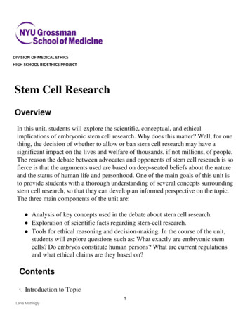
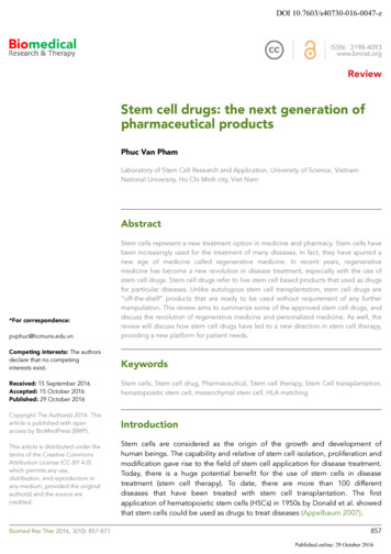
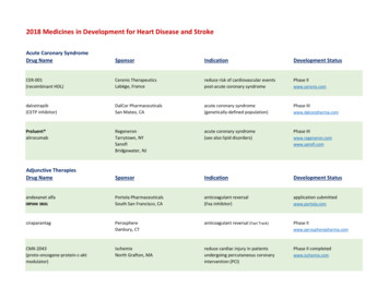
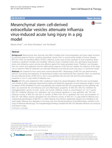
Khatri et al. Stem Cell Research & Therapy (2018) 9:17titration in MDCK cells as described [33]. Virus titerswere calculated by the Reed and Muench method.Detection of cytokines in lungsLung lysates from pigs were prepared and levels oftumor necrosis factor (TNF)α, CXCL10, and interleukin (IL)-10 in lung lysates were determined byenzyme-linked immunosorbent assay (ELISA) as described previously [8].Page 5 of 1337 ng/100 μl EVs (n 5) total RNA and 79 1 μg/100 μlEVs (n 4) total protein.Additionally, MSC-EVs showed the ability to incorporate into LECs. EVs stained red with PKH-26 dye werefound inside the cytoplasm of cells when examinedunder a fluorescent microscope. Incorporation of MSCEVs in LECs was also confirmed by flow cytometry(Fig. 3).MSC-EVs inhibit the HA activity of influenza virusesStatistical analysisIn vitro data on MSC-EV-mediated inhibition of SwIVreplication were analyzed by Student’s t test. Microscopic lung lesions, virus titers, and cytokine concentrations between groups of pigs were compared usingKruskal-Wallis test. P values 0.05 were consideredstatistically significant.ResultsCharacteristics of MSC-derived EVsWe isolated MSCs from the BM of 2- to 6-week-old pigsthat showed characteristic features of MSCs, such asadherence to a plastic surface, fibroblast-like morphology (Fig. 1), self-renewal potential, and high in vitroproliferation capacity and differentiation potential (datanot shown). Colony-expanded MSCs showed the expression of the mesenchymal markers CD29, CD44, CD90,and SLA-I, but SLA-II was not detected on these cells.MSC-EVs were round-shaped and, similarly to MSCs,EVs isolated from BM-MSCs also expressed mesenchymal markers; however, unlike MSCs, they lacked theexpression of both SLA-I and II (Fig. 1). MSC-EVs alsoexpressed EV-specific markers such as CD9, CD63, andCD81 (Fig. 2). We also determined the RNA and proteinconcentration in MSC-EVs. MSC-EVs contained 113 To determine if MSC-EVs possess anti-influenza activity,we first examined if EVs inhibited the HA activity of influenza viruses. Eight HA units of swine/TX/98; H3N2,swine/MN/08; H1N1, gull/MD/1995; H9N5, chicken/NY/H7N2, and human/CA/09; H1N1 influenza viruseswere incubated with different concentrations of MSCEVs and the HA activity of these viruses was examined.MSC-EV concentrations between 1.25 and 5 μg/ml completely inhibited the HA activity of these viruses(Table 1).MSC-EVs inhibit SwIV replication and virus-induced apoptosis in LECsWe examined MSC-EVs derived from BM-MSCs foranti-influenza activity. SwIV (Sw/MN/08; MOI of 1) wasincubated with MSC-EVs (10 μg/ml) for 20 min at roomtemperature. LECs were infected with EV-SwIV mixtureor virus only. At 8 h after infection, IAV-NP wasdetected by IFA (Fig. 4a and c). Significantly reduced (P 0.05) influenza replication was detected when cellswere infected with SwIV pretreated with MSC-EVs.Consistent with reduced influenza virus replication inLECs treated with MSC-EVs, MSC-EVs also significantly(P 0.05) inhibited the apoptosis of influenza-infectedLEC (Fig. 5a and b).Fig. 1 Characteristics of MSC-derived EVs. Extracellular vesicles (EVs) were isolated from the conditioned medium of porcine bone marrow-derivedmesenchymal stem cells (BM-MSCs) by ultracentrifugation. Morphology and size of MSC-EVs was examined by TEM. EVs were round-shaped andapproximately 100 nm in size ( 25 K). Expression of mesenchymal markers on MSCs and MSC-EVs was examined by flow cytometry. MSCs expressedthe mesenchymal markers CD29, CD44, and CD90, and swine leukocyte antigen (SLA)-I, but SLA-II was not expressed. Similarly to MSCs, EVs expressedthe mesenchymal markers but lacked the expression of SLA-I and SLA-II (black line: isotype staining; red line: specific staining)
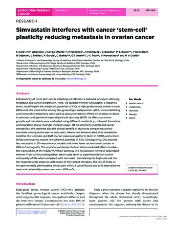
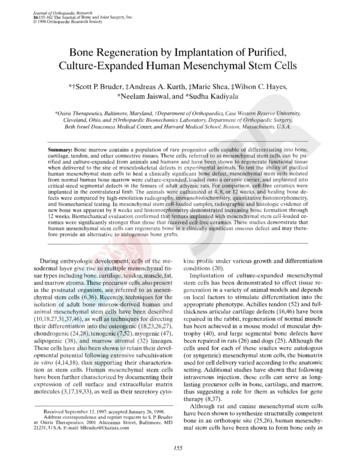
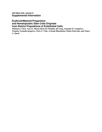
Khatri et al. Stem Cell Research & Therapy (2018) 9:17Page 6 of 13Fig. 2 Mesenchymal stem cell extracellular vesicles (MSC-EVs) express EV markers. EV-coated latex beads were examined for the expression of EVmarkers by flow cytometry. EVs expressed the specific EV markers CD9, CD63, and CD81 (broken line: isotype staining; solid line: specific staining)In another set of experiments, porcine lung epithelialLECs were infected with SwIV for 1 h, washed, and incubated with media without or with MSC-EVs (10 μg/ml).At 8 h after infection, IAV-NP was detected by IFA. Theaddition of MSC-EVs after virus entry significantly (P 0.05) reduced influenza replication in LECs (Fig. 6).MSC-EVs attenuate SwIV-induced ALI in pigsWe examined the anti-influenza and immunomodulatory effect of MSC-EVs in influenza virus-induced ALIin pigs. Eight-week-old pigs were infected intranasallywith 5 106 TCID50 of SwIV. Twelve hours after SwIVinoculation, DMEM or MSC-EVs (80 μg/kg) wereadministered intratracheally in influenza-infected pigs.At days 1 and 3 after EV administration, we collectedthe nasal swabs, and at day 3 after EV administration thepigs were euthanized and bronchoalveolar lavage (BAL)and lungs were collected. Lungs were homogenized tomake 10% lung lysate for determining virus titers, andcytokines were examined in the lung lysate.SwIV induced extensive lung lesions in infected pigsas determined by infiltration of inflammatory cells,thickened alveolar walls, and collapsed alveoli, whereaslungs of pigs administered with MSC-EVs showedminor infiltration of inflammatory cells. The meanmicroscopic lung lesion score in SwIV DMEM inoculated pigs was 7.3 1.5 compared with 2.6 1.5 in theSwIV EV administered group (Fig. 7a–d). ConsistentFig. 3 MSC-EVs incorporate into lung epithelial cells. PKH-26 labeled extracellular vesicles (EVs) were incubated with LECs for 24 h. Incorporationof EVs in LECs was examined by fluorescent microscope and flow cytometry. The inset in the middle panel shows the internalization of EVs in thecytoplasm of LECs. Cellular cytoplasm was stained using β-tubulin antibody (green); EVs (red) were found to be localized inside the LECs ( 200)
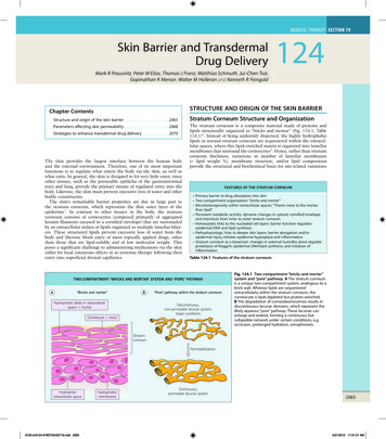
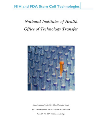
Khatri et al. Stem Cell Research & Therapy (2018) 9:17Page 7 of 13Table 1 MSC-EVs inhibit hemagglutination (HA) activity ofinfluenza virusesInfluenza virusHemagglutination at EV ; H3N2–– Human/CA/09; H1N1–––– Swine/MN/08; H1N1–– Gull/MD/1995; H9N5–– Chicken/NY/1995; H7N2–––– Eight HA units of swine, human, and avian origin influenza viruses wereincubated with different concentrations of mesenchymal stem cellextracellular vesicles (MSC-EVs) or medium control at room temperature for20 min; 50 μl 1% turkey red blood cells were then added to the wells andMSC-EV-mediated inhibition of HA by influenza viruses was examined. Resultsare representative of three different experiments using EVs derived from MSCsisolated from three different pigswith the inflammatory lesions in lungs, the levels oftotal protein in the BAL of SwIV EV (174 20 μg/ml)administered pigs were also lower than the levels inSwIV DMEM (270 34 μg/ml) inoculated pigs(Fig. 7e).We detected the influenza virus shedding in nasalswabs at 1 and 3 days after EV administration. Virusshedding was 100-fold lower in the EV-administeredgroup at 3 days post-EV administration as comparedwith SwIV DMEM inoculated pigs (Fig. 8). Similarly,virus titers were also 100-fold lower in the lung lysateof EV-administered pigs. These data suggest thatMSC-EVs inhibit influenza virus replication and shedding in pigs.MSC-EVs attenuate SwIV-induced inflammatory cytokinesNext, we examined if MSC-EVs also modulated theinflammatory cytokine production in lungs. In theMSC-EV administered group, the levels of TNFα were251 46 pg/g lung lysate compared with 386 40 pg/gin pigs inoculated with SwIV DMEM. Similarly, levelsof CXCL10 in SwIV EV administered pigs were 3259 469 pg/g compared with 4456 495 pg/g in lunglysates of SwIV DMEM inoculated pigs. The levels ofthe anti-inflammatory cytokine IL-10 were slightlyhigher in the SwIV EV administered group comparedwith the SwIV DMEM inoculated pigs (Fig. 9). Thesefindings suggest that MSC-EVs possess anti-influenzaand anti-inflammatory properties and attenuated influenza virus-induced ALI in a pig model.Fig. 4 MSC-EVs inhibit influenza virus replication in lung epithelial cells. Swine/MN/08; H1N1 (SwIV; MOI 1) was incubated with Dulbecco’s modifiedEagle’s medium (DMEM) or 10 μg/ml MSC extracellular vesicles (EVs) for 20 min at room temperature. After the incubation, LECs were inoculated withvirus-EV mixture or virus alone and incubated for 1 h at 37 C. Influenza virus NP was detected at 8 h after infection (a) and SwIV-induced cytopathologywas observed at 48 h after infection (b). SwIV-infected cells expressing NP were counted at 8 h after infection. Each bar represents mean SD of virusinfected cells in five microscopic fields (20 ) (c). Virus titers in supernatants of SwIV-infected and MSC-EV-treated cells at 48 h after infection weredetermined by titration in MDCK cells. Data are expressed as mean SD from three independent experiments using EVs derived from BM-MSCs from threedifferent pigs (d). *P 0.05
Khatri et al. Stem Cell Research & Therapy (2018) 9:17Fig. 5 MSC-EVs inhibit influenza virus-induced apoptosis. a Swine/MN/08; H1N1 (SwIV; MOI 1) was incubated with Dulbecco’s modified Eagle’s medium (DMEM) or 10 μg/ml MSC extracellular vesicles(EVs) for 20 min at room temperature. After the incubation, pig lungepithelial cells were incubated for 1 h with SwIV or SwIV preincubated with MSC-EVs. At 24 h after infection, apoptotic cells weredetected by TUNEL assay using the ApopTag Fluorescein ApoptosisDetection Kit (EMD Millipore). b TUNEL-positive cells in SwIV-infectedand MSC-EV-treated LECs were counted at 24 h after infection.Values are expressed as mean SD of apoptotic cells in fivemicroscopic fields (20 ). *P 0.05DiscussionIn this study, we isolated porcine BM-MSC-derived EVsand examined their anti-viral and immunomodulatoryproperties in a pig model of influenza virus. MSC-EVswere round-shaped and, similarly to BM-MSCs,expressed mesenchymal markers but, unlike MSC, EVslacked the expression of both SLA-I and II whereasMSCs expressed SLA-I. Importantly, MSC-EVs inhibitedthe HA activity of influenza viruses, suppressed thereplication of influenza virus in lung epithelial cells, andinhibited influenza virus replication and proinflammatory cytokine production in the lungs of virusinfected pigs.We isolated and characterized EVs from porcine BMMSCs using protocols as described for human MSCderived EVs [23, 29, 30]. As for human MSC-EVs, wealso used TEM to determine the size and morphology ofMSC-EVs. Size distribution of human MSC-EVs was alsodetermined by nanotracking but this technique was notPage 8 of 13used in this study. Human MSC-derived EVs expressedmesenchymal markers and lacked the expression ofMHC molecules [36]. In our study, pig MSC-derivedEVs also expressed mesenchymal markers and theexpression of both SLA-I and II was not detected inEVs, suggesting that pig MSC-EVs are similar t
markers (CD9, CD63, and CD81) by flow cytometry as described previously [31]. EVs (30 μgin50μl PBS) were incubated with 10 μlof4-μm diameter aldehyde/sulfate latex beads for 15 min at room temperature followed by the addition of 1 ml PBS and incubation was further . Cells and beads were acquired by C6 flow cytometer (BD Accuri Cytometers .