Transcription
Jorirrial of Orthopedic Xe.vearch16155-162 The Journal of Bone and .Joint Surgery, Tnc01998 Orthopaedic Research SocictyBone Regeneration by Implantation of Purified,Culture-Expanded Human Mesenchymal Stem Cells*?Scott P. Bruder, Andreas A. Kurth, Marie Shea, Wilson C. Hayes,*Neelam Jaiswal, and *Sudha Kadiyala*Osiris Therapeutics, Baltimore, Maryland, f Department of Orthopaedics, Case Western Reserve University,Cleveland, Ohio, and f Orthnpaedic Biomec-hanics Labarafory. Department of Urdiopaedic Surgery,Beth Israel Deaconess Medical Centel; arid Harvard Medical School, Boslon, Massachusetts, U.S.A.Summary: Bone marrow contains a population of rare progenitor cells capable of differentiating into bone,cartilage. tendon, and other connective tissues. These cells. referred to as mesenchymal stem cells, can be purified and culture-expanded from animals and humans and have been shown to regencrate functional tissuewhen delivered to the site of musculoskeletal defects in experimental animal . To test the ability of purifiedhuman mesenchymal stem cells to heal a clinically significant bone defect, mesenchymal stem cells isolatedfrom normal human bone marrow were culture-expanded, loaded onto a ceramic carrier, and implanted intocritical-sized segmental defects in the femurs of adult athymic rats. For comparison, cell-free ceramics wereimplanted in the contralateral limb. The animals were euthanized at 4,8, or 12 weeks, and healing bone defects were compared by high-resolution radiography, immunoliistochemistry. quantitative histomorphometry.and biomechanical testing. In mesenchymal stem cell-loaded samples, radiographic and histologic evidence ofnew bone was apparent by 8 weeks and histoniorphometry demonstrated increasing bone formation through12 weeks. Biomechanical evaluation confirmed that femurs implanted with mesenchymal stem cell-loaded ceramics were significantly stronger than those that received cell-lree ceramics. These studies demonstrate thathuman mesenchymal stern cells can regenerate bone in a clinically significant osscous defect and may therefore provide an alternative to autogcnous bone grafts.During embryologic development, cells of the rnesodermal layer give rise to multiple mesenchymal tissue types including bone, cartilage, tendon, muscle, fat,and marrow stroma. These precursor cells, also presentin the postnatal organism, are referred to as mesenchymal stem cells (6,36). Recently, techniques for theisolation of adult bone marrow-derived human andanimal mesenchymal stem cells have been described(10,18,27,31,37,46),as well as techniques for directingtheir differentiation into the osteogenic (18,23,26,27),chondrogenic (24,28),tenogcnic (7,52),myogenic (47),adipogenic (38), and marrow stromal (32) lineages.These cells have also been shown to retain their developmental potential following extensive subcultivationin vitro (4,14,18), thus supporting their charactcrixation as stem cells. Human mesenchymal stem cellshave been further characterized by documenting theirexpression of cell surface and extracellular matrixmolecules (3,17,19,33),as well as their secretory cytoReceived Septembcr 12,1YY7; accepted January 26,1998.Address correspondence and reprint requcsts to S . I? Bruderat Osiris Therapeulics, 2001 Aliceanna Street. Baltimore, MD21231.IJ.S.A. E-mail: SBruder@Osiristx.com155kine profile under various growth and differentiationconditions (20).Implantation of cul ture-expanded mesenchymalstem cells has been demonstrated to effect tissue regeneration in a variety of animal models and dependson local factors to stimulate diffcrcntiation into theappropriate phenotype. Achilles tendon (52) and fullthickness articular cartilage defects (16,46) have beenrepaired in the rabbit, regeneration of normal musclehas been achieved in a mouse model of muscular dystrophy (40), and large segmental bone defects havebeen rcpaired in rats (26) and dogs (25). Although thecells used for each of these studies were autologous(or syngeneic) mesenchymal stem cells, the biomatrixused for cell delivery varied according to the anatomicsetting. Additional studies have shown that followingintravenous injection. these cells can serve as longlasting precursor cells in bone, cartilage, and marrow,thus suggesting a role for them as vehicles for genetherapy (8,37).Although rat and canine mesenchymal stern cellshave been shown to synthesize structurally competentbone in an orthotopic site (25,26), human mesenchyma1 stcm cells have been shown to form bone only zn
S. P. B R U D E R E T AL.156vitro (4,23) and in ectopic implantation sites in immunodeficient mice (18). Because fracture healing andbonc repair depend on the ability to amass enoughcells at the defect site to form a repair blastema. onetherapeutic strategy is to directly administer the precursor cells to the site in need of repair. This approachis particularly attractive in the treatment of patientswho have fractures that are difficult to heal or whohave a decline in their mesenchymal stem ccll rcpository as a result of age (22,28,39,45):osteoporosis(43), or other metabolic derangement. The goal of thecurrent study was to determine whether purified,culture-expanded human mesenchymal stem cellscould regenerate bone at the site of a clinically significant osseous defect.MATERIALS AND METHODSCultivation and Manipulation ofHuman Mesenchymal Stem CellsIsolation and culture-expansion of human mesenchymal stemcells from a bone inarrow aspirate, obtained from a normal volunteer after informed consent. was conducted as previously described(15,20). Following the initial plating in Dulbecco's modified Eaglenicdiuni (Sigma Chemical, St. Louis, MU. U.S.A.) containing 10%fctal bovine serum (BioCell Laboi-atorics. Kancho Dominguez.CA, LJ.S.A.) from a sclcctcd lot (31). nonadhcrcnt cells were removed o n day 3 at the first changc of medium, and fresh mediumwas replaced twice wcckly thereafter. Adhcrcnt niesenchynialstem cells represent approxiniatcly oiic in lo5 nuclcated cells originally plated. When culture dishes becamc nearly confluent, thecells were detached and serially subcultured.Preparation of ImplantsPorous hydroxyapatitcip-tricalcium phosphate ccraniic blocks.with a mean porc size of 200-450 pm (generously supplied byZimmer. Warsaw, IN. U.S.A.), were shaped into cylinders approxiniatcly 4 mni in diameter and S inn1 in length with a 1 mincentral canal or were cut into cubes 3 mni per side. Mesenchymalstcm cell-loaded implants werc preparcd by incubating humanfihroncctin-coated hydroxyapatite/p-tricalcium phosphate cubesand cylinders in a 7.5 X lo6 cellinil suspension of first-passagemcscnchymal stem cells for 2 hours at 37 C. following the application of a brief vacuum to draw the cell suspension into the poresof the sample. as previously described (26). Uniform adherence ofcells deep within the pores was confirmed by scanning electronmicrography of representative samples, as published elsewhere(10). Ccll-free implants were prepared siniilarly by incubatingfibroncctin-coated cylinders in a suspension devoid of cells.Surgical Model and Experimental DesignThe fcmoral gap surgical model eiiiployed liere has been usedextensively in euthymic rats to study long-bone repair (11.13,26.30.35.42,44,18,49.5 I ). Briefly, both femurs of Harlan Nude(1Tsd:Rh-mu) rats (325 g) were exposed by an anterolateral approach. Athymic rats were used because they have an impairedimmune response against xcnogcncic implants. A polyethylcncfixation plate was attached to each fcinur by four Kirschner wiresand two cerclage wircs, and an 8 inm transverse segment of thccentral diaphysis. along with its adhcrcnt periosteum, was removedwith use of a rotary osteotomy burr undcr saline irrigation. Eachanimal then received a cell-frcc hydroxyapatiteiB-tricalciuniphosphate cylinder in one fcmoral defect. an identical cylinder loadedwith human mesenchymal stem cells in the contralateral defect,.I Orthop Res, Vol. 16. No. 2, 1998and a mesenchymal slem cell-loaded hydroxyapatitei -tricalciumphosphate cube in the subcutancous tissue of the back. The muscles wcrc apposed. and the fascia and skin wcrc closed in i routinelayei-ed fashion. Inimcdiately after cutlianasia at 4,X,or 12 wccks,all specimens were radiographed in a lateral position with use ofa high-resolution Faxitron Imaging system (Buffalo Grove, IL,U.S.A.) with an cxposure of 35 kVp for 30 scconds. At each of thethree time points, thc femurs froiii thrcc of the animals wcre usedfor quantitative Iiistol iorphoiiietryand saniplcs from one animalwere used for iniinunochemisti-y.'lhc fcmurs from the remainingseven animals wcrc harvested at thc 12-wcck time point for biomechaiiical testing and were subsequently pooled with thc thrcesamples originally destined for quantitative histoniorphomctry. Allsubcutaneous implants were processed for decalcified histology. Inaddition to the 19 animals that received implants of hydroxyapatitcip-tricalcium phosphate. three additional animals had bilateralfcmoral defects that were left empty to confirm the critical-siLediiaturc of this defect in atliymic rats. These three animals werekilled at 12 wccks, and the femurs wcrc proccssed for undecalcifiedhistology aftcr radiographic imaging. All procedures involving animals were pcrfoniicd in accordance with a protocol approved bythe liistitutiotial Animal Care and Use Committee.Quantitative Histomorphometry and ImmunochemistryFor quantitative histomorphometry, the limbs were fixed, dehydrated. cleared, embedded in polyinethylniethacrylate, cut. andground to a thickness of 100 pin for staining with Toluidinc blue-0.Quantitative assessnicnt of bone formation was pcrfornicd withuse of Quantimct 500MC (Leica Cambridgc. Cambridge, England)image-analysis software as previously described (26). Specifically,analysis was performed on the area of each scction correspondingto thc ncw cortical rcgions defined by thc dimcnsions of the original implant. Arcas of bone, ceramic, and soft tiscuc were easilydistinguished by the image-analysis software on the basis of thedifference i n color. At the magnifications used, approximately 10separate fields were required to encompass the entirety of eachlongitudinal section. The data are presented as the lractional areaof bonc as a percentage of available porc area, in keeping with thehistorical precedent for histomorphometry in this type ol orthopacdic implant (21.25.42,49).Liinbs from one animal at each tiinc point were also preparedfor routine immunostaining by monoclonal antibody 6E2. whichdistinguishcs human cells froni rat cells (18). Undecalcified cryosections were incubated with 6E2 supernatant or an irrclcvantprimary inonoclonal antibody control (SB-1) (1).followed by fluorescein isothiocyanatc-conjugated goat anti-niousc IgCi secondaryantibody (Gr tcoBKL. Gaithersburg, MD, U.S.A.) diluted 1:5Win phosphate-buffered saline. Subcutaneously iinplanted sampleswere fixed in formalin, decalcified, embedded in paraffin, scctioned, stained with Malloi-y-Heidcnliaiii dyc, and evaluated forthe presence of bonc in the pores of the implant.Biomechanical Testing' h e l v e weeks aftcr implantation. seven animals that had ccllfree and niesenchynial stem cell-loaded implants and six controlanimals that did not undergo the procedure were euthanizcd fortorsion testing of femurs as described in technical detail elsewhere(30).The lixation plate and adherent soft tissue were removed, andthe metaphyses of the bones were embedded. The specimens wereexternally rotated in a custom torsion test apparatus at 6" persecond until failure was observed. The displacement and load tofailure were recorded. and the values lor stiffness and strengthwere calculated with use of custom software.Statistical AnalysisTo analyze the histomor-phometric data, the mesenchymal stemcell-loaded and the cell-free samples Crom each animal were paired.
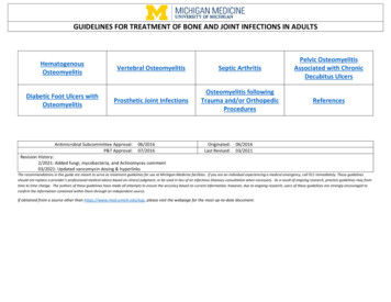
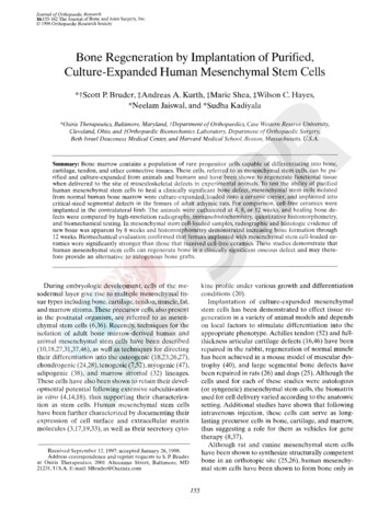
H U M A N M E S E N C H Y M A L S T E M CE LLS R E G E N E R A T E RONE157of the 8 mm diaphyseal dclect. Although a mild, naturally occurring varus deformity was sccn in theseathymic rats. no gross failure of fixation or otherpostoperative complications occurred throughoutthe course of study. However, compromised fixationat euthanasia was found in one animal implantedFIG. 1. Light micrograph of a representative histological sectionfrom a human mesenchynial stem cell-loaded hpdroxyapatiteiatricalciiim phosphate implant placed ectopically in the subcutancous tissue of an athyinic rat. The ceramic phase o f the implant (c),which was rcmoved during the decalcification step of histologicprocessing, can he seen as a granular matcrial. l'hc pores of thcimplant are filled with bone (b) 01.blood vessels and fibrous tissue(arrow). C:uboidal osteoblasts are seen lining the surface of thedeveloping bone. (Mallory-Heidenhain, X75.)and the data were analyzed by a paired r test at each of the threedifferent time points. Because the biomechanical analysis requiredcomparison of three different treatment groups (mesenchymalstein cell-loaded hydroxyapatite/ -tricalciuiii phosphate implants,cell-free hydroxyapatiteia-tricalciurn phosphate implants, and intact controls), one-way analysis of variance with p o s t hoc StudentNewman-Keuls tests were used to compare the values or torque.stiffness, and energy to failure for each group.RESULTSMesenchynial Stem Cell-Mediated Osteogenesisin Ectopic Hydroxyapatitelp-TricalciumPhosphate ImplantsHuman mesenchymal stem cell-loaded hydroxyapatite/p-tricalcium phosphate cubes implanted in thesubcutaneous space of athymic rats displayed evidence of osteogenesis by 4 weeks, but considerablymore bone was present within the pores at 8 arid 12weeks. A representative section from a mesenchymalstem cell-loaded cube harvested 12 weeks followingimplantation is shown in Fig. 1. Bone formation occurswithin the pores of the cubes and is associated withvascular elements that penetrate the implant. Suchangiogenesis is obligatory t o new bone formation,beca
Cultivation and Manipulation of Human Mesenchymal Stem Cells Isolation and culture-expansion of human mesenchymal stem cells from a bone inarrow aspirate, obtained from a normal volun- teer after informed consent. was conducted as previously described (15,20). Following the initial plating in Dulbecco's modified Eagle nicdiuni (Sigma Chemical, St. Louis, MU. U.S.A.) containing 10% fctal