Transcription
Endocrine-RelatedCancerS Kato et al.25:10Statins reduce metastasis inovarian cancer821–836RESEARCHSimvastatin interferes with cancer ‘stem-cell’plasticity reducing metastasis in ovarian cancerS Kato1, M F Liberona1, J Cerda-Infante2,3, M Sánchez2, J Henríquez2, C Bizama4, M L Bravo5,6, P Gonzalez5,R Gejman4, J Brañes1, K García1, C Ibañez2,6, G I Owen5,6, J C Roa4,6, V Montecinos2 and M A Cuello11Divisionof Obstetrics and Gynecology, Faculty of Medicine, Pontificia Universidad Católica de Chile (PUC), Santiago, Chileof Hematology and Oncology, Faculty of Medicine, PUC, Santiago, Chile3Department of Cellular and Molecular, Faculty of Biological Sciences, PUC, Santiago, Chile4Department of Pathology, Faculty of Medicine, PUC, Santiago, Chile5Department of Physiological Sciences, Faculty of Biological Sciences, PUC, Santiago, Chile6Millennium Institute on Immunology and Immunotherapy, PUC, Santiago, Chile2DepartmentCorrespondence should be addressed to M A Cuello: macuello@med.puc.clAbstractCell plasticity of ‘stem-like’ cancer-initiating cells (CICs) is a hallmark of cancer, allowingmetastasis and cancer progression. Here, we studied whether simvastatin, a lipophilicstatin, could impair the metastatic potential of CICs in high-grade serous ovarian cancer(HGS-ovC), the most lethal among the gynecologic malignancies. qPCR, immunoblottingand immunohistochemistry were used to assess simvastatin effects on proteins involvedin stemness and epithelial-mesenchymal cell plasticity (EMT). Its effects on tumorgrowth and metastasis were evaluated using different models (e.g., spheroid formationand migration assays, matrigel invasion assays, 3D-mesomimetic models and cancerxenografts). We explored also the clinical benefit of statins by comparing survivaloutcomes among statin users vs non-users. Herein, we demonstrated that simvastatinmodifies the stemness and EMT marker expression patterns (both in mRNA and proteinlevels) and severely impairs the spheroid assembly of CICs. Consequently, CICs becomeless metastatic in 3D-mesomimetic models and show fewer ascites/tumor burden inHGS-ovC xenografts. The principal mechanism behind statin-mediated effects involvesthe inactivation of the Hippo/YAP/RhoA pathway in a mevalonate synthesis-dependentmanner. From a clinical perspective, statin users seem to experience better survivaland quality of life when compared with non-users. Considering the high cost and thelow response rates obtained with many of the current therapies, the use of orally orintraperitoneally administered simvastatin offers a cost/effective and safe alternative totreat and potentially prevent recurrent HGS-ovCs.Key Wordsff ovarian cancerff metabolismff stemnessff therapyff statinsEndocrine-Related Cancer(2018) 25, 821–836IntroductionHigh-grade serous ovarian cancer (HGS-ovC) remainsthe deadliest gynecological cancer worldwide. Despiteachieving complete response, most patients will recur anddie from their disease. Unfortunately, less than 40% ofpatients will exceed 10-year survival (Baldwin et al. 2012).http://erc.endocrinology-journals.org https://doi.org/10.1530/ERC-18-0132 2018 Society for EndocrinologyPublished by Bioscientifica Ltd.Printed in Great BritainSuch a poor outcome is mainly explained by the latediagnosis when the disease has already disseminatedthroughout the whole abdominal cavity. Accordingly,most patients will first present with ascites andcarcinomatosis. For surgeons, reducing the disease to itsDownloaded from Bioscientifica.com at 07/14/2022 08:45:29PMvia free access
Endocrine-RelatedCancerS Kato et al.microscopic burden is considered a success. However, sucha load still presents numerous single or aggregated cancercells. By then, some cancer cells have already adapted,established cooperative metabolic cell relationships andexhibit properties that confer a survival advantage (Blatter& Rottenberg 2015).A key cellular phenotype during the metastatic wave,responsible for drug resistance and later recurrence, isthe so-called cancer-initiating cell (CIC) subpopulation(Blatter & Rottenberg 2015). These cells exhibit stemcell-like properties and have experienced epithelialmesenchymal transition or epithelial-mesenchymal cellplasticity (EMT). They grow slowly, are more resistantto apoptotic and cytotoxic stimuli and can reformulatea complete tumor in xenograft models (Yan et al. 2014).These cells are part of floating aggregates present in ascites(Yeung et al. 2015). Cell aggregates offer enhanced survivalwhile in transit to their metastatic niche, better resistanceto environmental menaces and increased attachment andfaster formation of metastatic foci compared to singlefloating cells (Yeung et al. 2015). Interestingly, in vitro,the expression of CIC stemness and EMT markers tend toincrease when CICs are exposed to chemotherapy pulses(Chisholm et al. 2015). They also tend to adopt a spheroidconformation and enhanced resistance, mimicking theaggregates observed in ascites (Shield et al. 2009). At thetime of recurrence, many cancer cells present the CICphenotype because of the chemotherapy-selecting effect.This phenomenon explains the lower response rates(less than 30%) to most of the consecutive treatments(Fuh et al. 2015).Changes in TP53 function are critical events forHGS-ovC progression (Iwanicki et al. 2016). In fact, TP53is mutated (mut-TP53) in nearly all HGS-ovCs (96%),predominantly by missense mutations (Network CGAR2011). Besides its classical tumor-suppressive functions,TP53 regulates metabolic pathways. Additionally, newevidence suggests that mut-TP53 is associated with cancermetastasis (Hu et al. 2013). Along with changes in TP53functions, HGS-ovCs seem to acquire a mevalonatepathway gene signature that prompts tumor growth andmetastasis (Hu et al. 2013, Greenaway et al. 2016). Thissignature is a consequence of metabolic re-programming,an evolutionary adaptation to satisfy the demand of lipidarising from fast dividing and moving HGS-ovC cells(Baenke et al. 2013).The mevalonate pathway facilitates multiple metaboliccell functions (Kobayashi et al. 2015). Intermediateproducts of this pathway include sterol isoprenoids suchas farnesyl pyrophosphate (FPP) and g https://doi.org/10.1530/ERC-18-0132 2018 Society for EndocrinologyPublished by Bioscientifica Ltd.Printed in Great BritainStatins reduce metastasis inovarian cancer25:10822pyrophosphate (GPP), responsible for prenylation,membrane localization and activation of different smallGTPases that regulate important cell functions, and manyare also well-established oncogenes (Cheng et al. 2010).The rate-limiting enzyme in the mevalonate pathwayis the hydroxymethyl-glutaryl coenzyme A reductase(HMGCR) that converts HMG-CoA into mevalonic acid(Kato et al. 2010). Interestingly, some mut-TP53 formsinteract with SREBP transcription factors and upregulatemevalonate genes, including HMGCR (Freed-Pastor et al.2012). Consistently, we showed that HGS-ovCs exhibithigher HMGCR levels compared to benign tissues(Kato et al. 2010).Simvastatin is a lipophilic statin that inhibitsHMGCR activity, thereby preventing mevalonate andcholesterol synthesis and blocking protein prenylation bydownstream depletion of FPP and GGP (Berndt et al. 2011).Treating mut-TP53 HGS-ovC cell lines with increasingconcentrations of statins induces autophagy, cell deathand drug sensitization in a mevalonate-dependent manner(Kato et al. 2010). Supporting a potential therapeutic roleof statins in ovarian cancer, two recent studies showeda decrease in hazards ratio of disease-specific death andlonger survival among statin users compared to non-users(Khan et al. 2015, Couttenier et al. 2017).Statin research in ovarian cancer has been limited totest its effects in vitro or in vivo models. To our knowledge,no publication addressed its effect on the CIC-derivedHGS-ovC subpopulation responsible for metastasisand recurrence. Thus, we investigated the effects ofsimvastatin on this subpopulation using different models.Herein, we identified new mechanisms by which statinscan reduce or prevent CIC-mediated progression and thusdisease recurrence.Materials and methodsReagentsSimvastatin was from EMD Millipore Corp. Carboplatin,etoposide, docetaxel, cisplatin, leptin, mevalonate, andgeranylgeranyl pyrophosphate (GGPP) were from SigmaAldrich, mafosfamide from Santa Cruz Biotechnology andpaclitaxel from Kampar Laboratories (Santiago, Chile).Cell lines and spheroidsHuman ovarian cancer cell lines, HeyA8 (harboring TP53P72R polymorphism, RRID: CVCL 8878), UCI101 (TP53 WT,high p-glycoprotein and high EGFR, RRID: CVCL 5380) andDownloaded from Bioscientifica.com at 07/14/2022 08:45:29PMvia free access
Endocrine-RelatedCancerS Kato et al.SKOV3 (TP53 null, RRID: CVCL 0532) were maintainedin RPMI 1640 medium. In particular, HeyA8 and UCI101cell lines were used for most of the experiments based ontheir capabilities of giving origin to tumors resemblingHGS-ovC behavior in mouse models (Buick et al. 1985,Fuchtner et al. 1993, Domcke et al. 2013, Mitra et al. 2015,Hernandez et al. 2016, Tanenbaum et al. 2017). Cell lines,once acquired, were periodically authenticated usingthe Genemarker 10 kit (Promega), routinely screenedfor mycoplasma infection by PCR and discard beforeachieving 30 passages. CIC-enriched spheroids wereisolated as previously described (Kato et al. 2015). Togrant purity and to confirm that effects were effectivelyon stem-like cells, we also established CIC-only derivedspheroids by combining limit dilution (single cell startingpoint) and stem-selecting media. To characterize CICs andassess statin effects, we studied two of the most probablestem-like cell markers (CD44 and ALDH1) described forovarian cancer (Klemba et al. 2018).Tissue culture, patient data and systemicimmune-inflammatory indexThis study was reviewed and approved by the institutionalreview board of PUC. All patients enrolled in this studysigned an informed consent before the beginning of thisstudy. Ascites were collected, and clinical records frompatients who signed informed consent (IRB-approvedprotocol) were reviewed. Ascites-derived cultureswere established as previously described (Kato et al.2010). Mesothelial cells (calretinin( )/claudin-4( ))were isolated by differential trypsinization. Systemicimmune-inflammatory index (SII) was calculated atthe time of recurrence as described elsewhere (Hu et al.2014). Briefly, the formula to calculate the score wasSII platelet * neutrophil/lymphocyte.Statins reduce metastasis inovarian cancer25:10823Quantitative PCRTotal RNA was extracted using TRIzol (Life Technologies).cDNA was generated from 1 µg RNA using iScript ReverseTranscription Supermix for RT-qPCR (Bio-Rad). HMGCR,CD44, POU5F1, NANOG, SOX2, ZEB2, CDH2 and CDH1mRNA levels were assessed by qPCR and normalized tothat of the porphobilinogen deaminase gene. Primersused in this study are listed in Supplementary Table 1 (seesection on supplementary data given at the end of thisarticle). The qPCR reactions were performed using SYBRGreen Master Mix (Applied Biosystems) in an ECO qPCRSystem (Illumina San Diego, CA, USA). Relative amountsof mRNAs were calculated using the 2 ct method.Immunoblotting (WB) and RhoA pull-down assayProtein pellets and electrophoresis were carried outas previously described (Kato et al. 2015). Overnightprimary antibody exposure was performed by usingantibodies against PARP and CD44, TWIST (SantaCruz Biotechnology), ZEB2, SOX2, E-cadherin, TAZ,YAP, phospho-YAP (Cell Signaling), OCT4, NANOG,ALDH1A1, and N-cadherin (Abcam), ACTB (β-actin)(Sigma-Aldrich) or TUBB (β-tubulin) (Thermo-Fisher).Peroxidase-conjugated goat anti-mouse/rabbit/goat IgGswere used as secondary antibodies (Bio-Rad). The reactionwas developed with chemiluminescence using WesternLightning ECL Pro (Perkin-Elmer) and the signal wasdetected by Imagequant LAS 500 system (GE HealthcareBio-Science AB). Densitometry analysis was made usingAdobe Photoshop Software Tools and protein levels werenormalized against β-actin protein levels.RhoA pull-down assays were performed using theRho activation assay kit according to manufacturer’sinstructions (Cell Biolabs, San Diego, CA, USA).Immunocytochemistry and immunofluorescenceXenograft mouse modelPathogen-free NOD-Cg-Prkdcscid-IL2rgtm1Wjl/Szj (NSG) micewere obtained from Jackson Laboratories. Six- to eightweek-old mice were used for experiments. All IRB-approvedexperiments were conducted under Conicyt-Chile andARRIVE/AMVA guidelines. For xenograft implantation,non-selected or CIC-isolated HeyA8 cells were injectedintraperitoneally (IP). Tumor formation was monitoreddaily by palpation and measurement of the abdominaldiameter. After 7 days of tumor growth, we treated themice (n 4 per group, three consecutive cohorts) withplacebo or simvastatin (2 mg/kg/day via IP) for 4 weeks.http://erc.endocrinology-journals.org https://doi.org/10.1530/ERC-18-0132 2018 Society for EndocrinologyPublished by Bioscientifica Ltd.Printed in Great BritainImmunocytochemistry (IC) was carried out as previouslydescribed (Kato et al. 2010). Primary antibodies, CD44(Santa Cruz Biotechnology), ALDH1A1 (Abcam), YAPand phospho-YAP (Cell Signaling) and TP53 (Dako) werediluted following the manufacturer’s instructions. Forthree-dimensional IC (3D-IC), spheroids were collectedand spread on Superfrost Plus microscope slides (ThermoScientific), fixed with methanol and treated as described for2D-IC (P-YAP, CD44, ALDH1A1). The immunofluorescence(IF) protocol has been described elsewhere (Kato et al.2015). Each picture consisted of z-series of 1024-pixelimages. Merge analysis was performed using ImageJ.Downloaded from Bioscientifica.com at 07/14/2022 08:45:29PMvia free access
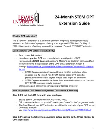
Endocrine-RelatedCancerS Kato et al.ImmunohistochemistryParaffintissue-sections(5 µmthickness)fromxenografts and HGS-ovC specimens were prepared forimmunohistochemistry (IHC) as described previously(Ohyagi-Hara et al. 2013). PAX8 (Roche), WT1 (Dako),TP53, KI67, CD44, ALDH1A1, YAP and p-YAP stainingwere quantified by using the Aperio Imaging AnalysisToolbox, under Aperio nuclear algorithm. Spheroids wereembedded in HistoGel and included in paraffin blocks.Five micrometer paraffin sections were prepared to carryout the IHC for TP53. Nuclei staining was quantified usingAdobe Photoshop counting tools and Aperio ImagingAnalysis Toolbox, under Aperio nuclear algorithm. Todetect the vasculature, a double staining was performedby using huCD34, huCD31 and msCd31 antibodies andthe EnVision G/2 Doublestain System (Dako), followingthe manufacturer’s instructions. The quantification ofthe vascular area was performed by using image colordeconvolution and particle analysis with ImageJ.Statins reduce metastasis inovarian cancer25:10824was calculated individually for each sphere using thefollowing formula ((the area at 24 h area at time 0)/areaat time 0) 100. The graphs presented reflect the averagescore of at least ten replicates for each condition in threedifferent experiments.3D organotypic mesomimetic assayThe assay was performed as described previously, withminimal modifications (Kenny et al. 2009). For invasionassays, six-well plates containing 8 µm pore size insertswere coated with a mix of Hs832 fibroblasts (ATCC),collagen I, and medium and incubated at 37 C for 4 h.Mesothelial cells isolated from primary cultures werecultured on top of the fibroblast layer and incubated at37 C for 18 h. Spheroids were then seeded on top of themesothelial layer. DMEM/F12 10% FBS was used as thechemoattractant stimulus.Statistical analysesMTS-based cell proliferation assayCancer cells from primary cultures were exposed for 48 hto different chemotherapeutic reagents. Cell viability wasassessed as described previously (Kato et al. 2010).Matrigel invasion assayTranswell inserts containing 8 μm isopore membranes(Nunc) were coated with Corning Matrigel Matrix GrowthFactor Reduced (GFR) diluted 1:10, as described elsewhere(Kato et al. 2015). Equal numbers of CICs suspended inthe stem-selecting medium were spread over and treatedas indicated for 48 h. The number of invading cells wasdetermined by IC for pan-cytokeratin (Sigma-Aldrich). Toquantify the number of cells trans-passing the isopores, theinserts were examined under a microscope (100 , Nikon)and 15 fields were counted per experiment.Spheroid migration assayFlat bottom 96-well plates were coated with 0.3% CorningMatrigel Matrix GFR for 30 min at 37 C. Spheroidsestablished from a single ‘stem-like’ cell (confirmed bystemness and EMT marker expression) were treated withsimvastatin 1 µM for 24 h and then transferred to thecoated flat bottom plates. Effects of simvastatin weremeasured at 24 h, and the images obtained by opticalmicroscopy were analyzed by ImageJ. A migration scorehttp://erc.endocrinology-journals.org https://doi.org/10.1530/ERC-18-0132 2018 Society for EndocrinologyPublished by Bioscientifica Ltd.Printed in Great BritainJMP13 (SAS) was used for data analyses. Statisticalsignificance (P values 0.05) was calculated by Student’st-test, Chi-squared test or Mann–Whitney. Survivalcurves were generated using the Kaplan–Meier methodand analyzed by Wilcoxon test. Cox proportional hazardregression model was carried out to assess the effect ofdifferent risk factors on survival.ResultsCancer cell line-derived spheroids exhibit amorphology and stemness/EMT expressionpattern resembling that in ascites aggregatesfrom HGS-ovCsAs observed in Fig. 1A, ovarian cancer aggregates collectedfrom ascites from HGS-ovC cases exhibit a similarmorphology compared with established ovarian cancercell lines (HeyA8 and UCI 101) when cultured undernormal (in 2D) or low-attachment conditions (in 3D).Under 2D conditions, all of them grew as attached cellgroups until reaching full confluency. In contrast, underlow-attachment, all formed 3D structures or spheroidssimilar in size, but different in cell density (as confirmedwhen visualized with magnification, identifying singlecell limits and measuring cell membrane overlapping),mimicking the aggregates found in ascites.Then,wetestedwhetherspheroidsfromcancer cell lines represented an enriched CIC orDownloaded from Bioscientifica.com at 07/14/2022 08:45:29PMvia free access
Endocrine-RelatedCancerS Kato et al.Statins reduce metastasis inovarian cancer25:10825Figure 1Identification, isolation and characterization of cancer-initiating cells (CICs) from HGS-ovC cell lines and primary tissue cultures both in vitro and in vivo.(A) Representative microphotography of morphologic aspects adopted by two ovarian cancer cell lines (HeyA8 (named as HEY in the figure) and UCI101) and a primary tissue culture (established from ascites from a HGS-ovC) when growing under normal (2D conditions) or low-attachment conditions(3D conditions) (40 ). (B and C) Relative mRNA and protein expression levels of stemness (NANOG, OCT4 and CD44) and EMT (N-cadherin, E-cadherin,TWIST and ZEB2) markers in HeyA8 cells growing under 2D and 3D conditions and measured by qPCR and WB. Graphs represent the results of threeindependent experiments. ACTB is shown as a loading control for WB. The bar graph at the right of blots shows the densitometry analyses. The asteriskindicates statistical significance (P 0.05, Mann–Whitney test). The vertical bars indicate s.e.m. (D) The TP53 expression level in HeyA8 cells growingunder 2D or 3D conditions and detected by IC. (E) Representative photographs of the whole abdominal cavities (upper panels) and segments of gut andmesentery of NSG mice 4 weeks after IP inoculation (1 106 cells) with either MOCK, non-selected, or HeyA8-derived CICs. The green arrows highlightmetastatic foci highly vascularized either at the omentum or the mesentery. (F) Representative microphotographs of TP53, PAX8 and WT1 pattern andintensity staining as found in HGS-ovC (positive control) and in metastasis generated in the omentum by HeyA8-derived CIC intraperitoneal injection inthe mouse model. The black bar in the microphotograph indicates 200 µm.http://erc.endocrinology-journals.org https://doi.org/10.1530/ERC-18-0132 2018 Society for EndocrinologyPublished by Bioscientifica Ltd.Printed in Great BritainDownloaded from Bioscientifica.com at 07/14/2022 08:45:29PMvia free access
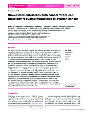
Endocrine-RelatedCancerS Kato et al.‘stem-like’ subpopulation. In Fig. 1B and C, we showthe findings in one of the cell lines tested, the HeyA8cell line. As shown there, cancer cells significantlymodified their stemness (e.g., CD44) and EMT(e.g., TWIST) expression patterns when forming spheroids,as measured by qPCR and WB. This pattern resembled thatdescribed by others as characteristic of CICs isolated fromHGS-ovCs (Shield et al. 2009). Additionally, we assessedTP53 expression. We hypothesized that spheroids alsopossessed a TP53( ) enriched population, favoring a prosurvival and metabolic adaptation within ascites (Hu et al.2013). Accordingly, TP53( ) cells were significantlyenriched in spheroids (Fig. 1D, left panel) as confirmedby quantification using Adobe Photoshop counting toolsand the Aperio Imaging Analysis Toolbox, under Aperionuclear algorithm (P 0.008, Fig. 1D, right panel).Cells forming spheroids generate tumor xenograftsmore rapidlyAs shown in Fig. 1E, both non-selected (cells growingin 2D conditions) and the CIC-enriched population(3D-spheroids) generated xenografts upon IP injectioninto NSG mice. However, the number of cells requiredto generate a tumor and the growth curve slope weresignificantly different. A lower number of CICs was requiredto generate a tumor compared with non-selected cells(5 104 vs 1 106 cells). Once tumors were identifiable, thegrowth curve slope was significantly steeper for xenograftsarising from CICs. Additionally, metastatic foci were morevascularized in tumors arising from CICs (as indicatedby green arrows in Fig. 1E). Supporting the resemblanceof the model with HGSOCs in humans, HeyA8-derivedCICs, injected intraperitoneally, disseminated in theabdominal cavity in a similar fashion in the mouse model(e.g. omentum, peritoneum, diaphragm, mesentery root,so on) and expressed markers commonly used to confirmthe serous histology and the tube-ovarian epithelialorigin (e.g. PAX8 and WT1, Fig. 1F) (Liliac et al. 2013,Di Palma et al. 2014, Mitra et al. 2015, Xiang et al. 2018).Simvastatin decreases CIC invasion and migration,induces spheroid disassembly and CIC cell deathStatins can induce cell death in vitro (Kato et al. 2010).Additionally, CICs from this malignancy are moreinvasive and resistant to commonly used chemotherapies(Kato et al. 2015). Therefore, we tested whetherone lipophilic statin, simvastatin, used at differentconcentrations (including those clinically achievable),http://erc.endocrinology-journals.org https://doi.org/10.1530/ERC-18-0132 2018 Society for EndocrinologyPublished by Bioscientifica Ltd.Printed in Great BritainStatins reduce metastasis inovarian cancer25:10826affected ovarian cancer spheroids. Varying concentrationsof simvastatin were added to the cultures either before orafter spheroid formation in single or repeated pulses.To mimic clinical settings, we used differentadministration schemes, always considering thesimvastatin achievable plasma concentrations (BjörkhemBergman et al. 2011, Lee et al. 2014). As shown in Fig. 2Aand Supplementary Fig. 1A, a single dose of simvastatin(up to 5 µM) did not affect morphology or cell deathin spheroids established from HeyA8 and UCI101 cellsor primary cultures (the chemo-naïve patient UC172).Although not significant, the number of spheroidsobtained from ascites tended to decrease upon simvastatinexposure and at higher concentrations. A significantdecrease in the number of migrating or invading cellswas detected upon a single pulse of simvastatin (1 µM) inmatrigel invasion assays, spheroid migration assays andmesomimetic 3D cultures in both HeyA8 (Fig. 2B, C andD) and UCI101 cells (Supplementary Fig. 1B and C) eitherusing isolated spheroids (CIC-enriched populations) orspheroids established from single stem-like cell (CIC-onlyderived spheroids). In contrast, upon repeated pulses,a significant spheroid disassembly was observed in aconcentration-dependent manner in both cell lines andprimary tissue cultures (Fig. 2E, F and SupplementaryFig. 1D.1). Meanwhile, cancer cell death significantlyincreased in a concentration-dependent manner asmeasured by detection of PARP cleavage in W-B (Fig. 2Gand Supplementary Fig. 1D.2). We also assessed theeffect of starting the pulses before spheroid formation.Regardless of the starting point, a decrease in spheroid celldensity and a reduction in spheroid numbers were found(Supplementary Figs 1D.3, 2A and B).Simvastatin impairs stem-cell plasticity byregulating the Hippo/YAP/TAZ pathwayand the RhoA activitySince repeated simvastatin pulses affected CICs, weassessed the underlying mechanisms. Our laboratory andothers showed that statins reduce cancer cell migrationand induce cell death by inhibiting the mevalonatepathway and prenylation of small GTPase familymembers (Horiuchi et al. 2008, Kato et al. 2010). TP53mutation is involved in controlling EMT, stemness, andmetabolic adaptations of cancer cells destined to formspheroids (Vousden & Ryan 2009, Freed-Pastor et al.2012). Interestingly, TP53 mutations correlate with theacquisition of a mevalonate gene signature in HGS-ovC(Greenaway et al. 2016, Cuello et al. 2018). Thus, weDownloaded from Bioscientifica.com at 07/14/2022 08:45:29PMvia free access
Endocrine-RelatedCancerS Kato et al.Statins reduce metastasis inovarian cancer25:10827Figure 2Effects of single and repeated doses of simvastatin on CIC-derived spheroid assembly, spheroid count, invasion and cell death. (A) Upper panels, HPFmicrophotograph (40 , contrast microscopy) showing the effect of a single pulse of vehicle (DMSO) or simvastatin (5 µM for 24 h) on HeyA8-derived andchemo-naïve primary culture-derived (patient UC172) spheroid integrity. Middle panels M, immunoblotting for PARP levels, as a cell death marker, uponthe single pulse of vehicle or simvastatin (1–5 µM for 24 h). Lower panels, the graph represents the results of three separate and consistent experimentsusing a single pulse of vehicle or simvastatin on the total spheroid count. The vertical bars indicate s.e.m. (B) Left panel, the effect of single simvastatinor vehicle pulse on HEY-derived CIC migration measured by Spheroid Migration Assay. The graph summarizes the results of three separate experiments(vertical bars indicate s.e.m.). The asterisk indicates statistical significance (P 0.05, Mann–Whitney test). Right panel, representative microphotographsof the spheroid appearance at 0 h and 24 h of incubation upon MOCK or simvastatin treatment. (C) Effect of single simvastatin or vehicle pulse onHEY-derived CIC invasion measured by Matrigel Assays. The graph in the left summarizes the results of three separated experiments (vertical barsindicate s.e.m.). The asterisk indicates statistical significance (P 0.05, Mann–Whitney test). At the right, a representative microphotography of cellstrespassing to the reverse of the insert. (D) Effects on spheroid adherence and invasion in 3D-mesomimetic inserts. Red flags point spheroids on theobverse of the insert. Green flags indicate CICs trans-passing to the obverse of the insert. (E) The effect of repeated simvastatin pulses (four pulses,added after spheroid formation) on the number of spheroids formed from HeyA8 cells and primary tissue cultured cells (chemo-naïve patient UC172)after a week of incubation. Graphs represent the results of three separate experiments using repeated simvastatin pulses on the total spheroid count.The vertical bars indicate s.e.m. The asterisk indicates statistical significance (P 0.05, Mann–Whitney test). (F) HPF microphotograph (40 , contrastmicroscopy) showing the effect of repeated pulses (up to four) of simvastatin (2–5 µM for 24 h) on HeyA8-derived and chemo-naïve primary culturederived (patient UC172) spheroid integrity. (G) Detection of PARP cleavage by WB upon repeated pulses of simvastatin in HeyA8-derived and chemonaïve primary culture-derived (patient UC172) spheroids. ACTB is shown as loading control of respective blots.postulated that the mevalonate pathway might be criticalfor stemness, EMT and spheroid formation. Thus, we firstassessed HMGCR mRNA levels in cancer cells growingunder 2D or 3D conditions. As shown in Fig. 3A, HMGCRlevels were higher in cells (HeyA8, UCI 101, SKOV3http://erc.endocrinology-journals.org https://doi.org/10.1530/ERC-18-0132 2018 Society for EndocrinologyPublished by Bioscientifica Ltd.Printed in Great Britain(upper panel) and primary cultures (lower panel)) formingspheroids, supporting the mevalonate dependence. Wethen assessed the effect of simvastatin on the expressionof EMT and stemness markers. As shown in Fig. 3B, C,and Supplementary Fig. 1E, a single simvastatin pulseDownloaded from Bioscientifica.com at 07/14/2022 08:45:29PMvia free access
Endocrine-RelatedCancerS Kato et al.(1 µM for 24 h) induced significant changes in EMT (e.g.,decrease in N-cadherin) and stemness (e.g., reduction inCD44) marker expression as measured by qPCR and WBin HeyA8 and UCI 101 cells. Different pathway interplay/crosstalks modulate EMT and stemness during themetastatic process, among them the Hippo/YAP pathwayand the activity of small GTPases (Chen et al. 2015, Sebio& Lenz 2015, Vieira & Paredes 2015). Interestingly, CD44levels are involved in this crosstalk and the expressionof EMT genes controlled by these pathways (Zhang et al.2014, Orian-Rousseau 2015). As shown in Fig. 3D, asingle pulse of simvastatin significantly decreased CD44levels in HeyA8 cells. This decrease correlated with theaccumulation of the inactive/un-prenylated form ofRHOA (confirmed by pull-down, Supplementary Fig. 2C).Both CD44 levels and RHOA activity were re-establishedStatins reduce metastasis inovaria
the so-called cancer-initiating cell (CIC) subpopulation (Blatter & Rottenberg 2015). These cells exhibit stem-cell-like properties and have experienced epithelial-mesenchymal transition or epithelial-mesenchymal cell plasticity (EMT). They grow slowly, are more resistant to apoptotic and cytotoxic stimuli and can reformulate