Transcription
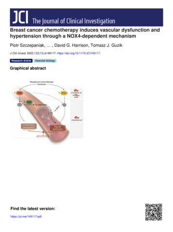
Breast cancer chemotherapy induces vascular dysfunction andhypertension through a NOX4-dependent mechanismPiotr Szczepaniak, , David G. Harrison, Tomasz J. GuzikJ Clin Invest. 2022;132(13):e149117. https://doi.org/10.1172/JCI149117.Research ArticleVascular biologyGraphical abstractFind the latest version:https://jci.me/149117/pdf
RESEARCH ARTICLEThe Journal of Clinical InvestigationBreast cancer chemotherapy induces vasculardysfunction and hypertension through a NOX4dependent mechanismPiotr Szczepaniak,1,2,3 Mateusz Siedlinski,1,2 Diana Hodorowicz-Zaniewska,4 Ryszard Nosalski,1,2 Tomasz P. Mikolajczyk,1,2Aneta M. Dobosz,5 Anna Dikalova,6 Sergey Dikalov,6 Joanna Streb,7 Katarzyna Gara,4 Pawel Basta,8 Jaroslaw Krolczyk,9Joanna Sulicka-Grodzicka,10 Ewelina Jozefczuk,1 Anna Dziewulska,5 Blessy Saju,2 Iwona Laksa,7 Wei Chen,6 John Dormer,11Maciej Tomaszewski,12,13 Pasquale Maffia,2,14,15 Marta Czesnikiewicz-Guzik,16,17 Filippo Crea,18 Agnieszka Dobrzyn,5 Javid Moslehi,19Tomasz Grodzicki,9 David G. Harrison,6 and Tomasz J. Guzik1,2Department of Medicine, Jagiellonian University Medical College, Krakow, Poland. 2Institute of Cardiovascular and Medical Sciences, University of Glasgow, Glasgow, United Kingdom. 3Centre for1Cardiovascular Science, Queen’s Medical Research Institute, University of Edinburgh, Edinburgh, United Kingdom. 4Breast Unit, Department of Surgery, Jagiellonian University Hospital, Krakow,Poland. 5Laboratory of Cell Signaling and Metabolic Disorders, Nencki Institute of Experimental Biology of Polish Academy of Sciences, Warsaw, Poland. 6Division of Clinical Pharmacology,Department of Medicine, Vanderbilt University, Nashville, Tennessee, USA. 7Department of Oncology, 8Department of Gynecology and Oncology, 9Department of Internal Medicine and Gerontologyand 10Department of Rheumatology, Jagiellonian University Medical College, Krakow, Poland. 11Department of Cellular Pathology, University Hospitals of Leicester, Leicester, United Kingdom. 12Divisionof Cardiovascular Sciences, Faculty of Medicine, Biology and Health, University of Manchester, Manchester, United Kingdom. 13Division of Medicine and Manchester Academic Health Science Centre,Manchester University NHS Foundation Trust Manchester, Manchester, United Kingdom. 14Institute of Infection, Immunity and Inflammation, University of Glasgow, Glasgow, United Kingdom.Department of Pharmacy, School of Medicine and Surgery, University of Naples Federico II, Naples, Italy. 16Department of Periodontology, Preventive Dentistry and Oral Pathology, Jagiellonian15University Medical College, Krakow, Poland. 17Department of Periodontology and Oral Sciences Research Group, University of Glasgow Dental School, Glasgow, United Kingdom. 18Departmentof Cardiovascular and Thoracic Sciences, Catholic University of the Sacred Heart, Largo A. Gemelli, Rome, Italy. 19Section of Cardio-Oncology and Immunology, Division of Cardiology and theCardiovascular Research Institute, UCSF, San Francisco, California, USA.Cardiovascular disease is the major cause of morbidity and mortality in breast cancer survivors. Chemotherapycontributes to this risk. We aimed to define the mechanisms of long-term vascular dysfunction caused by neoadjuvantchemotherapy (NACT) and identify novel therapeutic targets. We studied arteries from postmenopausal women who hadundergone breast cancer treatment using docetaxel, doxorubicin, and cyclophosphamide (NACT) and from women withno history of such treatment matched for key clinical parameters. We explored mechanisms in WT and Nox4–/– mice andin human microvascular endothelial cells. Endothelium-dependent, NO-mediated vasodilatation was severely impairedin patients after NACT, while endothelium-independent responses remained normal. This was mimicked by a 24-hourexposure of arteries to NACT agents ex vivo. When applied individually, only docetaxel impaired endothelial function inhuman vessels. Mechanistic studies showed that NACT increased inhibitory eNOS phosphorylation of threonine 495 in aRho-associated protein kinase–dependent (ROCK-dependent) manner and augmented vascular superoxide and hydrogenperoxide production and NADPH oxidase activity. Docetaxel increased expression of the NADPH oxidase NOX4 inendothelial and smooth muscle cells and NOX2 in the endothelium. A NOX4 increase in human arteries may be mediatedepigenetically by diminished DNA methylation of the NOX4 promoter. Docetaxel induced endothelial dysfunction andhypertension in mice, and these were prevented in Nox4–/– mice and by pharmacological inhibition of Nox4 or Rock.Commonly used chemotherapeutic agents and, in particular, docetaxel alter vascular function by promoting the inhibitoryphosphorylation of eNOS and enhancing ROS production by NADPH oxidases.Conflict of interest: JM has served on advisory boards for Pfizer, Novartis, BristolMyers Squibb, Deciphera, Audentes Pharmaceuticals, Takeda, Myokardia, AstraZeneca, GlaxoSmithKline, Boston Biomedical, ImmunoCore, Janssen, Myovant, SilverbackTherapeutics, Amgen, Kurome Therapeutics, Kiniska Pharmaceuticals, DaiichiSankyo, CRC Oncology, BeiGene, Star Therapeutics, ProteinQure, Pharmacyclics,Mallinckrodt Pharmaceuticals, Boehringer, and Cytokinetics.Copyright: 2022, Szczepaniak et al. This is an open access article published underthe terms of the Creative Commons Attribution 4.0 International License.Submitted: March 3, 2021; Accepted: May 19, 2022; Published: July 1, 2022.Reference information: J Clin Invest. 9117.IntroductionImprovements in breast cancer detection and therapy have significantly increased survival. Breast cancer mortality decreasedby 40% between 1989 and 2017 (1). As a result, millions of women alive today are breast cancer survivors. Despite this markedly improved survival, breast cancer survivors are at high risk foratherosclerotic cardiovascular disease (CVD) (2–4). In particular,among women with lower grades of breast cancer, the likelihoodof dying from CVD is substantially higher than that of either cancer death (5, 6) or breast cancer recurrence (7). Vascular disease1
RESEARCH ARTICLEThe Journal of Clinical Investigationunderlies most of these late comorbidities in cancer survivors (5,6, 8). A multi-hit hypothesis has been proposed to explain this,including risk factors at the time of diagnosis, lifestyle alterationscaused by the disease, and the toxicity of drugs and radiation(9). To date, much attention has been focused on anthracyclineanalogs, which have both immediate and delayed effects on cardiac function (2, 3, 10, 11). Other agents probably also contributeto cardiovascular morbidity in this population. Treatment withtamoxifen increases the risk of myocardial infarction by 1.7-fold(12). Radiation is also known to enhance coronary atherosclerosis,probably via the induction of endothelial injury (9, 13, 14). Giventhese direct and indirect influences, there is a pressing need toimprove cardiovascular outcomes in this patient population.A standard chemotherapy regimen, often used with surgery,includes taxanes, doxorubicin, and cyclophosphamide (15). Suchadjuvant chemotherapy has reduced breast cancer mortality byapproximately 15% (16). Given that these agents are commonlyused in this population of post-menopausal women, at high riskfor cardiovascular disease, it is imperative that we understandtheir impact on vascular health (17). In the current study, we showthat vascular function was markedly impaired in patients whohad received docetaxel, doxorubicin, and cyclophosphamide. Wefound that this impairment was primarily due to docetaxel and wasassociated with a shift in the balance between the vascular NO bioavailability and ROS production. The latter seems to result fromelevated expression and activation of the NADPH oxidase catalyticsubunits NOX2 and NOX4. These effects are partially dependenton increased Rho-associated protein kinase (ROCK) activation.Genetic or pharmacological inhibition of murine Nox4 preventsthe development of vascular dysfunction, implicating NOX4 as atarget in preventing the vascular consequences of chemotherapy.ResultsChemotherapy induces endothelial dysfunction in human arteries. Ininitial experiments, we studied arteries from healthy tissue margins that were unaffected by cancer and extracted during breastcancer surgery. Arteries, with a diameter of approximately 1 mmwere obtained from 40 postmenopausal women who had receivedneoadjuvant chemotherapy (NACT) before the surgery and 55 whohad not received such treatment (controls, no NACT). Arterieswere collected 1 month after the last dose of NACT. These patientsdid not differ with respect to other demographics or major risk factors known to affect vascular disease, pharmacotherapies, or radiotherapy, apart from the stage of their cancer (Table 1), and therewas no difference in blood vessel size or morphology between the2 groups (Supplemental Figure 1A; supplemental material availableonline with this article; https://doi.org/10.1172/JCI149117DS1).We used isometric tension studies to assess endothelium-dependent and endothelium-independent vasodilatation in response toacetylcholine (ACh) and sodium nitroprusside (SNP), respectively.We found that NACT significantly altered endothelium-dependentrelaxation response to ACh, while having minimal effect on endothelium-independent response to SNP (Figure 1C). Importantly,these changes were observed 1 month after the last cycle of chemotherapy (Figure 1, A and B). NACT-associated endothelial dysfunction was not dependent on the cancer stage, as it was observed ina subset of patients with stage 2 and 3 as well as in those with stage23 breast cancer (Supplemental Figure 1B). Moreover, the cancerstage was not associated with differences in endothelial functionwhen analyzed separately within the NACT and no-NACT groups(Supplemental Figure 1, C and D).In additional experiments, we established that a 24-hour ex vivoexposure of human arteries to a combination of docetaxel, doxorubicin, and cyclophosphamide (Figure 1D) also impaired ACh-inducedrelaxations, while not altering responses to SNP (Figure 1E). Usingthis ex vivo approach, we found that, individually, doxorubicin andcyclophosphamide did not significantly affect responses to eitherACh or SNP, however, exposure to docetaxel alone significantly impaired ACh-mediated vasodilatation but did not alter SNPevoked relaxations (Figure 1F and Supplemental Table 1).Effect of NACT on eNOS expression and phosphorylation. Inhuman arteries, endothelium-dependent relaxations can bemediated by NO, vasodilator prostaglandins, and/or endothelium-derived hyperpolarizing factors such as hydrogen peroxide (H2O2). To define the relative role of NO, we exposed humanarteries to the NO synthase inhibitor Nω-nitro-l-arginine methyl ester hydrochloride (l-NAME) before administration of ACh.l-NAME markedly inhibited the endothelium-dependent vasodilatation response to ACh in arteries from women who had notreceived NACT, while it had a minimal effect on these responsesin the arteries of women who had received NACT. This strongly suggests that the NO-mediated mechanisms of endotheliumdependent vasodilatation are impaired by NACT treatment (Figure 2A). As the morphological presence of the endothelial cell layerdid not differ between the study groups (Supplemental Figure 1A),reduced endothelial NO synthase (eNOS) expression could provide a potential explanation for endothelial dysfunction. However,we found that NACT was not associated with loss of eNOS mRNAor protein expression (Figure 2B). eNOS is regulated by phosphorylation, predominantly at serine 1177 (Ser1177), which is stimulatory, and at threonine 495 (Thr495), which is inhibitory. As shown inFigure 2C, there was no change in phosphorylation of Ser1177, butthere was an increase in the inhibitory phosphorylation of eNOS atThr495. This observation was confirmed in vivo in mice exposedto docetaxel for 3 weeks via i.p. injections every 5 days (Figure 2D)and in vitro in cultured human dermal microvascular endothelialcells (HDMECs) (Figure 2E), supporting the hypothesis that eNOSphosphorylation at Thr 495 is a crucial mechanism of impaired vascular function in patients with prior NACT.Thr495 can be phosphorylated by protein kinase C (PKC)and Rho kinase (ROCK) (18). Rho kinase activity was inducedin human arteries after NACT, and docetaxel increased theRho kinase activity in HDMECs (Figure 2E). To further explorethe role of ROCK and PKC, we exposed HDMECs in culture todocetaxel with and without the PKC inhibitor Go6976 or theRho kinase inhibitor Y27632. As shown in Figure 2F and Supplemental Figure 2A, ex vivo exposure of cultured endothelialcells and human vessel organ culture to docetaxel enhancedthe phosphorylation of eNOS on Thr495, mimicking the in vivoeffect of NACT. This was not changed by inhibiting PKC, but wasmarkedly reduced by the Rho kinase inhibitor. Thus, inhibitoryphosphorylation of eNOS via Rho kinase probably contributesto the loss of endothelium-dependent vasodilatation followingexposure to docetaxel. Rho kinase activity was induced by NACTJ Clin Invest. 2022;132(13):e149117 https://doi.org/10.1172/JCI149117
The Journal of Clinical InvestigationRESEARCH ARTICLEvascular segments to docetaxel or placebo for 24 hoursin organ culture experiments and analyzed global RNAexpression using RNA-Seq (Figure 3A). We found thatthe expression of 802 mRNA transcripts was significantNo NACT (n 55)NACT (n 40)PAly (FDR P 0.05) altered by docetaxel treatment (FigAge (yr)62 (57–69)60 (56–63)0.074Bure 3A and Supplemental Table 4), which contributed to28.2 5.327.3 3.90.390BMIAsignificant changes in gene sets and pathways relevant130 (120–140)128 (120–135)0.700SBP (mmHg)A80 (70–80)80 (73–83)0.405DBP (mmHg)to vascular function. These included endothelial andA72 (68–80)74.5 (66–80)0.663HR (min)smooth muscle cellular processes such as differentiaC0.828Obesity, n (%)tion, morphogenesis, migration, and signaling as well13 (32.5)20 (36.4)Not overweight, BMI 24.9as pathways involved in epigenetic regulation and DNA20 (50.0)24 (43.6)Overweight (25.0–29.9)methylation. Importantly, a broad range of vascular pro7 (17.5)11 (20.0)Obesity BMI 30Ccesses that were altered were redox sensitive. These proHypertension, n (%)28 (50.1)18 (45.0)0.569Ccesses included endothelial and smooth muscle cell bio9 (16.4)7 (17.5)0.884Diabetes mellitus, n (%)Cgenesis, proliferation, and migration, kinase signaling,10 (18.2)9 (22.5)0.603Smoking, n (%)Cblood pressure regulation, cellular response to oxidative16 (29.0)8 (20.0)0.314Hypercholesterolemia, n (%)Cstress, and oxygen sensing (Figure 3A). This is consis 0.001Breast cancer staging, n (%)0 (0.0)2 (3.6)0tent with the role of ROS in vascular dysfunction in wide1 (2.5)23 (41.8)1range of cardiovascular pathologies (19).6 (15.0)13 (23.6)2Role of ROS in altering vascular function in response to33 (82.5)17 (30.9)3CNACT.Increased production of ROS plays a major roleTumor size 5 cm, n (%)2 (3.6)14 (35.0) 0.001Cinregulatingvascular function in pathological states. To0 (0.0)0 (0.0)Radiotherapy, prior, n (%)Cinvestigatetheimportance of this mechanism, we exam0 (0.0)0 (0.0)Hormonal therapy, prior, n (%)Cined the effect of the antioxidant N-acetylcysteine (NAC)0.29015(37.5)15(27.3)ACE-I/ARB, n (%)C0.28011 (27.5)10 (18.2)Diuretics, n (%) Con ACh-evoked vascular relaxations. As shown in Figure0 (0.0)0 (0.0)Alpha blocker, n (%)C3B, NAC markedly improved endothelium-dependent0.71911 (27.5)17 (30.9)Beta blocker, n (%)Crelaxation responses to ACh in arteries of patients who0.2225 (12.5)3 (5.4)Ca blocker, n (%)Cpreviously received NACT, but had no effect in the non0.0784 (10.0)1 (1.8)Nitrates, n (%)CNACT group (Figure 3B). NAC can react with and scav1.0008 (20.0)11 (20.0)Statins, n (%) C0.0942 (5.0)0 (0.0)Opioids, n (%)Cenge several ROS, including superoxide, H2O2, and the0.6863 (7.50)3 (5.4)Aspirin, n (%)Chydroxyl radical. In additional experiments, we observed0.1733 (7.50)1 (1.8)Insulin, n (%) Cincreased vascular superoxide production by arteries0.3910 (0.0)1(1.8)NSAIDs, n (%)cfrom women who had received NACT compared with0.5536 (15.0)6 (10.9)Antidiabetes medication, n (%)those who had not (Figure 3C).SBP, systolic blood pressure; DBP, diastolic blood pressure; HR, heart rate; ACE-I,NADPH oxidases are major sources of ROS in humanangiotensin-converting enzyme inhibitors; ARB, angiotensin-receptor blockers. ADataarteries(19). We therefore used electron spin resonance toare presented as the median (range), and comparisons of the 2 groups were doneBexamineNADPH oxidase activity in homogenates of arterusing the Mann-Whitney U test; Data are presented as the mean SD, comparisonsCiesfrompatientswho had received NACT and from thosebetween 2 groups were done using the Student’s t test. Comparisons between 2who had not and found a significant increase in superoxgroups were done using the χ2 test.ide production in response to NADPH in those with priorNACT (Figure 3D). Amplex Red assays also demonstratedan increase in vascular production of H2O2 in arteries ofwomen who had undergone prior NACT (Figure 3E). Usingin human arteries, indicating its potential role in endothelialthe fluorescent dyes dihydroethidium (DHE) to detect superoxidedysfunction. Moreover, docetaxel increased Rho kinase activityand 2′,7′-dichlorofluorescein (DCF) to detect H2O2, we detectedin HDMECs (Figure 2E). In addition, the Rho kinase inhibitorthe production of both superoxide and H2O2 predominantly in theY27632 markedly improved endothelium-dependent relaxationmedia, with a less apparent contribution from the endothelium.responses to ACh in the arteries of patients who had previouslyThese signals were eliminated by the selective superoxide and H2O2undergone NACT (Supplemental Figure 2B).scavengers PEG superoxide dismutase (SOD) and PEG catalaseImportantly, the mechanisms regulating eNOS activity depen(PEG-CAT), respectively (Figure 3F and Supplemental Figure 2E).dence on the binding of HSP90α to eNOS were not changed inEffect of NACT on the vascular expression of NADPH oxidase catHDMECs exposed to docetaxel (Supplemental Figure 2C), andalytic subunits. There are 4 key NOX proteins in human vascularHSP90α expression was unaltered in NACT-exposed human artercells: NOX1, -2, -4, and -5. Using Western blotting (Figure 4A) andies (Supplemental Figure 1D).reverse transcription PCR (RT-PCR) (Figure 4B), we found thatDocetaxel modulates key pathways relevant to vascular functhere was no change in NOX1 or NOX5 expression and increasedtion. To gain further insight into the mechanisms responsibleexpression of NOX2 and NOX4 in the arteries of women whofor altered endothelium-dependent vasodilatation, we exposedhad undergone NACT. These increases in NOX2 and NOX4Table 1. Clinical characteristics of patients without prior NACT or withprior NACTJ Clin Invest. 2022;132(13):e149117 https://doi.org/10.1172/JCI1491173
RESEARCH ARTICLEThe Journal of Clinical InvestigationFigure 1. Effects of breast cancer neoadjuvant chemotherapy using docetaxel, cyclophosphamide, and doxorubicin (NACT) on vascular function. (A) Studydesign of human blood vessel collection in patients with breast cancer without NACT (No NACT) or after 6 3 courses of NACT prior to surgery. (B) Representative examples of endothelium-dependent vasorelaxations in response to increasing concentrations of ACh (1 nM–10 μM) in arteries from patients with orwithout NACT. (C) Average endothelium-dependent vasorelaxation responses to ACh (1 nM–10 μM) and endothelium-independent relaxation responsesto SNP (1 nM–10 μM) in arteries from patients without NACT (n 55) and with NACT (n 40). Two rings were studied per patient, and the values wereaveraged. Data are expressed as the mean SEM. ****P 0.0001 versus no NACT (C, left); *P 0.05 versus no NACT (C, right); 2-way, repeated-measuresANOVA. (D) Experimental design of an ex vivo organ culture study of the effects of NACT on vascular function in NACT-naive arteries. (E) Endothelium-dependent (ACh) and endothelium-independent (SNP) vasorelaxations in NACT-naive arteries after a 24-hour organ culture with the combined NACT componentsdocetaxel (100 nM), 4-hydroperoxycyclophosphamide (4-HC) (10 nM), and doxorubicin (100 nM) or vehicle (Veh, solvent) (paired arterial rings for each treatment; n 7 patients). *P 0.05 versus vehicle; 2-way, repeated-measures ANOVA. (F) Effects of individual components of NACT on endothelium-dependent(ACh) and endothelium-independent (SNP) vasorelaxations in NACT-naive arteries after a 24-hour organ culture with either docetaxel (100 nM), cyclophosphamide (100 nM), doxorubicin (100 nM), or vehicle (solvent) (paired vessel rings for each treatment; n 5). *P 0.05 versus vehicle; 2-way, repeated-measuresANOVA with Tukey’s test. Data in E and F are expressed as the mean SEM. DTX, docetaxel; DOX, doxorubicin; CP, cyclophosphamide; Phe, phenylephrine.were recapitulated by a 24-hour exposure of vascular segmentsto docetaxel (Figure 4C). Through additional experiments usingcultured endothelial and vascular smooth muscle cells (VSMCs),we found that docetaxel increased NOX2 and NOX4 expressionin endothelial cells and NOX4 expression in smooth muscle cells(Figure 4D). Immunofluorescence showed that NOX2 stainingwas predominantly increased in the vascular media, whereasNOX4 was increased in both the media and endothelial cells ofwomen who had undergone NACT (Figure 4E).Considering the prolonged effect of NACT observed 4 weeksafter its discontinuation, we considered that epigenetic mechanisms4might modulate NOX4 expression over the long term. Indeed,studies of the NOX4 promoter showed that 23 CpG sites exhibited decreased DNA methylation in arteries from women that hadundergone NACT compared with no-NACT donor arteries (Figure5, A and B). We found that 6% of the CpG sites located in the CpGisland of the NOX4 promoter were normally methylated in arteries of women who had not received NACT and that NACT therapy eliminated methylation of these CpG sites (Figure 5, A and B,and Supplemental Table 5). As a control, we studied methylationof the GTP cyclohydrolase gene (GCH1) and found that it wasunaltered by NACT (Figure 5C). To further understand the effectsJ Clin Invest. 2022;132(13):e149117 https://doi.org/10.1172/JCI149117
The Journal of Clinical InvestigationRESEARCH ARTICLEFigure 2. Effects of breast cancer neoadjuvant chemotherapy using docetaxel, cyclophosphamide, and doxorubicin (NACT) on the regulation of eNOS.(A) Endothelium-dependent vasorelaxation responses to ACh (1 nM to 10 μM) in arteries from patients without NACT and from patients with prior NACTafter preincubation with l-NAME (100 μM) (n 8/group). Data expressed as the mean SEM. ***P 0.001 versus no NACT; *P 0.05 versus no NACT;2-way, repeated-measures ANOVA with Tukey’s test. (B) Expression of eNOS mRNA (n 13–16/group; bottom left) and eNOS protein (Western blotting;n 12/group) in arteries from patients without NACT and from patients who underwent NACT. (C) Phosphorylation of eNOS at Ser1177 (left; n 12/group)and at Thr495 in arteries from patients with or without prior NACT (right; n 12/group), normalized to total eNOS. Data were derived from 3 independentexperiments and are expressed as the mean SEM. ****P 0.0001 versus no NACT; 2-tailed, unpaired Student’s t test. (D) Effects of in vivo exposureto docetaxel in C57BL/6J mice (i.p. injections; 10 mg/kg or placebo every 5 days for 3 weeks) on the inhibitory phosphorylation of eNos at Thr495, normalized to total eNOS in mouse aortas (n 5 mice/group). Data are expressed as the mean SEM. **P 0.01 versus WT; 2-tailed, unpaired Student’s t test.(E) Rho kinase activity in arteries from patients with or without NACT (n 10/group; left) and in HDMECs (n 5/group; right). Data are expressed as themean SEM. ***P 0.001 versus vehicle or no NACT; 2-tailed, unpaired Student’s t test. (F) Effects of docetaxel (100 nM) on eNOS at Thr495 phosphorylation in HDMECs and the modulating effect of docetaxel plus Go6976 (1 μM) or docetaxel plus Y27632 (5 μM) (n 6/group). Densitometric analysis ofproteins normalized to total eNOS is shown. Immunoblots are from 1 of 2 independent experiments (left) and are expressed as the mean SEM. **P 0.01 versus vehicle or docetaxel; *P 0.05 versus docetaxel; 1-way ANOVA with Tukey’s test.J Clin Invest. 2022;132(13):e149117 https://doi.org/10.1172/JCI1491175
RESEARCH ARTICLEThe Journal of Clinical Investigationof docetaxel on the NOX4 promoter, we performed an in vitropromoter activity assay in HEK293 cells. A 48-hour exposure todocetaxel induced NOX4 promoter activity in an in vitro promoteractivity assay in HEK293 cells (Figure 5D).Docetaxel induces hypertension and vascular dysfunction in micevia a Nox4-dependent mechanism. To gain additional insight intothe role of NOX4 in response to docetaxel, we treated WT andNox4 –/– mice with this agent (10 mg/kg every 5 days for 3 weeks,Figure 6A). Measurements using radiotelemetry showed thatblood pressure significantly increased following the first docetaxelinjection and was sustained throughout treatment with this agent(Figure 6B). In mice lacking Nox4, this hypertensive responsewas absent (Figure 6C). In agreement with our findings in humanarteries, isometric tension studies of the aorta showed that treatment with docetaxel impaired endothelium-dependent vasorelaxation responses to ACh (Figure 6D) but did not affect endothelium-independent vasodilatation responses to SNP (Figure6D). To confirm the role of Nox4-derived superoxide in impairingendothelium-dependent vasodilatation, we also treated arteriesin the organ chamber with GKT137831, a selective inhibitor ofNOX1 and NOX4. This agent normalized ACh-induced vasodilatation in mice previously treated with docetaxel (SupplementalFigure 3G). In line with our results in human arteries, docetaxeltreatment increased vascular H2O2 (Figure 6E) and superoxide(Figure 6F) production in WT mice, and these responses wereabsent in Nox4-deficient mice (Figure 6, E and F). DHE fluorescence confirmed an increase of superoxide production in themedia of WT mouse aortas treated with docetaxel that was absentin Nox4 –/– mice (Figure 6G). RT-PCR analysis revealed that aorticNox4 mRNA expression was increased in WT mice treated withdocetaxel and was absent in the Nox4 –/– mice (Figure 6H).As shown in Figure 2F, docetaxel treatment promotedROCK-dependent phosphorylation of eNOS at Thr495. SinceH2O2 can activate Rho kinase, we examined Thr495 phosphorylation on eNOS in WT and Nox4 –/– mice and found that this inhibitory phosphorylation increased by docetaxel treatment in WT butnot Nox4 –/– mice (Figure 6I).Microtubule stabilization is a key mechanism of action of taxanes (20) and is also needed for basal eNOS phosphorylation andsubcellular organization (21). One way that taxanes affect microtubule stabilization is the lysine acetylation of α-tubulin (AcK40)(20). We found that lysine acetylation was an unlikely mechanismof docetaxel-induced vascular dysfunction, as docetaxel treatment similarly increased Ack40–α-tubulin in WT and Nox4 –/–mouse aortas, despite striking differences in vascular and bloodpressure phenotypes (Supplemental Figure 3H). In line withincreased acetylation, however, changes in direct eNOS acetylation cannot be excluded.To confirm that Nox4 represents a therapeutic target for preventing the development of vascular dysfunction, we administereddaily i.p. injections of the Nox1/Nox4 inhibitor GKT137831 duringdocetaxel exposure (Figure 7A) and found that this prevented boththe increase in blood pressure (Figure 7B) and development of vascular dysfunction in mouse aortas (Figure 7C) caused by this taxane.In keeping with our observations in human arteries, ROCKinhibition by fasudil (Figure 7D) prevented the blood pressureincrease (Figure 7E) and vascular dysfunction caused by docetaxel6(Figure 7F) in mice. Fasudil decreased the docetaxel-inducedThr495 phosphorylation of eNOS (Figure 7G), further supportingthe role of ROCK in docetaxel-induced eNOS dysfunction.Other possible mechanisms of the blood pressure effects ofdocetaxel in mice were investigated. Docetaxel did not change theheart rate of mice after 3 weeks of treatment (Supplemental Figure3B). Treatment of mice with docetaxel did not affect aldosteronelevels (Supplemental Figure 3C), renal damage as assessed byhistopathology (Supplemental Figure 3D), urinary albumin (Supplemental Figure 3E), or urinary neutrophil gelatinase–associatedlipocalin (NGAL) (Supplemental Figure 3F).DiscussionIn the present study, we identify a mechanism by which the commonly used chemotherapeutic agent docetaxel promoted vascular dysfunction and loss of NO-mediated vasodilatation. This wasin part mediated by an increase in vascular production of ROSthrough the NADPH oxidase subunits NOX4 and NOX2. ROS produced by these enzymes likely have 2 major effects. First, superoxide reacts at a diffusion-limited rate with NO, limiting the vasodilatation caused by the latter and yielding the strong prooxidantperoxynitrite (22). Second, H2O2 stimulates Rho kinase, which inturn causes inhibitory phosphorylation of eNOS (23), reducing itsability to produce NO (18). Both effects appear to play a role in thevascular dysfunction caused by docetaxel.NACT-induced ROS overproduction was observed in bothendothelial cells and smooth muscle cells. Importantly, eithershort-lived superoxide or more stable H2O2, produced by VSMCshave been shown to affect endothelial function by direct scavenging of NO and by production of H2O2, which in turn regulates signaling and gene expression. Our findings were based on an analysisof human arteries, and we gained additional mechanistic insightfrom studies of WT and Nox4 –/– mice, providing possible mechanisms of long-term vascular disease in breast cancer survi
The Journal of Clinical Investigation RESEARCH ARTICLE 1 Introduction Improvements in breast cancer detection and therapy have sig-nificantly increased survival.