Transcription
INTRODUCTIONTO ULTRASOUNDEMERGENCYULTRASOUND.CA SJRHEM.CA@ECCULTRASOUND
Outline How does ultrasound work? When is it used in the ED? Binary questions AAA? Tamponade? IUP? Clinical integration
Sound Energy Ultrasound: non audible sound energy Produced by piezoelectric materials in the probe Sound waves are sent out and those that bounce backare interpreted
Generating Images The sound waves that are reflected back to the transducerproduce the image Brightness intensity of the reflection Depth time delay of the reflection
Acoustic Impedance The resistance of sound wave propagation through tissueis called acoustic impedance Each material (e.g. air, blood, bone, fat) has its ownacoustic impedance The differenced in acoustic impedance cause reflection ofsound waves as the tissue interface
Absorption More absorption limits how deep the waves can travel High frequency sound waves are more readily absorbed,and therefore produce shallower images
Ultrasound: Wavelengths and Probes frequency wavelength resolution depth frequency wavelength resolution depth
Probes
Ultrasound: Orientation Orient the transducer so that the transducer marker alignswith the marker on the screen
OrientationRadiologyCardiac
Transducer Movements
Planes
Sagittal Plane
Coronal Plane
Transverse Plane
Preparation Clean the machine and the transducer with antisepticwipe Position yourself on the patients right side and with themachine in front of you Select the appropriate transducer Enter patient’s information Select appropriate exam type Position patient appropriately
Preparation While maintaining proper draping technique and patientprivacy, expose the appropriate body area Place gel on the area to be scanned
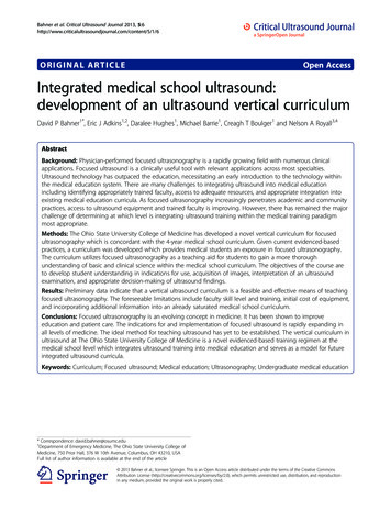
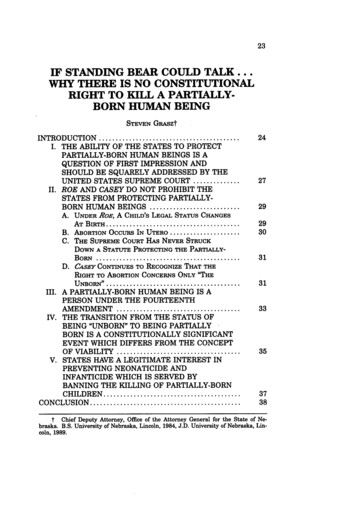
Exam Set the imagingmode Brightness mode Motion mode Doppler modeA, Apical four-chamber view of the heart instandard 2-D mode. B, M-mode view throughthe mitral valve. C, Color Doppler map of themitral valve. D, Spectral Doppler of the mitralvalve inflow.
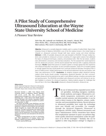
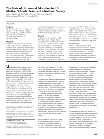
Exam Adjust depth for thestructure of interestImage of the heart in parasternal long window. A,Too much depth: the far field is empty (however,this can be a desired image when looking for aneffusion). B, Appropriate depth. C, Too little depth:the posterior wall and descending aorta are notvisualized.
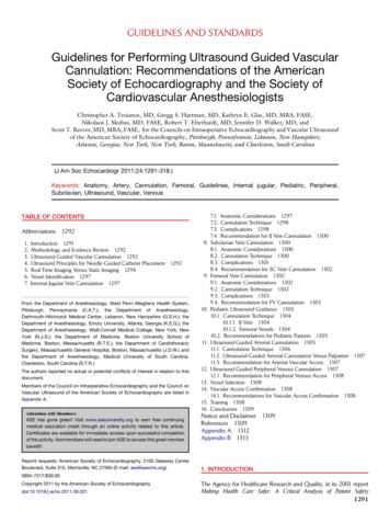
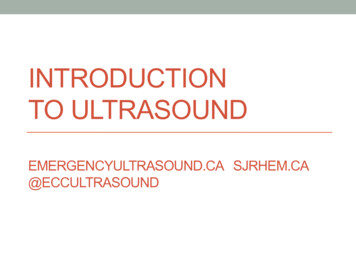
Exam Adjust the gain Controls how the signal is amplified upon return to the transducerExamples of the internal jugular vein demonstrating undergained(A), appropriately gained (B), and overgained (C) images.
Post Exam Clean the gel off the patient’s skin. Return the patient’sposition and bed to normal. End the exam on the machine Clean the transducer and probe with antiseptic Return machine to storage location
Ultrasound in Emergency Medicine The eFAST Exam Extended Focused Assessment with Sonography for Trauma Multi-view examination of the intra-abdominal space and the heart Cardiac, RUQ/Chest, LUQ/Chest, and Pelvic examsACES Abdominal and Cardiothoracic Evaluation with Sonography inShock Cardiac, RUQ/Chest, LUQ/Chest, IVC, Aorta, and Pelvic examsIndividual components
ReferencesSoni NJ, Arntfield R, and Kory P. Point of Care Ultrasound.2015. Philadelphia: Elsevier Saunders.Stone CK, and Humphries RL. Current EmergencyDiagnosis & Treatment 7e. 2011. New York: McGraw-Hill.
Ultrasound in Emergency Medicine The eFAST Exam Extended Focused Assessment with Sonography for Trauma Multi-view examination of the intra-abdominal space and the heart Cardiac, RUQ/Chest, LUQ/Chest, and Pelvic exams ACES Abdominal and Cardiothoracic Evaluation with Sonography in Shock Cardiac, RUQ/Chest, LUQ/Chest, IVC, Aorta, and Pelvic exams