Transcription
Global clinical dementia rating of 0.5 inMCI masks variability related to levelof functionY.-L. Chang, PhDM.W. Bondi, PhDL.K. McEvoy, PhDC. Fennema-Notestine,PhDD.P. Salmon, PhDD. Galasko, MDD.J. Hagler, Jr., PhDA.M. Dale, PhDFor the Alzheimer’sDiseaseNeuroimagingInitiativeAddress correspondence andreprint requests to Dr. Yu-LingChang, Department ofPsychology, National TaiwanUniversity, No. 1, Sec. 4,Roosevelt Road, Taipei, 10617Taiwanychang@ntu.edu.twABSTRACTObjective: To evaluate whether ratings on Clinical Dementia Rating (CDR) items related to instrumental activities of daily living (IADL) are associated with cognitive or brain morphometric characteristics of participants with mild cognitive impairment (MCI) and global CDR scores of 0.5.Methods: Baseline cognitive and morphometric data were analyzed for 283 individuals with MCI whowere divided into 2 groups (impaired and intact) based on their scores on the 3 CDR categories assessingIADL. Rates of progression to Alzheimer disease (AD) over 2 years were also compared in the 2 groups.Results: The impaired IADL MCI group showed a more widespread pattern of gray matter lossinvolving frontal and parietal regions, worse episodic memory and executive functions, and ahigher percentage of individuals progressing to AD than the relatively intact IADL MCI group.Conclusions: The results demonstrate the importance of considering functional information captured by the CDR when evaluating individuals with MCI, even though it is not given equal weight inthe assignment of the global CDR score. Worse impairment on IADL items was associated withgreater involvement of brain regions beyond the mesial temporal lobe. The conventional practiceof relying on the global CDR score as currently computed underutilizes valuable IADL informationavailable in the scale, and may delay identification of an important subset of individuals with MCIwho are at higher risk of clinical decline. Neurology 2011;76:652–659GLOSSARYACC anterior cingulate cortex; AD Alzheimer disease; ADL activities of daily living; ADNI Alzheimer’s DiseaseNeuroimaging Initiative; BNT Boston Naming Test; CDR Clinical Dementia Rating; FAQ Functional Activity Questionnaire; HC healthy control; IADL instrumental activities of daily living; LM II Logical Memory II; MANOVA multivariateanalysis of variance; MCI mild cognitive impairment; MMSE Mini-Mental State Examination; PCC posterior cingulatecortex; RAVLT Rey Auditory Verbal Learning Test; ROI region of interest; SB sum of boxes; WAIS-R Wechsler AdultIntelligence Scale–Revised.Mild cognitive impairment (MCI) is an established risk state for the development of Alzheimer disease(AD).1 The requirement that functional activities remain essentially intact was originally considered central for differentiating MCI from mild dementia. However, recent studies have shown that subtle changesin the ability to perform instrumental activities of daily living (IADL) occur in MCI.2-6The designation of MCI is often supported by a global rating of 0.5 on the Clinical DementiaRating (CDR)7 scale. The most heavily weighted component of the global CDR score is memoryfunction; nonmemory components, including the 3 IADL categories, receive less weighting. Consequently, 2 individuals with MCI receiving the same global CDR score can noticeably differ in IADLperformance. This lack of richness in characterizing IADL changes might prevent identification ofmeaningful clinical differences in individuals with MCI. To address this possibility, we comparedbaseline brain morphometry, cognition, and 2-year clinical outcome in subgroups of MCI particiSupplemental data atwww.neurology.orgFrom the Departments of Psychiatry (Y.-L.C., M.W.B., C.F.-N.), Radiology (L.K.M., C.F.-N., D.J.H., A.M.D.), and Neurosciences (D.P.S., D.G.,A.M.D.), University of California, San Diego, La Jolla; Veterans Affairs San Diego Healthcare System (M.W.B., D.G.), San Diego, CA; andDepartment of Psychology (Y.-L.C.), National Taiwan University, Taipei, Taiwan.The Alzheimer’s Disease Neuroimaging Initiative Coinvestigators are listed in appendix e-1 on the Neurology威 Web site at www.neurology.org.Study funding: Funding information is provided at the end of the article.Disclosure: Author disclosures are provided at the end of the article.Data used in the preparation of this article were obtained from the Alzheimer’s Disease Neuroimaging Initiative (ADNI) database(www.loni.ucla.edu{ADNI). As such, the investigators within the ADNI contributed to the design and implementation of ADNI and/or provideddata but did not participate in analysis or writing of this report. A complete listing of ADNI investigators is available online.652Copyright 2011 by AAN Enterprises, Inc.
pants who had global CDR scores of 0.5 butwho differed with regard to IADL items. Wepredicted that, relative to individuals with intactIADL, those with relatively impaired IADLwould show 1) a more widespread pattern ofcortical atrophy involving frontal regions in addition to the expected medial temporal lobe involvement, 2) poorer cognitive performance,especially on tests of executive function, and 3) ahigher rate of progression to probable AD.METHODS Raw data used in the current study were obtainedfrom the Alzheimer’s Disease Neuroimaging Initiative (ADNI)database (www.loni.ucla.edu{ADNI). ADNI was launched in2003 by the National Institute on Aging, the National Instituteof Biomedical Imaging and Bioengineering, the Food and DrugAdministration, private pharmaceutical companies, and nonprofit organizations, as a 60 million, 5-year public–private partnership. ADNI’s goal is to test whether serial MRI, PET, otherbiological markers, and clinical and neuropsychological assessment can be combined to measure the progression of MCI andearly AD. Determination of sensitive and specific markers ofvery early AD progression is intended to aid researchers and clinicians to develop new treatments and monitor their effectiveness, as well as lessen the time and cost of clinical trials.Standard protocol approvals, registrations, and patientconsents. This study was approved by an ethical standardscommittee on human experimentation at each institution. Written informed consent was obtained from all participants or authorized representatives participating in the study.Participants. ADNI eligibility criteria are described at ocolSummary.aspx. Briefly,participants were 55–90 years old, nondepressed, with a modifiedHachinski score of 4 or less, and a study partner able to provide anindependent evaluation of functioning. Individuals with a history ofsignificant neurologic or psychiatric disease, substance abuse, or metal intheir body other than dental fillings were excluded.Healthy control (HC) participants had Mini-Mental StateExamination (MMSE) scores of 24 –30, normal activities ofdaily living (ADL) as assessed with the Functional Activity Questionnaire (FAQ, clinical judgment without suggested cutoff), aglobal CDR score of 0, and normal memory function, as indicated by education-adjusted scores on the modified WechslerMemory Scale Logical Memory II (LM II, story A only) (i.e., ascore 8 for individuals with ⱖ16 years of education; 4 forindividuals with 8 –15 years of education; and 2 for individuals with 0 –7 years of education). MCI participants had a subjective memory complaint, objective memory loss as indicated byeducation-adjusted scores on the LM II (score ⱕ8 for individualswith ⱖ16 years of education; ⱕ4 for individuals with 8 –15years of education; and ⱕ2 for individuals with 0 –7 years ofeducation), a global CDR score of 0.5 and a score ⱖ0.5 on thememory box of the CDR, essentially preserved ADL primarilyassessed by the FAQ, and an absence of dementia.1This study included individuals classified as HC or MCIwithin ADNI with baseline MRI scans that passed local qualityinspection. We excluded 7 HC who converted to MCI at anyfollow-up visit to minimize the possibility of misclassification ofHC participants at baseline. The present study thus consisted of202 HC and 283 MCI participants. MCI participants were divided into 2 subgroups (intact IADL and impaired IADL) basedon their scores on the 3 CDR categories assessing IADL (i.e.,judgment and problem solving, community affairs, home andhobbies). Specifically, the intact IADL group (n 179) consisted of individuals with a rating of 0 on all 3 IADL categories ora rating of 0.5 on 1 of the 3 categories; the impaired IADL group(n 104) consisted of individuals with a rating of 0.5 on 2 ormore of the 3 IADL categories or a rating of 1 on any 1 of thecategories. The 2 MCI groups did not differ in age (t281 1.79,p 0.07), level of education (t281 0.15, p 0.88), sex distribution ( 2 (1) 0.65, p 0.42), history of hypertension( 2 (1) 0.67, p 0.41), use of hypertension medications( 2 (1) 0.08, p 0.78), or scores on the Geriatric DepressionScale (t281 1.67, p 0.10). The impaired IADL groupdemonstrated higher FAQ scores (t281 7.95, p 0.001) anda higher frequency of APOE 4 carriers relative to the intactIADL group ( 2 (1) 3.95, p 0.04; table 1). Two-yearfollow-up clinical outcome data (i.e., reversion to normal cognitive status, stable MCI, or progression to probable AD) wereavailable for 233 of the MCI participants. The determination ofprogression to probable AD was based on National Institute ofNeurological and Communicative Disorders and Stroke–Alzheimer’s Disease and Related Disorders Association criteria (see appendix e-2 on the Neurology Web site at www.neurology.org foroperational criteria).Neuropsychological assessment. All participants were administered a cognitive battery as previously described.8,9 Measuresincluded MMSE,10 Wechsler Adult Intelligence Scale–Revised(WAIS-R) Digit Span and Digit Symbol subtests, Boston NamingTest (BNT),11 animal fluency, Rey Auditory Verbal Learning Test(RAVLT),12 the LM II,13 and the Trail Making Test.14Magnetic resonance scanning and brain morphometry.Dual 3-dimensional T1-weighted volumes were downloadedfrom the public ADNI database (http://www.loni.ucla.edu/ADNI/Data/index.shtml). All image processing and analyses occurred at the Multimodal Imaging Laboratory, University ofCalifornia, San Diego. Images were corrected for gradient nonlinearities15 and intensity nonuniformity.16 The 2 images werealigned, averaged to improve signal-to-noise ratio, and resampled to isotropic 1-mm voxels. Methods based on FreeSurfersoftware were used to obtain cortical gray matter volume andthickness measures in distinct regions of interest (ROIs).17-22To limit the number of statistical comparisons, analyses onlyincluded regions assumed to be involved in early AD pathology,23 such as bilateral hippocampal formation (volumetric measures) and regions of temporal, frontal, parietal, and cingulatecortex (thickness measures) (see ROIs listed in table e-1). The caudal and rostral anterior cingulate regions were combined as anteriorcingulate cortex (ACC); the isthmus and posterior cingulate regionswere combined as posterior cingulate cortex (PCC).Statistical analysis. Group comparisons were performed withseparate independent sample t tests or 2 tests for demographic,cognitive, and clinical outcome variables. Cognitive test scoresfor MCI participants were converted to z scores based upon themean and SD of the HC group (means and standard deviationsof the raw test scores for MCI and HC groups are shown in tablee-2). Because the impaired IADL group had more APOE 4carriers than the intact group, analyses of cognitive variables werealso performed with separate one-way analyses of covariance,controlling for APOE genotype. Although some of the distribuNeurology 76February 15, 2011653
Table 1Demographic, clinical, and cognitive characteristics of the 2 MCI groupsIntact IADL,mean (SD)(n ⴝ 179)Impaired IADL,mean (SD)(n ⴝ 104)Age, y75.69 (6.97)74.12 (7.27)0.07—Education, y15.93 (2.92)15.88 (2.85)0.88—% Men65610.42—p ValueCohen d% APOE 4ⴙ48620.04*—% Hypertension history50550.41—% On hypertension meds72750.78—Geriatric Depression Scale1.47 (1.33)1.76 (1.45)0.10—Functional Assessment Questionnaire1.98 (2.82)5.83 (5.33) 0.001*—CDR sum of boxes0.96 (0.40)2.10 (0.58) 0.001*—MMSE (raw score)27.25 (1.76)27.34 (1.58)Boston Naming Test 0.83 (1.60) 0.97 (1.75)0.480.08Category fluency 0.58 (0.85) 0.73 (0.86)0.170.18 0.19 (1.02) 0.36 (1.01)0.180.170.690.05Language (z score)Executive function/attention/processing (z score)Digit Span forwarda 0.31 (1.25) 0.56 (1.84)0.180.16Digit symbol 0.59 (0.99) 0.82 (1.08)0.070.22Digit Span backward 0.39 (0.96) 0.61 (0.94)0.060.23Trail Making Test Ba 0.61 (1.44) 1.02 (1.76)0.03*0.25Immediate recall 1.78 (0.86) 1.90 (0.88)0.280.14Delayed recall 2.50 (0.75) 2.54 (0.75)0.570.05Trial 1–5 total 1.21 (1.07) 1.41 (0.87)0.100.21Trail Making Test ALearning and memory (z score)Logical memoryRAVLTShort-delay recall 1.10 (0.99) 1.38 (0.84)0.02*0.30Long-delay recall 1.07 (0.96) 1.33 (0.82)0.02*0.29Recognition discriminability 1.24 (1.39) 1.84 (1.72)0.001*0.38Abbreviations: CDR Clinical Dementia Rating; IADL instrumental activities of daily living; MCI mild cognitive impairment; MMSE Mini-Mental State Examination; RAVLT Rey Auditory Verbal Learning Test.aFor ease of interpretation, scores on the Trail Making Test were inverted so negative scores represent poorer performance, consistent with other tests.* Statistically significant results.tions of scores on cognitive tests were skewed, we did not transform the data since the sample size was sufficiently robust toenable appropriate use of the t statistic.24Differences in morphometric variables among HC, intactIADL MCI, and impaired IADL MCI groups were assessed withmultivariate analyses of variance (MANOVAs) followed by univariate analyses of variance with Bonferroni adjustments forType I error ( 0.001) (table e-1). Pairwise comparisons wereconducted through separate independent sample t tests ( 0.05). Effects of age and gender were regressed from all thicknessand volumetric measures and standardized residual values (i.e., zscores) were used for analyses. Hippocampal volumes were alsocorrected for differences in head size by regressing the estimatedtotal cranial vault volume.25 Results controlling for APOE statusare only reported if they differed from noncontrolled analyses.Effect sizes were calculated with Cohen d for significant group654Neurology 76February 15, 2011differences on cognitive and morphometric measures. All analyses were conducted in SPSS (version 17.0).RESULTS Neuropsychological differences. Performance did not differ between MCI groups on MMSE,Digit Span forward and backward, Trail Making TestPart A, Digit Symbol, LM immediate and delayed recall,RAVLT Trials 1–5 total learning score, animal fluency, orBNT. However, the impaired IADL group demonstratedpoorer performance than the intact IADL group on theTrail Making Test Part B (t281 2.13, p 0.03),RAVLT short (t281 2.45, p 0.02) and long (t281 2.39, p 0.02) delayed recall, and RAVLT recognitiondiscriminability (t281 3.22, p 0.001) (table 1).
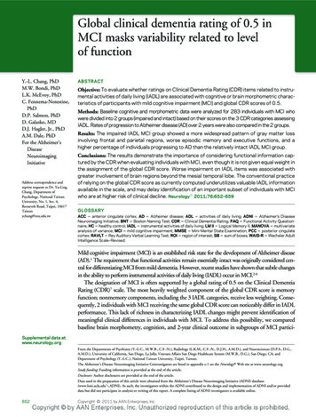
Table 2Standardized residual values (i.e., z scores and correspondingstandardized errors relative to healthy control individuals) ofregions that differed between the 2 MCI groupsIntact IADL,mean (SD)Impaired IADL,mean (SD)p ValueCohen d0.46 (0.07) 0.24 (0.10)0.020.28Left medial orbitofrontal 0.01 (0.07) 0.29 (0.09)0.020.28Right parahippocampus 0.01 (0.07) 0.34 (0.09)0.0070.33Right supramarginal 0.02 (0.07) 0.27 (0.09)0.030.27Right lateral orbitofrontalAbbreviations: IADL instrumental activities of daily living; MCI mild cognitive impairment.When APOE status was included as a covariate,the same pattern of findings was observed exceptthat Digit Symbol, which was marginally significant before, now reached significance (F1,271 4.73, p 0.03).Regional differences in morphometry. The overallMANOVA for group effects on all ROIs was significant (Wilks lambda 0.64, F72,896 3.06, p 0.001, partial 2 0.19). Follow-up univariate analyses comparing the MCI groups to the HC groupshowed that both MCI groups had smaller than normal hippocampal volumes bilaterally and thinnerthan normal cortex in frontal (i.e., bilateral caudaland rostral middle frontal, superior frontal, and rightlateral orbitofrontal areas), temporal (i.e., bilateralentorhinal cortex, parahippocampal, superior, middle, and inferior temporal, and temporal pole areas),and parietal (i.e., bilateral inferior parietal lobule andleft supramarginal) regions, as well as in the bilateralPCC. Moreover, the impaired IADL group— butFigurenot the intact IADL group—showed cortical thinning in bilateral medial orbitofrontal, pars orbitalis,and right supramarginal regions. Information onmagnetic resonance morphometric measures for the3 groups in all ROIs are presented in table e-1.The 2 MCI groups showed comparable hippocampal volumes and similar cortical thickness in entorhinal,lateral temporal, dorsolateral prefrontal, and cingulateROIs bilaterally. However, the impaired IADL grouphad reduced cortical thickness in left medial orbitofrontal (t281 2.29, p 0.02), right lateral orbitofrontal(t281 2.32, p 0.02), right parahippocampus(t281 2.71, p 0.007), and right supramarginal regions (t281 2.25, p 0.03) compared to the intactIADL group (table 2 and figure).Longitudinal progression rates. Two-year rate of pro-gression to AD was higher in the impaired IADLgroup (46%; 41/89 participants) than in the intactgroup (31%; 45/144 participants; 2(1, n 233) 5.19, p 0.02).MCI participants with or without clinical outcome data did not differ in age, education level, gender distribution, APOE 4 status, or MMSE scores(all p values 0.05; table 3).DISCUSSION When MCI participants with globalCDR scores of 0.5, CDR memory ratings of 0.5, andimpaired performance on the LM II were dividedinto groups with relatively intact or impaired ratingson the CDR IADL components, those with impairedIADL ratings exhibited poorer cognitive test perfor-Reconstructed cortical surface maps for the 2 mild cognitive impairment (MCI) groups relative to the healthy control (HC) groupReconstructed cortical surface maps representing the average mean difference in thickness (mm, p 0.001) for the 2 MCI groups, relative to the HCgroup, after controlling for the effects of age and gender. Blue and cyan indicate thinning. IADL instrumental activities of daily living.Neurology 76February 15, 2011655
Table 3Demographic and global cognitive characteristics of individuals with or without clinical outcome data (i.e., progression toAlzheimer disease) for the 2 MCI groupsIntact IADLClinical outcome dataYes(n ⴝ 144)Impaired IADLNo(n ⴝ 35)StatisticalcomparisonYes(n ⴝ 89)No(n ⴝ 15)StatisticalcomparisonAge76.00 (6.83)74.39 (7.48)t177 1.22, p 0.2273.97 (7.32)75.03 (7.12)t102 0.52, p 0.60Education16.01 (2.78)15.57 (3.46)t177 0.80, p 0.4215.92 (2.83)15.60 (3.04)t102 0.40, p 0.696253 2(1) 0.39, p 0.5827.34 (1.50)27.33 (2.06)t102 0.01, p 0.996064 2(1) 0.07, p 0.792% Men6374 MMSE27.31 (1.74)27.00 (1.86)t177 0.94, p 0.35% APOE 4ⴙ49462 (1)(1) 1.53, p 0.22 0.14, p 0.71Abbreviations: IADL instrumental activities of daily living; MCI mild cognitive impairment; MMSE Mini-Mental State Examination.mance, more widespread gray matter thinning infrontal and parietal lobe brain regions, and a higherrate of progression to probable AD over a 2-yearfollow-up period. Group differences in cognitive testperformance were apparent on tests of executivefunction (i.e., TMT-B and Digit Symbol test) andepisodic memory (i.e., RAVLT). Differences on episodic memory measures were somewhat unexpectedsince all participants had CDR memory ratings of0.5 and groups did not differ in degree of atrophy inmesial temporal lobe structures implicated in memory (i.e., hippocampus and entorhinal cortices), oron performance on the LM II. Although theRAVLT, an unstructured list-learning task, may simply be a more sensitive measure of episodic memorythan the LM II, a structured story-memory task, it ispossible that the worse performance of the IADLimpaired group on the RAVLT is related to theirgreater deficit in executive functions. Althoughmemory for complex narrative such as that requiredby the LM II is impaired in patients with frontal lobelesions,26 the RAVLT may place greater demands onexecutive abilities for organizing the unstructuredword-list material during encoding or for strategicsearch during retrieval.27 We previously found thatMCI participants with impaired executive functionperformed more poorly on the RAVLT, but not onthe LM II, than did MCI participants without executive dysfunction, and that thinning in frontal areascontributed to RAVLT performance beyond thewell-known contribution of medial temporalstructures. 27 These subtle distinctions betweenmemory measures are not likely to be made withinthe CDR because memory ability is assessed as anoverall rating based on the subjective judgment ofan informant and does not take into account various cognitive processes underlying objective memory performance.Morphometric analyses showed that the impairedIADL group had greater and more widespread atrophy than the intact IADL group in left medial orbitofrontal, right lateral orbitofrontal, right656Neurology 76February 15, 2011supramarginal, and right parahippocampal cortex.The bilateral involvement of the orbitofrontal cortexis consistent with a recent study that highlighted therole of ventromedial prefrontal regions in carryingout complex cognitive and emotional tasks encountered in everyday life.28 Greater cortical thinning infrontal regions in the impaired IADL group than inthe intact group is consistent with their poorer performance on measures of executive function. Thefinding that cortical thickness was thinner in theright supramarginal and parahippocampal areas inthe impaired IADL group than in the intact group isconsistent with recent results that suggest brain atrophy spreads from medial temporal lobe structures toparietal and frontal cortical regions as severity ofMCI increases,29 and with the typical distribution ofAD neuropathology early in the disease process.30Consistent with previous studies,2,3,5 MCI participants with impaired IADL were more likely thanthose with intact IADL to progress to a clinical diagnosis of probable AD within the next 2 years. The 2MCI subgroups may thus represent points along anMCI-to-AD continuum with the impaired IADLgroup having progressed farther toward AD than theintact IADL group. The higher scores on the FAQ inthe impaired than the intact IADL group is consistent with this. It could be argued that the presence ofdeficits in 2 or more areas of cognition (episodicmemory and executive function), coupled with impaired IADL, would support a diagnosis of mild dementia rather than MCI. However, the conventionalpractice of relying on summary screening measuresand rating scales such as the MMSE, LM, and CDRto differentiate MCI from AD suffers from a certain granularity31 and fails to capture the subtlebut significant cognitive and functional changes inearly dementia. Alternative approaches that incorporate more comprehensive neuropsychologicallybased methods for the diagnosis of MCI and ADhave recently shown advantage in improving thestability and reliability of a diagnosis that predictsclinical decline.32-35
Global CDR score is one of the most commonlyused measures for identifying and staging MCI orAD dementia. There are, however, a number ofproblems that have been pointed out36-39 with regardto the established methods for computing a globalCDR. These include inconsistency in ratings acrossfeatures (e.g., rating of memory vs other cognitivefeatures or function) and a lack of precision in detecting or scaling levels of impairment within a particularCDR category (e.g., 0.5) or with progression. Indeed, in the present study we found that, despitecomparable global CDR scores, MCI groups thatdiffered in IADL CDR subratings had meaningfuldifferences in baseline cognitive performance, brainmorphometry, and rate of progression to AD. Thissuggests that the global CDR score is not sensitiveenough to distinguish levels of severity or predictprogression within MCI cohorts. The present findings support studies that propose alternative algorithms38,39 to overcome this lack of sensitivity. Onesuch algorithm is the CDR sum of boxes (SB), whichhas been used in several recent studies to increase theability to discriminate MCI from very early AD andtrack disease progression.36,39,40 Though conceptuallydifferent from the CDR-SB, our results coincidewith studies that show higher CDR-SB scores predicthigher rates of progression to AD.36,39A limitation of the current study is that 2-yearclinical outcome data were available for only 82% ofindividuals with MCI at the time we conducted thisstudy. However, MCI participants with clinical outcome data did not significantly differ from thosewithout outcome data on any baseline demographicor global cognitive characteristic, making it unlikelythat selective attrition biased the results. Anotherlimitation inherent to the CDR is that changes inIADL abilities are based solely on an informant’s report and may be subject to reporter bias. Objectiveassessment of functional abilities such as subjectperformed tasks might provide better discriminationbetween MCI subgroups and have greater predictiveability. Additionally, histopathologic verification ofdisease is lacking; some MCI participants may havedisorders other than or in addition to AD, whichmight explain, in part, some of the differences observed between the 2 MCI groups. Finally, diagnosticians had access to all CDR data when making adetermination of conversion to dementia, and thuscould conceivably have used baseline CDR IADLswhen making the diagnosis of dementia at follow-up.However, since the same inclusion/exclusion criteria(i.e., global CDR of 0.5 and CDR memory of 0.5)were applied to all MCI participants at baseline, it ismore likely that the determination of functional impairment of sufficient severity to interfere with dailylife was based on follow-up FAQ and global CDRscores.The present findings demonstrate that the readilyavailable informant-based information on IADL incorporated in a baseline administration of the CDRis useful for identifying subgroups of individuals withMCI who have more widespread neurodegenerationand are more likely to progress to probable AD.AUTHOR CONTRIBUTIONSStatistical analysis was conducted by Dr. Yu-Ling Chang.ACKNOWLEDGMENTThe authors thank Alain Koyama, Robin G. Jennings, Michele Perry,Chris Pung, and Elaine Wu for downloading and preprocessing theADNI MRI data.STUDY FUNDINGSupported by the NIH (NIA R01 AG012674, NIA R01 AG031224, P30AG010129, and K01 AG030514) and the Dana Foundation. Data collection and sharing for this project was funded by the Alzheimer’s DiseaseNeuroimaging Initiative (ADNI) (NIH U01 AG024904). ADNI isfunded by the National Institute on Aging, the National Institute of Biomedical Imaging and Bioengineering, and through generous contributions from the following: Abbott, AstraZeneca, Bayer Schering PharmaAG, Bristol-Myers Squibb, Eisai Inc., Elan Corporation, Genentech, Inc.,GE Healthcare, GlaxoSmithKline, Innogenetics, Johnson & Johnson, EliLilly and Company, Medpace Inc., Merck and Co., Inc., Novartis, Pfizer Inc.,Roche, Schering-Plough Corp., SYNARC, Inc., and Wyeth, as well as nonprofitpartners the Alzheimer’s Association and Alzheimer’s Drug Discovery Foundation, with participation from the US Food and Drug Administration. Privatesector contributions to ADNI are facilitated by the Foundation for the NationalInstitutes of Health (www.fnih.org). The grantee organization is the NorthernCalifornia Institute for Research and Education, and the study is coordinated bythe Alzheimer’s Disease Cooperative Study at the University of California, SanDiego. ADNI data are disseminated by the Laboratory for Neuro Imaging at theUniversity of California, Los Angeles.DISCLOSUREDr. Chang receives research support from Alzheimer’s Association YoungInvestigator Award and the Stein Institute for Research on Aging, University of California San Diego (UCSD). Dr. Bondi serves as served as anAssociate Editor for the Journal of the International Neuropsychological Society; and receives research support from the Alzheimer’s Association andthe NIH/NIA. Dr. McEvoy receives research support from the NIH; andher spouse is president of and holds stock and stock options in CortechsLabs, Inc. Dr. Fennema-Notestine receives research support from theNIH, the Department of Veterans Affairs, and the Alzheimer’s Association. Dr. Salmon has received a speaker honorarium from KaiserPermanente San Diego; serves as a consultant for CHDI Foundation,Novartis, and Bristol-Myers Squibb; and receives research support fromthe NIH. Dr. Galasko serves on a scientific advisory board for Janssen/Elan Corporation; serves as Co-Editor for Alzheimer’s Research and Therapy; serves as a consultant for United BioSource Corporation; and receivesresearch support from Eli Lilly and Company and the NIH/NIA. Dr.Hagler has a patent pending re: Automated method for labeling whitematter fibers from MRI; and receives research support from the NIH. Dr.Dale receives research support from the NIH; receives funding to hislaboratory from General Electric Medical Systems as part of a MasterResearch Agreement with UCSD; and is a founder of, holds equity in, andserves on the scientific advisory board for CorTechs Labs, Inc.Received May 14, 2010. Accepted in final form October 22, 2010.Neurology 76February 15, 2011657
REFERENCES1. Petersen RC, Doody R, Kurz A, et al. Current concepts inmild cognitive impairment. Arch Neurol 2001;58:1985–1992.2. Tabert MH, Albert SM, Borukhova-Milov L, et al. Fu
baseline brain morphometry, cognition, and 2-year clinical outcome in subgroups of MCI partici-Supplementaldataat www.neurology.org Address correspondence and reprint requests to Dr. Yu-Ling Chang, Department of Psychology, National Taiwan University, No. 1, Sec. 4, Roosevelt Road, Taipei, 10617 Taiwan ychang@ntu.edu.tw