Transcription
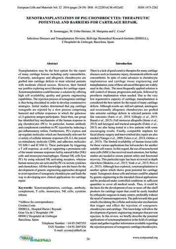
The effect and mechanism of miRNA-140 inexosomes of hypoxic bone marrow mesenchymalstem cells on articular chondrocytesYuezheng HuYuying Children’s Hospital of Wenzhou Medical UniversityHaiXiao LiuYuying Children’s Hospital of Wenzhou Medical UniversityDaoliang XuYuying Children’s Hospital of Wenzhou Medical UniversityXinhe XueYuying Children’s Hospital of Wenzhou Medical UniversityXinxian Xu ( a379806041@163.com )Yuying Children’s Hospital of Wenzhou Medical UniversityResearch ArticleKeywords: exosome, HIF-1α, articular chondrocyte, miR-140, bone marrow mesenchymal stem cellPosted Date: June 22nd, 2022DOI: : This work is licensed under a Creative Commons Attribution 4.0 International License.Read Full LicensePage 1/17
AbstractThis study aimed to investigate the effects and mechanisms of miRNA-140 in exosomes of hypoxic bonemarrow mesenchymal stem cells on articular chondrocytes. Articular chondrocytes were stimulated withIL-1β (10 μg/ml) to give them an inflammatory state. Exosomes were extracted by differential ultra-highspeed centrifugation, and their morphology was identified by transmission electron microscopy. The CellCounting Kit-8 (CCK-8) assay, cell scratch assay and flow cytometry method were utilized to assess theproliferation, migration and apoptosis of chondrocytes respectively. Expressions of miR-140, HIF-1αmRNA and mRNA associated with chondrocytes were investigated using quantitative reversetranscription PCR (qRT-PCR). Western blot was applied to assess the chondrocyte associated proteinsCaspase3, Collagen II, HIF-1α, SOX-9 and BMSC-Exo surface protein markers CD9, CD63, TSG101. Toobserve effects of inflammatory chondrocytes, immunohistochemistry was adopted to detect the stainingof Collagen I and Collagen II. Eventually, exosomes’ shape was almost round, and the scratch healingwas significantly increased in BMSC-Exo treated groups compared with the IL-1β group (P 0.001).Additionally, it was found that exosomes in the hypoxic state (Hypoxia-Exo) resulted in higher cellularactivity, less apoptosis and enhanced protein expression in inflammatory chondrocytes compared to thenormoxic state (Normoxia-Exo), while weakening adipose differentiation and enhancing chondrogenicdifferentiation. Furthermore, the healing effect of exosomes on inflammatory chondrocytes under hypoxicconditions was produced by a rise in miR-140 expression within them. In conclusion, under hypoxia,miRNA-140 in bone marrow mesenchymal stem cell exosomes promotes proliferation and migration ofchondrocytes and reduces apoptosis of chondrocytes.IntroductionOsteoarthritis (OA) is one of the leading causes of pain and disability worldwide and it affects more than250 million people worldwide[1, 2]. Osteoarthritis of the fingers, hip and kneecap are all considered to bethe major burden of the disease, which has a serious impact on people’s activities of daily living[3, 4].There are few cures for osteoarthritis and the focus is on reducing physical disability and managingpain[5–7].Mesenchymal stem cells (MSCs), with the potential of self-renewal and directed differentiation, can repaircartilage tissue and inhibit the secretion of inflammatory factors by chondrocytes[8]. Bone marrowmesenchymal stem cells (BMSCs) is one kind of the MSCs and BMSCs are superior in phenotype,morphology, function, and potential therapeutic applications compared with extraskeletal MSCs[9]. Themethod of obtaining BMSCs is very convenient and common, and they have the ability to repair bonetissue damage[10]. Activation of endogenous or exogenous BMSCs can repair long bone and vertebraefractures caused by osteoporosis or trauma and BMSCs can be used in preclinical and clinical Settings totreat bone-related diseases such as osteogenesis imperfecta[11, 12].Exosomes are a subset of extracellular vesicles that play a key role in normal and disease physiology, butexosomes are so small that it is difficult to do investigations[13]. Exosomes contain mRNAs, miRNAs,Page 2/17
enzymes, and lipids that, in addition to their structural roles and their function as vectors, play specificroles in this mode of cellular communication[14–16]. Studies have proved that normal cells and cancercells shed exosomes contain a large number of related proteins, nucleic acids, can be analyzed by thesesubstances to determine the type of cancer[17]. Moreover, exosomes improve heart function, reducemyocardial fibrosis, improve vascular remodeling, and are a new player in diabetes andcardiomyopathy[18–20]. Additionally, BMSC-derived exosomes alleviate osteoarthritis by promotingchondrocyte[8], which suggested that BMSCs-Exo have similar biological functions to BMSCs, that is,promoting bone regeneration. Therefore, this study will focus on the effect of BMSC-EXO on articularchondrocytes and preliminarily explore the mechanism of its effect.Hypoxia-inducible factor-1α (HIF-1α) is a subunit of hypoxia-inducible factor-1 (HIF-1), which can mediatehypoxia signal and regulate a series of compensatory responses to hypoxia through different signalingpathways with multiple upstream and downstream proteins, and play an important role in the growth anddevelopment of the body as well as physiological and pathological processes[21, 22]. HIF-1α can induceBMSCs to differentiate into chondrocytes, which can treat early cartilage defects caused byosteoarthritis[23]. However, the mechanism and process of miRNA-140's action with HIF-1α has not beenfurther investigated. In this study, we explored the effect of BMSC-Exo on articular chondrocytes underhypoxic conditions and attempted to investigate whether hypoxia-mediated upregulation of miR-140expression occurs in dependence on HIF-1α, thus further elucidating the mechanism of miR-140 effect onchondrocytes.Materials And MethodsAnimals4-week-old male SD rats with SPF grade, body weight about 100g, purchased from Changzhou CavensExperimental Animal Co., Ltd. The rats were free to move in the cage and eat normally at (25 2) .Cell extraction and cell cultureCulture of chondrocytes. The rats were killed by neck removal, and the knee joint of the rats wasseparated aseptic. The connective tissue of cartilage surface was removed, and hyaloid cartilage wastaken, washed with PBS (Thermo Fisher Scientific), and the tissue was cut into 1 mm3 pieces. 0.25%trypsin was predigested at 37 for 30 minutes, and the discard solution was added with 0.2% type IIcollagenase for digestion at 37 for 4h. Chondrocytes collected from digestion were suspended inDMEM medium (Thermo Fisher Scientific) containing 10% PBS and cultured at 37 and 5% CO2 for 8-10days.[24, 25].Culture of BMSCs. The rats were sacrificed by neck removal, disinfected with 75% alcohol and rinsed withPBS. The right tibia and femur of the rats were taken, the bone ends were removed, and the bone marrowcavity was exposed. The bone marrow cavity was fully rinsed with DMEM medium and the bone marrowPage 3/17
was collected, centrifuged at 1500 r/min for 10 minutes. Cells were collected and suspended again, andcultured in DMEM medium containing 15% FBS.Extraction and identification of ExosomesThe cultured BMSCs grew to about 90% of the surface area of the culture dish. The culture medium wasdiscarded, washed with PBS for 3 times, and the serum-free medium containing 1% penicillomycin DMEMwas added. After 24 hours of culture, the conditioned culture medium was collected and stored at 4 .Cell conditioned culture medium was collected to 300ml and centrifuged by supernatant centrifuge. Aftersupernatant was removed, the cell conditioned culture medium was cleaned and suspended by PBS. 10μlexosome suspension was dropped onto the copper wire, and the samples were dried at roomtemperature. Morphological characteristics of exosome were observed under transmission electronmicroscope. The exosomes added in this experiment were all Exo-100 μg/ml.CCK-8Cell Counting Kit-8 (CCK-8) was performed to test cell viability. The chondrocytes were inoculated on a 96well plate, and the cells were treated in groups after adherence, and cultured for 48 h. Then CCK-8 reagentwas added and incubated for another 4h. The absorbance value (OD value) of the cells was measured at560 nm of the microplate.Wound healing assayThe chondrocytes were cultured in 6-well plates to 70-80% confluency and were wounded with a 200-μlsterile pipette tip. After washing with PBS, the cells were cultured in serum-free medium. Images wereacquired at each time point (0 and 24 h).Flow cytometryThe apoptosis rates were determined by using Pharmingen annexin V-FITC Apoptosis Detection Kit I (BD,USA) according to the manufacturer’s instructions. Chondrocytes were inoculated with 6-well plates.When the cell density was about 80%, corresponding complete culture medium was added to each group,and cells were collected after 24 and 48 hours of culture. FITC and PI staining were performed accordingto the instructions of the kit, and the cell death was analyzed and quantified by flow cytometry within 1hour (BD Biosciences, USA).Quantitative real-time PCR (qRT-PCR)Total RNA was extracted using Trizol reagent (Thermo Fisher, USA) and reversely transcribed into cDNAaccording to manufacturer’s instructions (Takara Biological, Japan). Real-time PCR with gene-specificprimers was performed on the resulting cDNA using a Step One Plus real-time PCR system (AppliedBiosystems) in the presence of SYBR Green PCR Master Mix (Applied Biosystems). The relativeexpression level of mRNA was calculated using 2-ΔΔCt method.Page 4/17
Oil red O stainingThe saturated solution of oil red O (O8010; Solarbio, China) isopropyl alcohol was mixed with distilledwater in a ratio of 3:2 to prepare the dyeing solution and soaked for 20 minutes. The treated cells werewashed twice by PBS and fixed with 4% paraformaldehyde at room temperature for 30 min. For lipididentification, cells were stained with the prepared solution for 30 minutes and the absorbance wasmeasured at 490 nm.Toluidine blue stainingThe slides were sterilized, treated with polylysine, placed on plates and inoculated into 6-well plates withlow density chondrocytes. The cells were placed in a 5% CO2 37 C incubator and then fixed with 4%paraformaldehyde for 30 minutes. The fixate was rinsed with double steam water and dyed with 0.04%toluidine blue for 2h. The dye was removed with anhydrous ethanol and the cells were rinsed 3 times withPBS, followed by xylene cleaning and sealing. Tissue is viewed and photographed under a lightmicroscope.Expression and transfection of HIF-1αThe third generation rat BMSC with cell density of 1 106/ml were transfected with 5 ml human mediumand adenovirus vector carrying HIF-1α, and the medium was changed every 24 h after transfection. Thetransfection efficiency was observed by fluorescence microscope 3 days after transfection.Western blotChondrocytes were cultured in 6-well plates, treated with inflammatory factors and exosomes, andproteins were extracted from cell lysates RIPA buffer (Thermo Fisher, USA). BCA protein assay kit (P15111) (Applygen Technologies Inc. China) was applied to measure the protein concentration in lysatesaccording to the manufacturer’s instructions. Electrophoresis was performed by sodium dodecyl sulfatepolyacrylamide gel (SDS-PAGE) and then transferred to a polyvinylidene fluoride (PVDF) membrane(Millipore, USA). Then, the primary antibody was added and incubated overnight at 4 , and themembrane was washed 3 times every other day. The second antibody was added and incubated for 1h atroom temperature, and then the membrane was washed for 3 times before exposure. β-actin and GAPDHwere used as internal reference in the detection of proteins on exosomes and chondrocytes individually.Image J software was adopted to measure the gray value of protein bands.Immunohistochemistry (IHC)Formalin fixed tissue, paraffin-embedded tissue or paraformaldehyde fixed cells were stained byimmunohistochemistry. The fixed tissues or cells were infiltrated by blocking solution (0.1% Triton-x), thenincubated with primary antibody overnight at 4 C, and washed with PBS for 3 times before incubatingwith secondary antibody. The photos were collected with a digital camera (Nikon).Page 5/17
Statistical analysisAll data was analyzed by SPSS 20.0 and GraphPad Prism 6.0 (GraphPad Software, San Diego, CA). Theexperimental results are presented as the means standard deviation (SD). Student’s t-test and one-wayanalysis of variance were used to assess differences between groups. P 0.05 was consideredstatistically significant.ResultsEffects of BMSC on inflammatory articular chondrocytes and extraction and identification of exosomesUnder transmission electronic microscope, BMSC-Exo were round or oval in shape and had a completemembrane structure (Figure 1A). Besides, the expressions of surface specific markers CD9, CD63 andTSG101 were proved to be positive by Western blot (Figure 1B-C). Therefore, we proved that the extractionof exosomes was successful.CCK-8 experimental results showed that compared with the control group, the chondrocyte cell viabilitywas significantly lower under IL-1β inflammatory condition, while the survival of the BMSC-Exo groupwas higher than that of the IL-1β group (Figure 1D). Moreover, Western blot was also used to detect thechanges of apoptosis indicator Caspase3. The content of Caspase3 in IL-1β inflammatory condition wassignificantly higher than that in the control group, while Caspase3 levels in the BMSC-Exo group werelower than in the IL-1β group (Figure 1E-F). These results both suggested that BMSC can alleviate adversesymptoms of chondrocytes induced by IL-1β. In addition, we also detected the effect of BMSC-Exo onchondrocyte migration by wound healing assay. Compared with the control group, the cell migration wasinhibited in the IL-1β group and was significantly increased when Exo was added.(Figure 1G).Effect of Hypoxia-Exo on inflammatory chondrocytesBy CCK-8, we found that both normoxic and hypoxic conditions of Exo can lead to increased chondrocyteviability,compared with PBS IL-1β group, in which the IL-1β hypoxia-Exo group had a better effect.(Figure 2A). Identically, the expression levels of Collagen Type II on the surface of inflammatorychondrocytes also showed the same trend. The protein expression in the IL-1β Hypoxia-Exo group wassignificantly higher than that in the IL-1β Normoxia-Exo group compared with the PBS IL-1β group(Figure 2B-C). Furthermore, flow cytometry showed that the apoptosis rate of chondrocytes in the IL-1β Normoxia-Exo group was lower than that in the PBS IL-1β group, while the apoptosis rate ofchondrocytes in the IL-1β Hypoxia-Exo group was even lower than that in the IL-1β Normoxia-Exo group(Figure 2D). In addition, lipid differentiation ability and chondrogenic differentiation ability ofchondrocytes were detected by oil red O staining and toluidine blue staining respectively. Compared withthe IL-1β group, the IL-1β Exo group significantly reduced lipid differentiation and promotedchondrogenic differentiation of chondrocytes, and the effect of IL-1β Hypoxia-Exo group was moresignificant. (Figure 2E-F).Page 6/17
Effects of HIF-1α on BMSC-ExosTo further investigate the effect of hypoxia on chondrocytes, we evaluated miR-140 expression in BMSCand BMSC-exo under normoxic and hypoxic conditions, respectively. The experiments showed that therelative expression of miR-140 was much higher in hypoxia than in normoxia both in BMSC and BMSCExo (Figure 3A, p 0.001). In addition, we knocked down the HIF-1α gene in chondrocytes for experiments,and the results showed that the relative expression of miR-140 decreased dramatically in both hypoxicgroups after knocking down the HIF-1α gene, while the normoxic group showed essentially no change.(Figure 3B). Thus, Hypoxia-mediated upregulation of miR-140 expression occurs through HIF-1αEffect of miRNA-140 in BMSC-Exos on chondrocytesTo further investigate the effect of miR-140 on chondrocytes, we constructed OE-miR-140 mimics and
The slides were sterilized, treated with polylysine, placed on plates and inoculated into 6-well plates with low density chondrocytes. The cells were placed in a 5% CO2 37 C incubator and then xed with 4% paraformaldehyde for 30 minutes. The xate was rinsed with double steam water and dyed with 0.04% toluidine blue for 2h.