Transcription
Acta Medica OkayamaVolume 19, Issue 41965Article 2AUGUST 1965Histochemical studies on the red, white andintermediate muscle fibers of some skeletalmuscles. I. Succinic dehydrogenase activityand physiological function of intercostalmuscle fibersAkira Nishiyama Okayama University,Copyright c 1999 OKAYAMA UNIVERSITY MEDICAL SCHOOL. All rights reserved.
Histochemical studies on the red, white andintermediate muscle fibers of some skeletalmuscles. I. Succinic dehydrogenase activityand physiological function of intercostalmuscle fibers Akira NishiyamaAbstractFrom the histochemical study of the intercostal muscles of cat, the following results wereobtained. 1. Three different types of muscle fibers have been clearly distinguished in intercostalmuscles by histochemical demonstration of succinic dehydrogenase; namely, the white fibers arestained faintly, while the red fibers deep blue and the intermediate fibers purple or bluish purple.2. The difference in these stains is due to the degree of the enzyme activity, i. e., the areas ofhigh SDH activity are stained deep blue while those of relatively low SDH activity are stainedpurple. 3. At oil immersion magnification, the differences among the three types of fibers areclearly distinguishable by the amount, size, distribution pattern and subsarcolemmal precipitationof Nitro-BT formazan particles. 4. Concerning the spatial distribution of these three types of fibersin each intercostal muscle, the muscles in the cranial and caudal parts of thorax (I-IV, VIII-XII)show a higher proportion of red fibers, while those in the middle thorax show a higher proportionof white fibers. 5. The vertebral portion of the first internal intercostal muscle is composed of onlytwo types of fibers, red and intermediate ones, and their diameters are almost the same in size asin soleus muscle. In the middle intercostal muscle (V-VII), an intimate relationship can clearly beobserved between the size and the enzyme activity of muscle fibers as in the gastrocnemius muscle. 6. In comparison with the anatomy of thorax and the distribution of muscle fibers, it may bepresumed that there is a close relationship between the distribution and the scope of thorax movements, however, no definite relation between the distribution pattern and respiratory participationof muscle fibers. 7. Hence, it appears that the intercostal muscles in the cranial and caudal partsof thorax perform original respiratory movements, while the muscles in the middle thorax mainlyperform voluntary respiratory movements, perhaps display their function during forced breathing.The intermediate fibers may usually have some tonus and carry out the role of resisting ribs fromfalling inside by negative pressure of the thoracic cavity. PMID: 4223027 [PubMed - indexed for MEDLINE] Copyright c OKAYAMA UNIVERSITYMEDICAL SCHOOL
Nishiyama: Histochemical studies on the red, white and intermediate muscleActa Med. Okayama 19, 177-189 (1965)HISTOCHEMICAL STUDIES ON THE RED, WHITE ANDINTERMEDIATE MUSCLE FIBERS OF SOMESKELETAL MUSCLESI. SUCCINIC DEHYDROGENASE ACTIVITY ANDPHYSIOLOGICAL FUNCTION OF INTERCOSTALMUSCLE FIBERSAkira NISHIYAMADepartment of Surgery, Okayama University Medical School, Okayama, Japan(Director: Prof. S. Tanaka)Received for publication, August 2, 1965Since the classic work of RANVIER2lI, it is well known that the striatedmuscles of the mammals are composed of two different kinds of muscle; namely,red and white muscles. The former performs slow contraction and the latterdoes rapid one. DENNY-BROWNs showed that the red muscle is concerned withtonic postural adjustment and the white with phasic contraction. In electromyographic field NUMOTd6 and TOKIZANESG indicated that the kinetic motor unit wasinnervated by the pyramidal system, while the tonic motor unit was controlledby the extrapyramidal system.Since SELIGMAN and RUTENBERG28 published the histochemical technique forsuccinic dehydrogenase using Blue tetrazolium as an electron accepor, histochemical studies of the enzyme system in mammalian striated muscles have beenreported by several investigators. lS s:1 OGATA18 19 ,20 of our laboratory reported thatthe limb muscle fibers of the vertebrate can be divided into three types from thedifference in the activities of their histochemically demonstrable oxidative enzymes.On the other hand, two different opinions have been presented concerningfunctional differences of the intercostal muscles. PATON and ZAIMIS23 , studyingthe effect of neuromuscular blocking agents (D-tubocurarine and decamethonium), considered that the intercostal muscles of cat resembled the diaphragmand slow types of muscle such as soleus, rather than the fast types such astibialis anterior. However, BISCOE1 concluded that the intercostal muscles werefast muscle in cat from the observation of isometric contraction response to indirect stimulation. Regarding functional differences between the internal andexternal intercostal muscles, the opinions are also divided among physiologists.In the previous paper21 it has been demonstrated that three different typesof muscle fibers are clearly distinguished in the intercostal muscles of mammals177Produced by The Berkeley Electronic Press, 19651
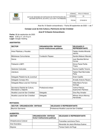
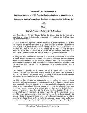
Acta Medica Okayama, Vol. 19 [1965], Iss. 4, Art. 2178A.NISHIYAMAby the difference in oxidative enzyme activities. In the present study detailedstructural differences in individual intercostal muscle fibers and spatial distribution of these three types of fibers in each intercostal muscle of cat were studiedby the histochemical demonstration of succinic dehydrogenase.MATERIALS AND METHODSThe intercostal muscle of healthy adult cats served as materials. Themuscle was removed immediately after death of the animals anesthetized withpentobarbital sodium 20 mg/kg body weight. Without fixation, cross-sectionswere made of muscle belly through a whole muscle and cut into 20-40 p. thickin a - 20 C cryostat and mounted on the slide glass. For the histochemicaldemonstration of succinic dehydrogenase, the sections were dried at room ternparature for about 5 minutes and incubated for 20-40 minutes at 37 C in substrate solution consisted of MI5 sodium succinate 5 ml, M/10 phosphate buffer,pH 7.4, 5 ml, Nitro-blue tetrazolium (-BT) 5 mg/3 ml, 6 ml and distilled water,10 ml. Then the sections were mounted with glycerine jelly.For the calculation of spatial distribution of the muscle fibers, respectivenumbers of three types of the fibers within a certain lens field were countedunder the light microscope and the average was taken of the percentagescounted in 12 adult cats. The diameter of the fibers was determined with theaid of micrometer scale.RESULTSIn transverse sections of the intercostal muscle of cat, three types of fiberswere clearly distinguished, i. e., generally small red fibers showed a higher SDHactivity while large white fibers a lower activity, and the third type of fibers,'intermediate fibers', showed a moderate activity between the red and whitefibers (Fig. 1). Furthermore, a careful observation at oil immersion magnification revealed the following characteristics of these three types of fibers individually, that is, white fibers: Fibers of this type were stained pale showingFig. 1 Intercostalis externus of cat, cross-section, succinic dehydrogenase. Note three types offibers, namely, the small fiOOr with a higher enzyme activity, the large fiOOr with a loweractivity and the third type of fiber, intermediate fiOOr, being intermediate in size with amoderate activity. X 125Fig. 2 Intercostalis externus of cat, longitudinal section, succinic dehydrogenase. x 125Fig. 3 Intercostalis externus of cat, cross-section, succinic dehydrogenase. Note three types offiOOrs, namely, the small fiber (R) containing a large number of diformazan granules andwith an active subsarcolemmal SDH activity. The large white fiOOr (W) with a few granulesreveals fine streaks in sarcoplasm. The intermediate fiOOr (M) contains a moderate numberof diformazan granules which compose a uniform network. X ol19/iss4/22
Nishiyama: Histochemical studies on the red, white and intermediate muscleSuccinic Dehydrogenase ActivityProduced by The Berkeley Electronic Press, 19651793
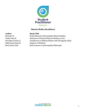
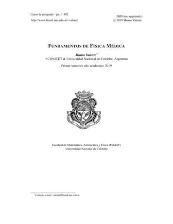
Acta Medica Okayama, Vol. 19 [1965], Iss. 4, Art. 2A.180NISHIYAMAa slight or no enzyme activity and a few granules, contammg numerous finestreaks in their sarcoplasm (Fig. 3, W). Red fibers: these were stained deepblue, showing a higher SDH activity with a large number of diformazan granules. These granules could be divided into two groups from their size, largerand smaller ones, and the granules were preferentially distributed toward theperiphery of the fibers. Particularly, this fiber showed a strong subsarcolemmalSDH activity with distinct accumulation of large diformazan particles (Fig. 3, R).Intermediate fibers: Fibers of this type were stained purple or bluish purple,showing an intermediate enzyme activity between the red and white fibers. Di·(%(%)100100505000357911- .","'''" 3-------.57.---911(c)7(d)Fig. 4 Spatial distribution of three types of fibers in intercostal muscles of the cat. Ordinateindicates the percentage of three types of fibers and abscissa indicates each intercostal spaces.(a) sternal region of intercost. ext.(b) vertebral region of intercost. ext.(c) sternal region of intercost. into(d) vertebral region of intercost. intosolid line: red fiberdotted line: white fiberbroken line: intermediate ol19/iss4/24
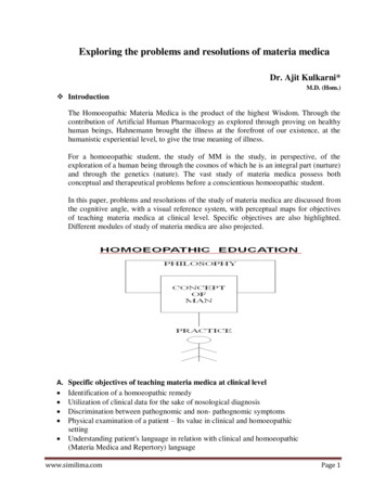
Nishiyama: Histochemical studies on the red, white and intermediate muscle1 1Succinic Dehydrogenase Activityf ormazan granules were generally smaller in size than those observed in the redfiber, and constituted a uniform network (Fig. 3, M).It has been further observed that the spatial distribution and the diameterof these three types of fibers vary considerably with respective intercostalmuscles. Therefore, the percentages of the fibers contained in a unit area ofthe muscle were continuously calculated. Anatomically, internal and externalintercostal muscles are distributed into two distinct regions, namely, intercartilagineous (sternal) and interosseous (vertebral). As a specimen of each region,parasternal and paravertebral parts of the intercostal muscles were chosen.As shown in Figures 4 and 5, the muscles in cranial and caudal parts of(u.)(fl)150150,.,.'.,,11,11 .100 .' ----.---- //100/505013759311(a)(p.)Co.)150'150100.--- . 5(b)7. .10050 .,. --.-5711.50399113-57911(d)Fig. 5 Diameters of three types of fibers in intercostal muscles of the cat. Ordinate indicatesthe diameter of three types of fibers and abscissa indicates each intercostal spaces.(a) sternal region of intercost. ext.(b) vertebral region of intercost. ext.(d) vertebral region of intercost. into(c) sternal region of intercost. intosolid line: red fiberdotted line: white fiberbroken line: intermediate fiber(c)Produced by The Berkeley Electronic Press, 19655
Acta Medica Okayama, Vol. 19 [1965], Iss. 4, Art. c.jp/amo/vol19/iss4/26
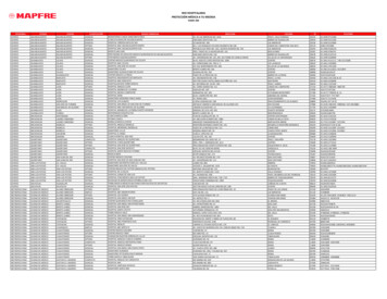
Nishiyama: Histochemical studies on the red, white and intermediate muscleSuccinic Dehydrogenase Activity183the thorax (I-IV and VIII-XII intercostal) show a higher ratio of red fibers andlower ratio of white ones. Particularly, the vertebral region of the first internalintercostal muscles is composed of only two types of fibers, red and intermediate,as soleus muscle (Fig. 4, d). Their diameters are almost the same (Fig. 6). Whilethe muscles in the middle thorax (V-VII intercostal) show a higher proportion ofwhite fibers. In this region, an intimate relationship between the size and theenzyme activity of muscle fibers can clearly be recognized, i. e., the fiber witha low enzyme activity has a larger diameter, while that with a high enzymeactivity a smaller diameter, and that with an intermediate degree of the activityan intermediate diameter (Figs. 7,8). However, there is no great differencein the distribution pattern of the intermediate fibers in respective intercostalmuscles, and as for the size and distribution of three types of fibers there is nomarked difference between the internal and external intercostal muscles orsternal and vertebral portions of the same intercostal spaces.DISCUSSIONHeterogeneity of the fibersWACHSTEIN and MEISELss first stated that succinic dehydrogenase showeddifferent activities depending upon different muscles of mammals. The relationship between this enzyme activity and the size of the fiber was first reported byNACHLAS and PADYKULA13 Concerning the other oxidative enzymes, similarrelationship has been reported by several workersu , i. e., small fibers with highoxidative enzymes are stained intensely, while large fibers with a low activityof these enzymes stained faintly.In the present investigation, hues of the staining varied when Nltro-BTwas used as a hydogen acceptor. The red fibers were stained deep blue, whileintermediate fibers purple or bluish purple, and the white fibers faintly. Generally, the observation of both red and blue stainings with the methods employingmany ditetrazoles gives rise to some confusion. SELIGMAN and R UTENBERd8 explained that the areas with a low enzyme activity are only able to reduce onehalf of ditetrazole and to yield a monoformazan which will be expected to becolored red. However, BURTNER et al.2 stated that this red compound is a monotetrazole as an impurity in commercial ditetrazoles. PEARSE 4 also indicated that1.Fig. 6 The vertebral portion of the first intercostalis internus of cat, cross-section, succinic dehydrogenase. One type of fiber CR) shows a higher enzyme activity while the other CM)a lower activity X 330Fig. 7 The VIth intercostalis externus of cat, cross·section, succinic dehydrogenase. Threetypes of fibers are clearly distinguished by their diameters and the enzyme activities. X 125Fig. 8 The VIth intercostalis externus of cat, cross-section, succinic dehydrogenase. Note threetypes of fibers (R, W, M). X 400Produced by The Berkeley Electronic Press, 19657
Acta Medica Okayama, Vol. 19 [1965], Iss. 4, Art. 2184A.NISHIYAMAboth red and purple compounds were consistently observed when using NitroBT. However, in this experiment no red stain in tissue sections has been notedwith our samples of pure Nitro-BT, in agreement with a study reported byNACHLAS et al. I'. Therefore, it may be assumed that the difference in stainability of these muscle fibers is attributable to the degree of enzyme activity,i. e., the areas with a higher SDH activity are stained deep blue, while thosewith a relatively low SDH activity are stained purple.It is well known that mitochondria contain many enzymes of TeA cycle.According to GREEN 9 mitochondria contain a complex electron transfer system,and one of these is the succinic dehydrogenase system. ODAl7 has demonstrated,using the electron microscope, that cytochemical reaction of the succinic dehydrogenase system is limited i mitochondria. NOVIKOFF et al. l6 also stated thatNitro-BT formazan was deposited inside the mitochondria. Thus, it is acceptedby most workers that this enzyme demonstrated by the tetrazolium techniquegives an accurate picture of mitochondria. Hence, it is obvious that the redmuscle fibers which participate in tonic contraction are rich in mitochondria,and supplied with abundant energy by the oxidative metabolism. Whereas thewhite muscle fibers which participate only in phasic movement have largeamounts of glycogen and phosphorylase',7,26 and supplied with energy for contraction by anearobic glycolysis.On the other hand, recent electron microscopic studies have revealed differences between the red and the white muscles with regard to the amount andthe distribution pattern of mitochondria and sarcoplasmic reticulm. As alreadymentioned in this study on the cat intercostal muscles, three types of musclefibers were clearly distinguished, especially the differences between the red andintermediate fibers were distinct by not only the size and enzyme activity of thefibers but also by the amounts, distribution pattern and subsarcolemmal precipitation of Nitro-BT formazan particles. Similar ohservations have been reportedby STEIN and PADYKULA 29 in limb muscle of rat. Recently OGATA22 has describedsimilar findings in limb muscles of mice using the electron microscope. That is,in the red fiber generally there are distinctly outlined accumulations of largemitochondria beneath the sarcolemma and in intrafibrillar space, while in theintermediate fiber usually mitochondria are sporadically dispersed in the subsarcolemmal space, and in the intrafibrillar space a pair of mitochondria areusually observed on both sides of Z-line. Sarcoplasmic reticulm is considerablywell developed in the white fibers but that of the red and intermediate fiberspoorly.It is well recognized that the muscle tonus can be classified into plastic andcontractile tonus, and that the red fibers participate in plastic tonus, while thewhite fibers in contractile tonus. However, the muscle which performs a vol19/iss4/28
Nishiyama: Histochemical studies on the red, white and intermediate muscleSuccinic Dehydrogenase Activity185ful contraction and requires contractile tonus contains denser sarcosomes in thered fiber (for example, diaphragm), while the muscle which performs isotoniccontraction contains small amount of sarcosomes in it. Furthermore, sarcoplasmic reticulm presumed to be a transmitter of the excitatory impulses to themuscle fiber, as described by OGATA22, is well developed in the white fiber butless in the red and the intermediate fibers. Thus, it may be assumed that theintermediate fiber is a slow contracting fiber and has a function similar to thered fiber. As described in KONDO'S report10, the white, intermediate and redmuscle fibers might be associated with phasic contraction, plastic tonus andcontractile tonus, respectively.2. Distribution of the fibersRegarding the spatial distribution of the three types of fibers, OGATA18 statedthat generally the muscle located near the body surface is rich in the white fiber,while deep muscle is rich in the red and intermediate fibers. TSUKAMOTO andMORI82 pointed out using the limb and jaw muscles of rat, rabbit and cat, thatthe white fibers are situated rather at the periphery and the red fibers rather atthe central part of the muscle. However, the length of the intercostal musclesof cat is generally short in the direction of muscle fiber, and there is no strikingdifference in the distribution of each type of fibers between the periphery andcentral parts of the muscle.In this study, it has been demonstrated that the intercostal muscles in cranialand caudal parts of the thorax show a higher proportion of red fibers, whilethat in the middle thorax a higher proportion of white fibers. These resultssuggest some functional differences under different situations of the intercostalmuscles in thorax.The movement of thorax during quiet breathing are managed by the cooperation of ribs, sternum, vertebra and diaphragm. In the case of man, thethorax enlarges downwards by contraction of diaphragm, forwards by elevatingribs but not backwards. However, as several workers have already pointed out,there is no concrete evidence that the physiological functions of respiratorymuscles of man and those of the quadrupedal animals are exactly identical.Considering anatomical relation of the thorax, the cranial part of the intercostal muscles is covered with many accessory respiratory muscles, namely, M.scalenus, M. transversus costarum and M. serratus anterior. Furthermore, inthis region the intercostal spaces are relatively narrower than that in otherregions. The action of intercostal muscles is closely related with the angle formedby the direction of muscle and rib6 The angles in the first and second intercostalmuscles are extremely small. On the other hand, the muscles in the middlethorax are considerably well developed and both the angle and the intercostalspace are wide. The muscles in the lower thorax, resembling those in the upperProduced by The Berkeley Electronic Press, 19659
Acta Medica Okayama, Vol. 19 [1965], Iss. 4, Art. 2186A. NISHIYAMAones, are poorly developed. Consequently, in comparison with the anatomy ofthorax described above and the spatial distribution of muscle fibers, it appearsthat there is a close relationship between the distribution pattern and the scopeof thorax movements.Though the function of the intercostal muscles in respiration has been thesubject of contentionl2, 27, these muscles are generally considered to have thefollowing functions, namely, the external intercostal muscles and intercartilagineous portion of the internal intercostal muscle are engaged in inspiratoryenlargement of the thoracic cavity and the interosseous portion of the internalintercostal muscle in its exspiratory diminution.According to GESSEL'S experimentS, using electromyography on dog, theinspiratory action potentials of external intercostal muscles are confined mostlyto the anterior half or two-thirds of the chest and dorsal aspects, approximatelyfrom the insertion of M. serratus anterior to the back, while the muscles belowthe VIth intercostal space show occasionally the opposite function, that is, theexspiratory participation. The intercartilagineous portion of the internal intercostal muscles in the 1st to Vth intercostal spaces actively participate in inspiration, while those below the VIth intercortal spaces mostly exspiration. TOKlZANEet a131 have also reported similar observations about man.Thus, the action of intercostal muscles is considerably complicated. Concerning this problem, KOTANIll postulates that the internal and external intercostal muscles are each functionally consisted of an elaborate mixture of bothinspiratory and exspiratory fibers, which are distributed with a density of beingable to manage harmonious respiratory movement. In the present study, therewas no essential difference in the distribution pattern of muscle fibers betweenthe internal and external intercostal muscles. Hence, we cannot find any definiterelation between the distribution and respiratory participation of different typesof muscle fibers.All these observations lead us to suggest that the intercostal muscles in thecranial and caudal parts of thorax, showing a higher proportion of red fibers,perform the tonic contraction, perhaps original involuntary respiratory movements. On the contrary, the muscles in the middle thorax, showing a higherproportion of white fibers, may perform mainly the phasic contraction, perhapsvoluntary respiratory movements, for instance, they may display their functionwith assistance of many other accessory respiratory muscles during forcedbreathing. While, intermediate fibers, correlating the plastic tonus, show aneary regular proportion in each intercostal muscle. So it may be consideredthat this fiber usually has some tonus and carries out the role in defense of theribs from caving in by negative pressure of the thoracic cavity. This hypothesismay give a clue to explain the fact that the respiratory action is endowed l19/iss4/210
Nishiyama: Histochemical studies on the red, white and intermediate muscleSuccinic Dehydrogenase Activity187the dual character of automaticity of the heart and the voluntary character ofthe skeletal muscle.SUMMA,RYFrom the histochemical study of the intercostal muscles of cat, the followingresults were obtained.i. Three different types of muscle fibers have been clearly distinguishedin intercostal muscles by histochemical demonstration of succinic dehydrogenase;namely, the white fibers are stained faintly, while the red fibers deep blue andthe intermediate fibers purple or bluish purple.2. The difference in these stains is due to the degree of the enzyme activity, i. e., the areas of high SDH activity are stained deep blue while those ofrelatively low SDH activity are stained purple.3. At oil immersion magnification, the differences among the three typesof fibers are clearly distinguishable by the amount, size, distribution pattern andsubsarcolemmal precipitation of Nitro-BT formazan particles.4. Concerning the spatial distribution of these three types of fibers in eachintercostal muscle, the muscles in the cranial and caudal parts of thorax (I-IV,VIII-XII) show a higher proportion of red fibers, while those in the middlethorax show a higher proportion of white fibers.5. The vertebral portion of the first internal intercostal muscle is composedof only two types of fibers, red and intermediate ones, and their diameters arealmost the same in size as in soleus muscle. In the middle intercostal muscle(V-VII), an intimate relationship can clearly be observed between the size andthe enzyme activity of muscle fibers as in the gastrocnemius muscle.6. In comparison with the anatomy of thorax and the distribution ofmuscle fibers, it may be presumed that there is a close relationship between thedistribution and the scope of thorax movements, however, no definite relationbetween the distribution pattern and respiratory participation of muscle fibers.7. Hence, it appears that the intercostal muscles in the cranial and caudalparts of thorax perform original respiratory movements, while the muscles inthe middle thorax mainly perform voluntary respiratory movements, perhapsdisplay their function during forced breathing. The intermediate fibers mayusually have some tonus and carry out the role of resisting ribs from fallinginside by negative pressure of the thoracic cavity.ACKNOWLEDGEMENTGrateful acknowledgement is made to Prof. S. TANAKA for his guidance and painstakingproofreading of this manuscript. The author also expresses his gratitude to Dr. T. OGATA andDr. T. KAWASHIMA for their valuable instructions and advices during the investigation.Produced by The Berkeley Electronic Press, 196511
Acta Medica Okayama, Vol. 19 [1965], Iss. 4, Art. 2188A. NISatYAMAREFERENCES1. BISCOE, T. J.: The isometric contraction characteristics of cat intercostal muscle.]. Physiol.164, 189, 19622. BURTNER, H. J. BAHN, R. C. & LONGLEY, J. B.: Observations on the reduction of tetrazolium salts in biological system. ]. Histochem. Cytochem. 4, 428, 19563. DENNY'-BROWN, D. E.: The histological features of striped muscle in relation to its functional activity. Proc. of Royal soc. of London 104B, 371. 19294. DUBOW ITZ, V. & PEARSE, A. G. E.: A comparative study of oxidative enzyme and phosphorylase activity of skeletal muscle. Histochemie 2, 105, 19605. FENNELL, R. A. & WEST, W. T.: Oxidative and hydrolytic enzymes of homozygousdystrophic and- heterozygous muscles of the house mouse. ]. Histochem. Cytochem. 11,374, 19636. Fl'KUDA, K.: Human physiology. Nanzando, Tokyo, 1948 (in Japanese)7. GEORGE, J. C. & NAIK, R. M.: Relative distribution and chemical nature of the fuelstore of the two types of fibers in the pectralis major muscle of the pigeon. Nature 181.709, 19588. GESSEL. R.: Individuality of breathing. Amer. ]. Physiol. 115, 168, 19369. GREEN, D. E.: Enzymes in metabolic sequences in Chemical Pathway of Metabolism,edited by Greenberg, D. M., Academie Press, New York. 195410. KONDO, M.: Histological studies on correlation between function and structure of skeletalmuscle. Part 1. Histological studies on correlation between function and structure of striatedmuscle fiber (in Japanese). Okayama I ggakai Zasshi 71. 3569. 195911. KOTANI, S.: On the participation of the intercostal muscles in the normal breathing (inJapanese). ]. Physiol. Soc., Japan 21. 146, 195912. MARTIN. H. N. & HARTWELL. E M.: On the respiratory function of the internal intercostal muscles. ]. Physiol. 2. 24. 187913. NACHMIAS. V. T. & PADYKULA. H. A.: A histochemical study of normal and denervatedred and white muscles of the rat. ]. Biophys. Biochem. Cytol. 4, 47, 195814. NACHLAS, M. M. & SELIGMAN, A. M.: Cytochemical demonstration of succinic dehydrogenase by the use of a new p-nitrophenyl substituted ditetrazole. ]. Histochem. Cytochem.5, 420, 195715. Nov IKOFF, A. B., SHIN, W. Y. & DRUCKER, J.: Mitochondrial localization of oxidativeenzymes: Staining results with two tetrazolium salts. ]. Biophys. Biochem. Cytol. 9, 47.196116. NUMOTO, M.: Electromyographycal studies on the regulation of the contraction of musclesin the central nervous system (in Japanese). Okayama Iggakai Zasshi 64. 1610. 195217. ODA, T.: Cytochemical and biochemical studies on the terminal electron transport system.H. Cytochemical demonstration of the succinic dehydrogenase system and cytochrome oxidase in JIlitochondria with the electron microscope (in Japanese). Symposia of tke Societyfor Cellular Chemistry 8. 195818. OGATA, T.: A histochemical study of the red and white muscle fibers. Part 1. Activityof the succinoxydase system in muscle fibers. Acta Medicinae Okayama 12, 216, 195819. OGATA, T.: A histochemical study of the red and white muscle fibers. Part H. Activityof cytochrome oxidase system in muscle fibers. Acta Medicinae Okayama 12. 228. 195820. OGATA, T.: A histcchemical study of the red and white muscle fibers. Part Ill. Activityof diphosphopyridine nucleotide and triphosphopyridine nucleotide diaphorase in musclefibers. Acta Medicinae Okayama 12, 233. 195821. OGATA, T., KAWASHIMA. T. & NISHIYAMA. A.: Histochemical demonstration of threetypes of muscle fibers of the intercostal muscles. Acta Medicinae Okayama 17. 257. l19/iss4/212
Nishiyama: Histochemical studies on the red, white and intermediate muscleSuccinic Dehydrogenase Activity18922. OGATA. T.: An electron microscope stud
were clearly distinguished, i. e., generally small red fibers showed a higher SDH activity while large white fibers a lower activity, and the third type of fibers, 'intermediate fibers', showed a moderate activity between the red and white fibers (Fig. 1). Furthermore, a careful observation at oil immersion magnifica