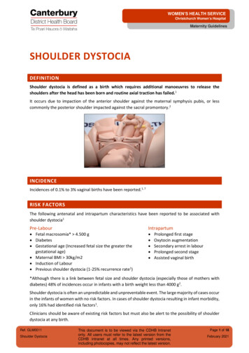
Transcription
Shoulder Dystocia: Managingan Obstetric EmergencyShoulder dystocia is an obstetric emergency in which normal traction on the fetal head does not lead to delivery of theshoulders. This can cause neonatal brachial plexus injuries, hypoxia, and maternal trauma, including damage to the bladder,anal sphincter, and rectum, and postpartum hemorrhage. Although fetal macrosomia, prior shoulder dystocia, and preexisting or gestational diabetes mellitus increases the risk of shoulder dystocia,most cases occur without warning. Labor and delivery teams should always beprepared to recognize and treat this emergency. Training and simulation exercises improve physician and team performance when shoulder dystocia occurs.Unequivocally announcing that dystocia is happening, summoning extra assistance, keeping track of the time from delivery of the head to full delivery of theneonate, and communicating with the patient and health care team are helpful.Calm and thoughtful use of release maneuvers such as knee to chest (McRobertsmaneuver), suprapubic pressure, posterior arm or shoulder delivery, and internal rotational maneuvers will almost always result in successful delivery. Whenthese are unsuccessful, additional maneuvers, including intentional clavicularfracture or cephalic replacement, may lead to delivery. Each institution shouldconsider the length of time it will take to prepare the operating room for general inhalational anesthesia and abdominalrescue and practice this during simulation exercises. (Am Fam Physician. 2020; 102(2): 84-90. Copyright 2020 AmericanAcademy of Family Physicians.)Shoulder dystocia is an obstetric emergency in whichgentle downward traction of the fetal head does not lead todelivery and additional maneuvers are required to deliverthe fetal shoulders.1 Shoulder dystocia is usually attributedto impaction of the anterior shoulder against the maternalsymphysis after delivery of the fetal head; less commonly, itis caused by impaction of the posterior shoulder against thesacral promontory.2Shoulder dystocia complicates 0.3% to 3% of all vaginal deliveries.3,4 The exact incidence can be difficult todetermine because the diagnosis is subjective and thereare no agreed upon diagnostic criteria for shoulder dystocia. Objective criteria of a head-to-body delivery intervalof 60 seconds or the need for additional delivery maneuvers are proposed based on the incidence of significantlymore birth injuries and lower Apgar scores during thesedeliveries.5CME This clinical content conforms to AAFP criteria forCME. See CME Quiz on page 81.Author disclosure: No relevant financial affiliations.Illustration by Christy KramesD. Ashley Hill, MD, AdventHealth Graduate Medical Education Program, Orlando, FloridaJorge Lense, MD, AdventHealth Orlando Hospital, Orlando, FloridaFay Roepcke, MD, MPH, AdventHealth Orlando Family Medicine Residency Program, Orlando, FloridaRisk Factors and PreventionRisk factors for shoulder dystocia include fetal macrosomia (odds ratio 16.1), prior shoulder dystocia (oddsratio 8.25), and preexisting or gestational diabetes mellitus (odds ratio 1.8).6-8 Other risk factors include maternalobesity, excessive maternal weight gain during pregnancy,oxytocin (Pitocin) use, prolonged first or second stagelabor, and operative vaginal delivery (forceps or vacuum); however, these are poorly predictive of shoulder dystocia.9There are no accurate models to predict or prevent shoulderdystocia.10,11Fetal macrosomia is difficult to accurately predict. Atterm, fetal sonography has at least a 10% margin of errorfor diagnosis of macrosomia.11 Although the incidence ofshoulder dystocia increases with increasing fetal weightand maternal diabetes, one study of pregnancies complicated by shoulder dystocia found that half of the neonatesweighed less than 4,000 g (8 lb, 13 oz) and that only 20% ofthe patients had diabetes.12 Results from studies evaluatinglabor induction for suspected macrosomia are inconsistent,and induction is not recommended to prevent shoulder dystocia.13 Given the increased risk of shoulder dystocia withDownloadedfromthe AmericanFamily Physician website at www.aafp.org/afp.Copyrightof Family102,Physicians.For2the afp 2020 American Academy VolumeNumber15,non2020commercial use of one individual user of the website. All other rights reserved. Contact copyrights@aafp.org for copyright questions and/or permission requests. Downloaded for Betty Burns (bburns@nccnet.org) at National Certification Corporation from ClinicalKey.com by Elsevier on August 11, 2020.For personal use only. No other uses without permission. Copyright 2020. Elsevier Inc. All rights reserved.
SHOULDER DYSTOCIAincreasing fetal weights,the American College ofObstetricians and Gynecologists (ACOG) recommendsconsideration of cesareandelivery for a patient whodoes not have diabetes andis carrying a fetus with anestimated fetal weight of5,000 g (11 lb). ACOG alsorecommends considerationof cesarean delivery for apatient who has diabetesand is carrying a fetus withan estimated fetal weight of4,500 g (9 lb, 15 oz).10ACOG and the AdvancedLife Support in Obstetricsprogram recommend thatlabor and delivery teamsconduct regular team training drills that include identification and managementof shoulder dystocia.10,14SORT: KEY RECOMMENDATIONS FOR PRACTICEBLongitudinal study of a mandatoryshoulder dystocia training programAnnounce unequivocally that there isa shoulder dystocia when it occurs.18BLongitudinal study of a mandatoryshoulder dystocia training programElevate both knees to the chest (McRoberts maneuver) as the first therapeuticmaneuver during shoulder dystocia.10BRetrospective analysis of shoulderdystocia casesConsider posterior arm delivery ifMcRoberts maneuver and suprapubicpressure are unsuccessful.10,14,21CClinical guidelines based on consensus, computer modeling, and aretrospective analysis of shoulderdystocia casesDocument precisely the head-to-bodydelivery interval and maneuvers performed after every shoulder dystocia.10CConsensus-based clinical guidelinesA consistent, good-quality patient-oriented evidence; B inconsistent or limited-quality patient-orientedevidence; C consensus, disease-oriented evidence, usual practice, expert opinion, or case series. Forinformation about the SORT evidence rating system, go to https:// w ww.aafp.org/afpsort.TABLE 1Complications of Shoulder DystociaMaternalLacerations of the bladder, urethra, vagina, anal sphincter,or rectumLateral femoral cutaneous neuropathyPostpartum hemorrhageSymphyseal separationUterine ruptureNeonatalFetal deathFetal hypoxic ischemic encephalopathyFracture of the clavicle or humerusNeurologic: brachial plexus injury, diaphragmatic paralysis, facial nerve injuries, Horner syndromeJuly 15, 2020 Volume 102, Number 2CommentsConduct team training simulation drillsthat include identification to improveperformance during actual shoulderdystocia emergencies.18ComplicationsShoulder dystocia can cause several maternal and neonatal complications (Table 1).10 The most common maternalcomplications are postpartum hemorrhage (11%) andobstetric anal sphincter injuries (3.8%).15 The most commonInformation from reference 10.EvidenceratingClinical recommendationneonatal injuries are brachial plexus injuries and clavicularor humeral fractures.16 Transient brachial plexus injuriesmay occur in up to 20% of deliveries complicated by shoulder dystocia.3 Most resolve without permanent disability,although approximately 10% may result in permanent neurologic injury.17 The head-to-body delivery interval doesnot predict fetal asphyxia or death.10 However, due to thepotential for serious maternal or neonatal harm, a systematic approach to expeditious delivery is necessary.10,15Initial ResponsePhysicians should announce delivery of the fetal head sothat an assistant can start a timer. If the fetus fails to deliverusing normal traction or if retraction of the fetal headagainst the perineum (turtle sign) occurs, the physicianshould announce that there is a shoulder dystocia, and thedelivery team should call for additional team members toassist. A longitudinal study of a shoulder dystocia simulationprogram found a significant reduction in neonatal brachialplexus injuries at discharge (7.6% to 1.3%) when the deliveryteam performed specified actions during shoulder dystociadeliveries.18 These included an unequivocal announcementof the shoulder dystocia, calling for additional assistancefrom qualified personnel, and having an assistant announcethe time from delivery of the fetal head every 30 seconds.www.aafp.org/afp American Family Physician 85Downloaded for Betty Burns (bburns@nccnet.org) at National Certification Corporation from ClinicalKey.com by Elsevier on August 11, 2020.For personal use only. No other uses without permission. Copyright 2020. Elsevier Inc. All rights reserved.
TABLE 2FIGURE 1Delivery Maneuvers for Shoulder DystociaCALL FOR HELPAdditional nurses, surgical obstetrics specialist, operating room team, anesthesia provider, and neonatal staffMcRoberts maneuverElevate both knees to chestSecondary maneuvers*Rubin II rotationalWoods corkscrew rotationalReverse Woods corkscrew rotationalGaskin all-foursSuprapubic pressureAssistant pushes above pubic bonetoward the side the fetus is facingConsider episiotomy for exposureDeliver posteriorarm or shoulderRotational maneuversRubin II: Place two to fourfingers along back of anteriorshoulder and rotate 30 degreesPlace hand between perineumand fetus and slide across fetalchest, grasp posterior arm,and sweep arm across chestWoods: Continue Rubin II andadd fingers to front of posteriorshoulder to rotate 180 degreesorPlace fingers from both hands,or a suction catheter, underthe posterior axilla and deliverthe posterior shoulder and armReverse Woods: Place fingerson the front side of theanterior shoulder and backside of the posterior shoulder and rotate 180 degreesGaskin all-fours maneuverConsider preparing operating roomorRepeat above maneuversConsider catastrophic maneuvers (abdominal rescue,cephalic replacement, intentional clavicular fracture)Management algorithm for shoulder dystocia.Physicians should also obtain assistance from a physician qualified to perform cesarean delivery and someone toresuscitate the neonate. Additional helpful actions that havenot been studied include communicating with the patientso that she knows when to push, lowering the bed, usinga stool for the assistant applying suprapubic pressure, andhaving someone record events for precise documentation.Delivery ManeuversIf the fetus does not deliver using gentle traction, releasemaneuvers can be used in a thoughtful and sequentialmanner to deliver the impacted shoulder (Figure 1). Aggressive lateral or downward traction on the fetal head andneck should be avoided because it can injure the brachial86 American Family PhysicianInitial maneuvers*McRobertsSuprapubic pressureDelivery of the posterior armMenticoglou posterior shoulder deliveryPosterior axillary sling tractionCatastrophic maneuversAbdominal rescue under general anesthesiaCephalic replacement (Zavanelli)Intentional clavicular fracture*—Listed in the suggested order that they should be performed.Information from references 10, 18, and 19.plexus.4 There are no randomized trials comparing thevarious maneuvers used to release an impacted shoulder 10 (Table 210,18,19). ACOG, the Royal College of Obstetricians and Gynaecologists, and the Advanced Life Supportin Obstetrics program recommend using the McRobertsmaneuver first, followed by suprapubic pressure if necessary.10,14,19 The McRoberts maneuver, performed by flexingthe hips and bringing both knees toward the chest, rotatesthe symphysis pubis cephalad and further opens the pelvic outlet (Figure 2). This is a simple and proven method tomanage shoulder dystocia, with a success rate of up to 42%as the sole maneuver.10,15If delivery does not occur, firm, steady suprapubic pressure should be performed concurrently with the McRobertsmaneuver. An assistant should apply firm downward oroblique pressure just above the symphysis pubis toward theside the infant is facing. This decreases the distance betweenthe infant’s shoulders (bisacromial distance), potentiallyassisting anterior shoulder dislodgement (Figure 3). Fundalpressure increases the risk of uterine rupture.20If the McRoberts maneuver and suprapubic pressure areunsuccessful, delivery of the posterior arm should be considered10,14,21 (Figure 4). A retrospective review revealed thatthe combination of the McRoberts maneuver, suprapubicpressure, and posterior arm delivery resulted in successfuldelivery within four minutes in 95% of cases.21 Computermodeling suggests that delivery of the posterior arm resultsin the least amount of brachial plexus stretch comparedwith other maneuvers.22 Delivery of the posterior armrequires patience and communication to keep the patientcalm. Training with a birth simulator will likely improveoperator confidence and performance of this procedure.www.aafp.org/afp Volume 102, Number 2 July 15, 2020Downloaded for Betty Burns (bburns@nccnet.org) at National Certification Corporation from ClinicalKey.com by Elsevier on August 11, 2020.For personal use only. No other uses without permission. Copyright 2020. Elsevier Inc. All rights reserved.
FIGURE 2SHOULDER DYSTOCIAMcRoberts maneuver. Flex the patient’s hips and bring her knees to her chest, which causes cephalad rotation of thematernal pelvis and flattening of the sacrum.Illustration by Christy KramesAn episiotomy may help depending on the size of the physician’s hands and ability to enter the posterior vagina, but itis not mandatory for this or any release maneuver.23 The physician should apply lubricant, compress all five fingers fromthe appropriate hand into a “duck-bill” shape, and gentlymaneuver the hand into the posterior vagina, under the baby(see video at https://www.aafp.org/afp/2020/0715/p84.html).The physician should then slide the hand along the fetal chest,not the back, up to the fetal hip, or until the posterior hand isidentified. Grasping the wrist by forming an OK sign with thephysician’s thumb and index finger (Figure 4), he or she shouldflex the fetal elbow and slide the arm along the fetal chest todeliver from the posterior vagina. Using the operator’s fifthfinger as a fulcrum by placing it along the fetal elbow may help.If the posterior arm is tight against the vaginal sidewalland cannot be delivered, other methods of delivering theposterior shoulder can be used. The Menticoglou maneuver involves placing one finger from each hand under theposterior axilla and applying gentle traction along the curveof the pelvis to deliver the posterior shoulder.24 After theshoulder delivers, it should be easier to deliver the entireposterior arm. The posterior axilla sling traction maneuver uses a suction catheter or urinary catheter placed underthe posterior shoulder axilla to apply downward traction todeliver the posterior shoulder.25 Alternatively, the physiciancan use the sling to rotate the posterior shoulder 180 degreesto anterior, similar to the Woods maneuver. A descriptionand video of this technique can be accessed at https:// w .July 15, 2020 Volume 102, Number 2Additional maneuvers include rotational methods (e.g.,Rubin II, the Woods or reverse Woods [corkscrew] maneuvers) and rolling the patient to her hands and knees (GaskinFIGURE 3Suprapubic pressure. An assistant applies firm downward or oblique pressure just above the symphysispubis toward the side the infant is facing.Illustration by Christy Krameswww.aafp.org/afp American Family Physician 87Downloaded for Betty Burns (bburns@nccnet.org) at National Certification Corporation from ClinicalKey.com by Elsevier on August 11, 2020.For personal use only. No other uses without permission. Copyright 2020. Elsevier Inc. All rights reserved.
FIGURE 4Elbow1. Grasp wristwith thumb andindex fingerand flex elbow2. Sweep handacross chestand deliverPosterior arm release. The physician’s hand enters thepelvis posteriorly and travels along the fetal chest tograsp the fetal posterior wrist using an OK sign. Theoperator’s hand should slide along the fetal chest,not the back, which may involve using the physician’snondominant hand depending on the direction thefetus is facing. Hooking the little finger around thefetal elbow may facilitate the maneuver. The arm isthen swept across the fetal chest.Illustration by Christy Kramesall-fours maneuver). To perform the Rubin II maneuver,the physician places two fingers into the vagina to push thescapula of the anterior fetal shoulder toward the fetal face toattempt to rotate the fetus 30 degrees (Figure 5; see video athttps://www.aafp.org/afp/2020/0715/p84.html).The Woods maneuver combines the hand placement for theRubin II maneuver with two fingers on the anterior aspectof the posterior fetal shoulder with the intent of rotating thefetus 180 degrees (Figure 5; see video at https://www.aafp.org/afp/2020/0715/p84.html). For the reverse Woods maneuver,fingers or hands are placed on the front side of the anteriorshoulder and back side of the posterior shoulder to rotatethe fetus 180 degrees. An episiotomy may be helpful for theWoods maneuvers to be able to gain access with two hands.The Gaskin all-fours maneuver requires the patient to rollonto her hands and knees. This had an 83% success rate asthe sole maneuver used in one series.26 This maneuver maybe more difficult if the patient is fatigued or has neuraxialanesthesia.Maneuvers for Catastrophic Shoulder DystociaIf these maneuvers do not result in delivery, options includeperforming the maneuvers again (Figure 1) or enlistingassistance from another experienced physician who mighttry the previously attempted maneuvers again or who canFIGURE 530 180 Rubin II maneuver (left): Place two fingers on the back (posterior) side of the anterior shoulder and rotate 30 degreestoward the fetal face. Woods maneuver (right): Continue the finger placement for the Rubin II maneuver and add twofingers to the front (anterior) of the posterior shoulder and rotate the fetus 180 degrees.Illustration by Christy Krames88 American Family Physicianwww.aafp.org/afp Volume 102, Number 2 July 15, 2020Downloaded for Betty Burns (bburns@nccnet.org) at National Certification Corporation from ClinicalKey.com by Elsevier on August 11, 2020.For personal use only. No other uses without permission. Copyright 2020. Elsevier Inc. All rights reserved.
SHOULDER DYSTOCIATABLE 3The AuthorsDelivery Maneuvers for CatastrophicShoulder DystociaAbdominal rescuePerform a hysterotomy incision and then manipulate thefetus to dislodge the shoulders, allowing vaginal delivery. 27Cephalic replacement (Zavanelli maneuver)Rotate the fetal head to a direct occiput anterior position,flexing the neck so the chin presses against the perineum,then push the head gently into the vagina.28 Apply continuous pressure to hold the head in place while anotherphysician performs a cesarean delivery to extract the babyabdominally. Relaxing the uterus with oral or intravenousnitroglycerin or inhalational anesthetics will likely make thisprocedure more successful, although this is unproven.Intentional clavicular fracturePull the clavicles outward to fracture one or both, collapsing the shoulders inward and allowing delivery. However,this can be difficult to perform because of the strength ofthe clavicles, and it may damage underlying vasculature.Blunt manipulation is recommended; avoid the use ofscissors or other sharp instruments.10D. ASHLEY HILL, MD, is the chair of the Department ofObstetrics and Gynecology at the AdventHealth Graduate Medical Education Program, Orlando, Fla.; a professorof obstetrics and gynecology at the University of CentralFlorida College of Medicine, Orlando; and the medical director of the Multiprofessional Obstetrics Simulation Training(M.O.S.T. ) program at AdventHealth Orlando.JORGE LENSE, MD, is the medical director of the AdventHealth Obstetric Hospitalists at the AdventHealth Altamonte,Celebration, Orlando (Fla.) and Winter Park Hospitals andAdventHealth Gynecology Hospitalists at AdventHealthOrlando Hospital.FAY ROEPCKE, MD, MPH, is a fellow at the Women’s HealthJunior Faculty Fellowship at AdventHealth Orlando Family Medicine Residency and is an Advanced Life Support inObstetrics (ALSO) course instructor.Address correspondence to D. Ashley Hill, MD, 235 East Princeton St., Ste. 200, Orlando, FL 32804 (email: D.Hill.MD@ AdventHealth.com). Reprints are not available from the authors.References1. Resnik R. Management of shoulder girdle dystocia. Clin Obstet Gynecol. 1980; 23(2): 559-564.Information from references 10, 27, and 28.2. Hankins GD, Clark SL. Brachial plexus palsy involving the posterior shoulder at spontaneous vaginal delivery. Am J Perinatol. 1995; 12(1): 4 4-45.collaborate to attempt less proven maneuvers, such asabdominal rescue, cephalic replacement (Zavanelli maneuver), and intentional clavicular fracture (Table 3).10,27,28 Eachinstitution should consider the length of time it will taketo prepare the operating room for general inhalationalanesthesia and abdominal rescue and practice this duringsimulation exercises.DocumentationPrecise documentation is extremely important after ashoulder dystocia to inform the clinical team of the delivery events, including the head-to-body delivery intervaland maneuvers used. ACOG has provided a checklist fordocumenting the occurrence of shoulder dystocia 12/08000/Patient Safety Checklist No 6 Documenting.43.aspx).29This article updates a previous article by Baxley and Gobbo. 30Data Sources: A PubMed search was completed in Clinical Queries using the key terms shoulder dystocia, shoulder, brachialplexus, and abnormal labor. The search included meta-analyses,randomized controlled trials, clinical trials, and reviews. Wealso searched Ovid, Clinical Key, Cochrane Library, Web of Science, the Agency for Healthcare Research and Quality evidencereports, and Essential Evidence Plus. Search dates: September 5,2019, and April 13, 2020.July 15, 2020 Volume 102, Number 23. Gherman RB, Chauhan S, Ouzounian JG, et al. Shoulder dystocia: theunpreventable obstetric emergency with empiric management guidelines. Am J Obstet Gynecol. 2006; 195(3): 657-672.4. American College of Obstetricians and Gynecologists; Task Force onNeonatal Brachial Plexus Palsy. Neonatal Brachial Plexus Palsy. American College of Obstetricians and Gynecologists; 2014.5. Beall MH, Spong C, McKay J, et al. Objective definition of shoulderdystocia: a prospective evaluation. Am J Obstet Gynecol. 1998; 179(4): 934-937.6. Tsur A, Sergienko R, Wiznitzer A, et al. Critical analysis of risk factors forshoulder dystocia. Arch Gynecol Obstet. 2012; 285(5): 1 225-1229.7. Bingham J, Chauhan SP, Hayes E, et al. Recurrent shoulder dystocia: a review. Obstet Gynecol Surv. 2010; 65(3): 183-188.8. Zhang C, Wu Y, Li S, et al. Maternal prepregnancy obesity and the risk ofshoulder dystocia: a meta-analysis. BJOG. 2018; 1 25(4): 407-413.9. Ouzounian JG, Gherman RB. Shoulder dystocia: are historic risk factorsreliable predictors? Am J Obstet Gynecol. 2005; 192(6): 1933-1935.10. Committee on Practice Bulletins—Obstetrics. Practice bulletin no. 178: shoulder dystocia. Obstet Gynecol. 2017; 1 29(5): e123-e133.11. Gupta M, Hockley C, Quigley MA, et al. Antenatal and intrapartum prediction of shoulder dystocia. Eur J Obstet Gynecol Reprod Biol. 2010; 151(2): 1 34-139.12. Ouzounian JG, Korst LM, Miller DA, et al. Brachial plexus palsy andshoulder dystocia: obstetric risk factors remain elusive. Am J Perinatol.2013; 30(4): 303-307.13. American College of Obstetricians and Gynecologists; Committee onPractice Bulletins—Obstetrics. Practice bulletin number 173: fetal macrosomia. Obstet Gynecol. 2016; 1 28(5): e195-e209.14. Shields SG, Ratcliffe S. Chapter F: labor dystocia. In: Leeman L, QuinlanJD, Dresang LT, et al. Advanced Life Support in Obstetrics Provider Manual. 8th edition. American Academy of Family Physicians; 2017: 1-14.www.aafp.org/afp American Family Physician 89Downloaded for Betty Burns (bburns@nccnet.org) at National Certification Corporation from ClinicalKey.com by Elsevier on August 11, 2020.For personal use only. No other uses without permission. Copyright 2020. Elsevier Inc. All rights reserved.
SHOULDER DYSTOCIA15. Gherman RB, Goodwin TM, Souter I, et al. The McRoberts’ maneuver for the alleviation of shoulder dystocia: how successful is it? AmJ Obstet Gynecol. 1997; 176(3): 656-661.16. Gherman RB, Ouzounian JG, Goodwin TM. Obstetric maneuvers forshoulder dystocia and associated fetal morbidity. Am J Obstet Gynecol.1998; 178(6): 1 126-1130.17. Gherman RB, Ouzounian JG, Miller DA, et al. Spontaneous vaginaldelivery: a risk factor for Erb’s palsy? Am J Obstet Gynecol. 1998; 178(3): 423-427.18. Grobman WA, Miller D, Burke C, et al. Outcomes associated with introduction of a shoulder dystocia protocol. Am J Obstet Gynecol. 2011; 205(6): 513-517.19. Royal College of Obstetricians and Gynaecologists. Shoulderdystocia (green-top guideline No. 42). March 28, 2012. Updated February 2017. Accessed March 11, 2020. https:// w delines/gtg42/20. Sturzenegger K, Schäffer L, Zimmermann R, et al. Risk factors of uterinerupture with a special interest to uterine fundal pressure. J Perinat Med.2017; 45(3): 309-313.21. Leung TY, Stuart O, Suen SS, et al. Comparison of perinatal outcomes ofshoulder dystocia alleviated by different type and sequence of manoeuvres: a retrospective review. BJOG. 2011; 1 18(8): 985-990.22. Grimm MJ, Costello RE, Gonik B. Effect of clinician-applied maneuverson brachial plexus stretch during a shoulder dystocia event: investiga-90 American Family Physiciantion using a computer simulation model. Am J Obstet Gynecol. 2010; 203(4): 339.e1-339.e5.23. Sagi-Dain L, Sagi S. The role of episiotomy in prevention and management of shoulder dystocia: a systematic review. Obstet Gynecol Surv.2015; 70(5): 354-362.24. Menticoglou SM. A modified technique to deliver the posterior arm insevere shoulder dystocia. Obstet Gynecol. 2006; 108(3 pt 2): 755-757.25. Cluver CA, Hofmeyr GJ. Posterior axilla sling traction for shoulder dystocia: case review and a new method of shoulder rotation with thesling. Am J Obstet Gynecol. 2015; 212(6): 784.e1-784.e7.26. Bruner JP, Drummond SB, Meenan AL, et al. All-fours maneuver forreducing shoulder dystocia during labor. J Reprod Med. 1998; 43(5): 439-443.27. O’Shaughnessy MJ. Hysterotomy facilitation of the vaginal delivery ofthe posterior arm in a case of severe shoulder dystocia. Obstet Gynecol.1998; 92(4 pt 2): 693-695.28. Sandberg EC. The Zavanelli maneuver: 12 years of recorded experience. Obstet Gynecol. 1999; 93(2): 312-317.29. American College of Obstetricians and Gynecologists. Patient safetychecklist no. 6: documenting shoulder dystocia. Obstet Gynecol. 2012; 120(2 pt 1): 430-431.30. Baxley EG, Gobbo RW. Shoulder dystocia. Am Fam Physician. 2004; 69(7): 1707-1714. Accessed March 11, 2020. https:// w fp Volume 102, Number 2 Downloaded for Betty Burns (bburns@nccnet.org) at National Certification Corporation from ClinicalKey.com by Elsevier on August 11, 2020.For personal use only. No other uses without permission. Copyright 2020. Elsevier Inc. All rights reserved.July 15, 2020
Shoulder dystocia is an obstetric emergency in which normal traction on the fetal head does not lead to delivery of the shoulders. This can cause neonatal brachial plexus injuries, hypoxia, and maternal trauma, including damage to the bladder, anal sphincter, and rectum, and postpartum hemorrhage. Although fetal macrosomia, prior shoulder .