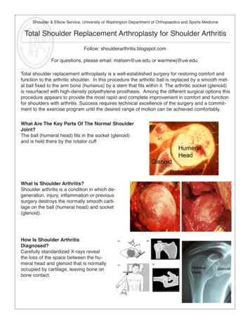
Transcription
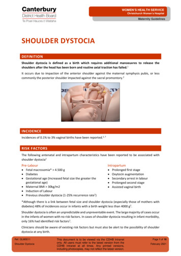
WOMEN’S HEALTH SERVICEChristchurch Women’s HospitalMaternity GuidelinesSHOULDER DYSTOCIADEFINITIONShoulder dystocia is defined as a birth which requires additional manoeuvres to release theshoulders after the head has been born and routine axial traction has failed.1It occurs due to impaction of the anterior shoulder against the maternal symphysis pubis, or lesscommonly the posterior shoulder impacted against the sacral promontory.2INCIDENCEIncidences of 0.1% to 3% vaginal births have been reported.1, 7RISK FACTORSThe following antenatal and intrapartum characteristics have been reported to be associated withshoulder dystocia1Pre-Labour Fetal macrosomia* 4.500 g Diabetes Gestational age (Increased fetal size the greater thegestational age) Maternal BMI 30kg/m2 Induction of Labour Previous shoulder dystocia (1-25% recurrence rate1)Intrapartum Prolonged first stageOxytocin augmentationSecondary arrest in labourProlonged second stageAssisted vaginal birth*Although there is a link between fetal size and shoulder dystocia (especially those of mothers withdiabetes) 48% of incidences occur in infants with a birth weight less than 4000 g2.Shoulder dystocia is often an unpredictable and unpreventable event. The large majority of cases occurin the infants of women with no risk factors. In cases of shoulder dystocia resulting in infant morbidity,only 16% had identified risk factors1.Clinicians should be aware of existing risk factors but must also be alert to the possibility of shoulderdystocia at any birth.Ref. GLM0011Shoulder DystociaThis document is to be viewed via the CDHB Intranetonly. All users must refer to the latest version from theCDHB intranet at all times. Any printed versions,including photocopies, may not reflect the latest version.Page 1 of 18February 2021
WOMEN’S HEALTH SERVICEChristchurch Women’s HospitalMaternity GuidelinesCOMPLICATIONSShoulder dystocia is an obstetric emergency that may lead to complications for both the woman andher baby including1:MATERNAL Postpartum haemorrhage (11%)Increased rates of 3rdand 4th degree tears (3.8%)Uterine ruptureBladder ruptureSymphyseal separationSacro-iliac joint dislocationPostpartum haemorrhagePsychological distress/traumaLateral femoral cutaneous neuropathyNEONATAL Brachial plexus injuries (BPI) (eg. Erb’s, Klumpke’s palsies, Total BPI) (2.3- 16%)1Clavicular and humeral fracturesHypoxic ischaemic encephalopathy and cerebral palsyPneumothoracesFetal/neonatal deathPREVENTIONInfants of mothers with diabetes have a 2-4 fold increased risk of shoulder dystocia compared withinfants of the same birth weight born to mothers without diabetes1.A decision-analysis model estimated that in women with diabetes with an EFW 4.5kg, 443 caesareansections would need to be performed to prevent one permanent BPI. In comparison, 3695 Caesareansections would be required to prevent one permanent BPI in the non-diabetic population1.The following recommendations are for specific situations:In cases of suspected macrosomia: With diabetes and estimated fetal weight 4.500 g or 95th Centile on Customised Growth Chart:elective caesarean section may be considered following discussion with Consultant Obstetrician1 Without diabetes: consider offering elective caesarean section if EFW is 5 kg or 99th centile onCustomised Growth Chart, following discussion with Consultant ObstetricianIn cases of maternal diabetes: Maintain tight glycaemic control Growth scans to estimate fetal weight, and interval growth at (28) 32, 36 weeks gestation to beplotted on customized growth chart. Monitor weight gain during preconception and pregnancy careRef. GLM0011Shoulder DystociaThis document is to be viewed via the CDHB Intranetonly. All users must refer to the latest version from theCDHB intranet at all times. Any printed versions,including photocopies, may not reflect the latest version.Page 2 of 18February 2021
WOMEN’S HEALTH SERVICEChristchurch Women’s HospitalMaternity GuidelinesIn cases of previous shoulder dystociaA discussion with a Consultant Obstetrician should be offered (as per Ministry of Health 2012 referralguidelines code 301619). A decision for either caesarean section or vaginal birth may be appropriateafter taking into consideration: The severity of previous shoulder dystocia Manoeuvres required Maternal trauma Maternal incontinence Neonatal morbidity Fetal size in this pregnancy Maternal choice Maternal mental health particularly PTSD (Post Traumatic Stress Disorder) Risk of repeat shoulder dystocia reported rates are 1- 25%1 (rates may be underestimated ascaesarean sections are performed for many women with prior shoulder dystocia or BPI’s).Regular training for all health professionals attending births is essential for the prevention andreduction of the complications from shoulder dystocia1. Training needs to not just be knowledge basedand should include recognizing that shoulder dystocia is an emergency, calling for appropriate helpand participating in hands on skills and team work practice.RECOGNITIONTimely management requires prompt recognition of the shoulder dystocia. These warning signs mayoccur: Difficulty with birth of face and chin When the head is born it remains tightly applied to the vulva or retracting into the perineal body(turtle-neck sign) Delay or failure of restitution of fetal headHowever, shoulder dystocia should be declared when gentle routine, axial* traction does not free theshoulder.*axial traction: in line with the fetal spineRef. GLM0011Shoulder DystociaThis document is to be viewed via the CDHB Intranetonly. All users must refer to the latest version from theCDHB intranet at all times. Any printed versions,including photocopies, may not reflect the latest version.Page 3 of 18February 2021
WOMEN’S HEALTH SERVICEChristchurch Women’s HospitalMaternity GuidelinesMANAGEMENTFlowchart for the management of shoulder dystocia is included below.This flowchart begins with simple measures and leads to more invasive manoeuvres. The order of theseactions may differ according to clinical circumstances (e.g. maternal obesity, maternal ability to changeposition, existing or available analgesia/anaesthesia). However, as a general principle do the simplemeasures first and do them well as the incidence of maternal and fetal trauma is higher in those birthswhere internal manoeuvres are performed.Do not delay. Fetal hypoxia will worsen the longer the delay between the birth of the head and thebirth of the body (approximate drop in blood pH of 0.01 per minute).Studies have demonstrated an increasing head to body interval time and drop in cord arterial pH wasrelated to prior abnormal fetal heart rate and subsequent increased incidence of hypoxic-ischaemicencephalopathy (HIE) 9, 10.The three crucial manoeuvres for the resolution of shoulder dystocia can be summarised as below. Itis recommended that we instigate the least intervention manoeuvres first, then progress through tointernal manoeuvres. Repeat the cycle if necessary.‘The 3 P’s’: Position, Pressure, Per Vaginum (PV)PositionPressurePVRef. GLM0011Shoulder DystociaThis document is to be viewed via the CDHB Intranetonly. All users must refer to the latest version from theCDHB intranet at all times. Any printed versions,including photocopies, may not reflect the latest version.Page 4 of 18February 2021
WOMEN’S HEALTH SERVICEChristchurch Women’s HospitalMaternity GuidelinesFLOWCHART FOR THE MANAGEMENT OF SHOULDER DYSTOCIACALL FOR HELP: Activate Emergency BellAnnounce: ‘shoulder dystocia’ as help arrives Midwifery Obstetric Neonatal Discourage pushingAvoid excessive tractionPrepare for newborn resusPrepare for PPHPOSITION Knees to shoulders (McRoberts) & repeatmovement if necessary Roll over onto ‘all fours’ (hands and knees/thighsto abdomen/chest) ‘Running start’ (flex hip, bend leading knee) Side lying (lie on side with leg raised)PRESSURE Stand on same side as fetal backGet above abdomenPlace ‘CPR hands’ just above symphysis pubisRocking or constant motionConstant and/or rocking pressurePushing direction is towards floor and away fromyourselfWith each new manoeuvre: Pull slowly & gently Pull in line with fetal spine (axial,not down or up)PV (PER VAGINUM) -INTERNAL MANOEUVRESWhole hand enters the pelvis posteriorly. Order ofmanoeuvres depends on which fetal part is available: Axillary traction Delivery of posterior arm Rotation of either shoulder into the obliquePost birth Document all actions on proforma (Ref.2407295)Arrange neonatal review and follow-upDebrief of parents and team by ObstetricianComplete ‘Safety First’ incident formMore help may be needed.Inform consultants: Obstetric Anaesthetic NeonatalConsider: Cleidotomy Zavenelli manoeuvre SymphysiotomyRefer to guideline detail below (GLM0011)Ref. GLM0011Shoulder DystociaThis document is to be viewed via the CDHB Intranetonly. All users must refer to the latest version from theCDHB intranet at all times. Any printed versions,including photocopies, may not reflect the latest version.Page 5 of 18February 2021
WOMEN’S HEALTH SERVICEChristchurch Women’s HospitalMaternity GuidelinesMANAGEMENT OF SHOULDER DYSTOCIA DETAIL (FLOWCHART ABOVE)CALL FOR HELP/INITIAL ACTIONS Activate red bell in room/ Birthing Suite emergency bellCall the Obstetric and Neonatal teamsNote the time of birth of the headInform the woman and her support people of the situationDiscourage maternal pushingAvoid excessive traction on fetal headDeclare the emergency: “This is a shoulder dystocia” to teams entering roomRequest a timekeeper/ documenter- use proforma in drawer in every birthing roomPrepare for neonatal resuscitationPrepare for postpartum haemorrhageCall for advanced help- obstetric, anaesthetic, if not arrived with emergency bellIn secondary/tertiary unit Activate red bell ‘in room’ /Birthing Suite emergency bell, to immediately call those on same floor Obstetric Emergency and Neonatal Emergency: Dial 777/Inform operator: “obstetric emergency” and “neonatal emergency” Request obstetric emergency team and neonatal team Advise location e.g. Birthing Suite, Room number #. This will bring: Obstetric Registrar, Anaesthetic Registrar, Birthing Suite ACMM, Maternity WardACMM, Obstetric House Officer, Neonatal ACNM and Neonatal Registrar or Clinical NurseSpecialist. Neonatal Consultant to be requested as required and as decided by Neonatal team present.In primary unitFast page on 777 – ACMM and/or obstetric teamDial 111 for ambulance and state “Code One” (lights and siren)Ref. GLM0011Shoulder DystociaThis document is to be viewed via the CDHB Intranetonly. All users must refer to the latest version from theCDHB intranet at all times. Any printed versions,including photocopies, may not reflect the latest version.Page 6 of 18February 2021
WOMEN’S HEALTH SERVICEChristchurch Women’s HospitalMaternity GuidelinesTHE 3 P’S:1P OSITION P RESSURE P V ( PER VAGINUM)P OSITIONAim To Increase functional size of bony pelvis Dislodge fetal shoulder due to pelvic movementOnce the position has been changed use of axial traction in any of the following positions must beslow, gentle and in line with the fetal spine.The choice of which position to use to relieve the shoulder dystocia will depend on what position thewoman has adopted to birth and whether or not she has had a regional block.i)Knees to shoulders (McRoberts' Manoeuvre) (Figure 1)Lie the bed flat, ask and/or assist the woman to move her buttocks to edge of bed and to bendher knees towards her shoulders so her buttocks are tilted up and off the bed.Practitioner must avoid excessive pushing back of the woman’s legs, particularly if she has adense epidural/spinal block and cannot report hip pain.Can be used with suprapubic pressure to increase success“Repeat McRoberts” may also assist: take the woman’s legs into a straight position and thenreturning knees to shoulders, as further movement of the maternal pelvis may dislodge a fetalshoulder that did not move at first McRoberts attempt.Figure 1Special note Some practitioners also perform prophylactic McRoberts. Whilst this does increase the woman’spelvic dimensions there is no evidence that doing it ‘in anticipation’ is of benefit. It is important to alert the attending team to be ready to perform a McRoberts’ Manoeuvre in theevent that restitution does not follow, and a shoulder dystocia occurs.Ref. GLM0011Shoulder DystociaThis document is to be viewed via the CDHB Intranetonly. All users must refer to the latest version from theCDHB intranet at all times. Any printed versions,including photocopies, may not reflect the latest version.Page 7 of 18February 2021
WOMEN’S HEALTH SERVICEChristchurch Women’s HospitalMaternity Guidelinesii)Roll over to all fours(hands and knees or facing prone move thighs to abdomen/chest)Ask the woman to roll over facing the floor so that she is supporting herself on her hands withher hips flexed and knees bent.Or lean forward so chest and head are on the floor and hips and knees flexed and splayed.Buttocks are raised in the air.Apply routine axial traction to attempt to deliver the baby.Figure 2: All foursiii)Figure 3: Thighs to abdomen/chestRunning startWhile in hands and knees position, ask the woman to place one foot flat in front of her, withknee bent and hip flexed. This is a modified ‘one-sided’ McRoberts manoeuvre.iv)Side lyingAsk the woman and/or assist her into a side-lying (lateral) position with upper leg raised andpulled back towards her shoulderRef. GLM0011Shoulder DystociaThis document is to be viewed via the CDHB Intranetonly. All users must refer to the latest version from theCDHB intranet at all times. Any printed versions,including photocopies, may not reflect the latest version.Page 8 of 18February 2021
WOMEN’S HEALTH SERVICEChristchurch Women’s HospitalMaternity Guidelines2P RESSUREApplied suprapubically – above the symphysis pubisAimTo decrease the bisacromial (shoulder to shoulder) diameter by adduction of shoulders. To rotate theanterior shoulder to the wider oblique diameter of the pelvis.To free the anterior shoulder to slip underneath the symphysis pubis with the aid of routine axialtraction. Mother’s legs maintained in the “knee to shoulder” (McRoberts) position Determine position of fetal back by abdominal palpationApply pressure on same side as fetal back to allow the shoulders to ‘scrunch’ (adduct) in towardsthe fetal chest. Position yourself above the abdomen to be able to use your upper body effectively. Place hands in “CPR position” just above the symphysis pubis, onto the back of baby’s anteriorshoulder Apply direct firm pressure downwards to shift the baby’s shoulder towards the maternal spinewhich may be enough to slide the shoulders through the pelvic brim. Simultaneously direct firm pressure laterally (towards the floor and away from yourself) to reducethe bisacromial diameter by curling the baby’s shoulder over towards its chest and also by rotatingthe shoulder away from the anterior pelvic position and into a more oblique position to allow abetter diameter for birthing. Provide firm constant and/or rocking pressure (no evidence one is more successful than the other). A second practitioner to perform gentle axial traction to the baby’s head and if the shoulder hasdislodged to safely deliver the baby.Ref. GLM0011Shoulder DystociaThis document is to be viewed via the CDHB Intranetonly. All users must refer to the latest version from theCDHB intranet at all times. Any printed versions,including photocopies, may not reflect the latest version.Page 9 of 18February 2021
WOMEN’S HEALTH SERVICEChristchurch Women’s HospitalMaternity Guidelines3P ER VAGINUM (PV) FOR INTERNAL MANOEUVRESAimChange relationship of bisacromial diameter with the bony pelvis (i.e. rotate)Allow more space for internal manoeuvres to be performed Advise woman of need to perform internal manoeuvres and gain verbal consent. The decision as to which manoeuvre to make will depend on the clinical circumstances. Make hand as small as possible, as though inserting hand into a tube or putting on a solidbracelet/bangle. Figures 4, 5 and 6 Consider a second practitioner to gently tilt the baby’s head up and out of the way allowing morespace to get a hand into the vagina. Take care to avoid excessive strain on nerves and tissues.Fig 7 Insert whole hand per vaginum entering posteriorly. If unable to insert whole hand, consider episiotomy. A systematic review11 showed no evidencesupporting the routine use of episiotomy in preventing or managing a shoulder dystocia. Nor doesit decrease the risk of brachial plexus injuries (BPI).12 The only indication for use would be tofacilitate access to the pelvis in the setting of a tight perineum. Inserting the whole hand is more likely to allow reach of the axilla or posterior arm rather thanusing the two fingers most commonly used for vaginal examination. There is likely to be ample space once whole hand is in the sacral hollow. Once hand rests in sacral hollow, reach along posterior vaginal wall to find fetal landmarks.Ref. GLM0011Shoulder DystociaFigure 4Figure 5Figure 6Figure 7This document is to be viewed via the CDHB Intranetonly. All users must refer to the latest version from theCDHB intranet at all times. Any printed versions,including photocopies, may not reflect the latest version.Page 10 of 18February 2021
WOMEN’S HEALTH SERVICEChristchurch Women’s HospitalMaternity GuidelinesDepending on what is found on PV proceed to either removal of the posterior arm or axillary traction.If neither of these works then proceed to internal rotational manoeuvres.Posterior arm removala) If baby’s arm is lying flexed across its chest ‘, grasp hand and wrist and gently sweep the arm acrossthe baby’s chest and face.b) If the hand is not lying across the chest, i.e. arm is straight by fetal side; slide your hand along thefetal arm and bend it at the elbow, by pressing on the antecubital fossa (front of elbow). Sweepthe baby’s arm across chest and then grasp wrist and remove.Ref. GLM0011Shoulder DystociaThis document is to be viewed via the CDHB Intranetonly. All users must refer to the latest version from theCDHB intranet at all times. Any printed versions,including photocopies, may not reflect the latest version.Page 11 of 18February 2021
WOMEN’S HEALTH SERVICEChristchurch Women’s HospitalMaternity GuidelinesAxillary traction15 (photos used with permission by Lesley Ansell6)Enter posteriorly – slide hand along fetal head toneckGrasp posterior shoulderGrasp – circle first finger and thumb around axilla2nd finger placed on top of arm – keep arm firmlyagainst chest and apply traction through axilla onlyApply axillary traction to follow the curve of thesacrumFirm traction may be neededAnterior shoulder ‘PIVOTS’ around symphysis –posterior shoulder delivered firstRef. GLM0011Shoulder DystociaThis document is to be viewed via the CDHB Intranetonly. All users must refer to the latest version from theCDHB intranet at all times. Any printed versions,including photocopies, may not reflect the latest version.Page 12 of 18February 2021
WOMEN’S HEALTH SERVICEChristchurch Women’s HospitalMaternity GuidelinesSpecial notes Another manoeuvre called posterior axillary sling traction (PAST9) has been proposed forintractable shoulder dystocia. This manoeuvre which involves the use of a sling (often suctiontubing) is not recommended by RANZCOG 20208 as “there is no real reliable evidence to support itswide use”. It is also known to cause significant morbidity, one study showed one third of liveborncases sustained a brachial plexus injury employing this technique and 15% sustained a fracturedhumerus9. There is insufficient evidence for CDHB to recommend the practice of ‘follow through’ or deliveringthrough’ in anticipation of shoulder dystocia, where the body is delivered immediately after thehead without waiting for restitution to prevent shoulder dystocia. It is possible that by interruptingthe normal mechanism of birth this manoeuvre may actually lead to impaction of the shoulderswhich may not have otherwise occurred.Internal rotation4&5Apply pressure to fetal shoulder(s) in order to rotate in a clockwise or anticlockwise direction into thelarger oblique diameter of the pelvis. The posterior shoulder will be most accessible.PLEASE NOTE: a manoeuvre called ‘shoulder shrug’ has been published in 201917. However, thepaper was based on 3 cases only with no reports of severe outcomes i.e. only 1st & 2nd degreeperineal tears and a brachial plexus injury which resolved by 2 weeks. There is insufficient evidenceto recommend this manoeuvre as there is no clear evidence for safety and efficacy.Ref. GLM0011Shoulder DystociaThis document is to be viewed via the CDHB Intranetonly. All users must refer to the latest version from theCDHB intranet at all times. Any printed versions,including photocopies, may not reflect the latest version.Page 13 of 18February 2021
WOMEN’S HEALTH SERVICEChristchurch Women’s HospitalMaternity GuidelinesIf position, pressure and PVmanoeuvres have not beensuccessful, consider ‘last resort’manoeuvres and call for seniorhelp to attend (if on site) or fortelephone assistance (incommunity setting), includingobstetric, anaesthetic andneonatal practitioners.Continue to reattempt the “3 P’s”in a cycle whilst awaitingadvanced help arrival or transfer.FURTHER INTERVENTIONThere are several ‘last resort’ manoeuvre options. They are rarely used in a high resource setting. In aconsecutive series of 17,000 births in the UK all shoulder dystocia’s were resolved, using the nationalguideline, without the need for last resort manoeuvres18.Some manoeuvres are more common in low resource countries, especially when there is no readyaccess to caesarean section.CleidotomyThis internal pelvic manoeuvre is the only non-surgical manoeuvre in this ‘last resort’ group It is thequickest to perform and least traumatic for the mother, so could be attempted whilst set up for thesurgical manoeuvres. Upward pressure with fingers onto the baby’s clavicle to cause a fracture This decreases the bisacromial diameterSymphysiotomyThis manoeuvre which divides the symphysis pubis to increase the diameter of the pelvic outlet isprimarily used in low resource countries Local anaesthetic injected over symphysisInsert a Foley catheterVaginal hand displaces urethra laterallySkin incision down to symphysisScalpel blade to divide the anterior fibres of the symphyseal ligamentAssistant should support the pelvis on either side to prevent excessive tractionSymphysis will then spread, allowing delivery.Ref. GLM0011Shoulder DystociaThis document is to be viewed via the CDHB Intranetonly. All users must refer to the latest version from theCDHB intranet at all times. Any printed versions,including photocopies, may not reflect the latest version.Page 14 of 18February 2021
WOMEN’S HEALTH SERVICEChristchurch Women’s HospitalMaternity Guidelines Support pelvis postpartum with a binder or support belt. High incidence of serious maternal morbidity and poor neonatal outcome16Zavanelli manoeuvreReplacement of the fetal head back into the vagina followed by emergency caesarean delivery Acute tocolysis will be required. Options are; terbutaline 0.25 mcg sub cutaneous injection,sublingual glyceryl trinitrate Flex the fetal head to replace. Place fingers of one hand over fetal maxilla (cheek bones) and the other on the fetal occiput. Push the maxillae towards and up into the vagina whilst flexing the occiput. Replacement success rates vary High proportion of fetuses will have irreversible hypoxic acidosis by this stage This manoeuvre may not reduce the incidence of BPI’s.POSTPARTUM CARE1.2.3.4.5.6.7.Active management of 3rd stage.Be prepared for postpartum haemorrhageBe prepared for newborn resuscitation.Perform thorough examination for perineal, vaginal and cervical injuries, with adequate painrelief.Debrief the family/ whanau and team members involved.Manage neonatal injuries in accordance with neonatal handbook (GLN0001, Ref: 235777)Do not transfer out of the tertiary/ secondary facility if maternal or fetal injury has occurred.This is to ensure appropriate post event care including opportunities for debrief andarrangement of follow-up.DOCUMENTATION Complete the shoulder dystocia documentation form (Ref.2407295) (see Appendix 1) Complete a Safety First incident report.SUMMARYShoulder dystocia is a life-threatening situationRecognise risk factors and be alert at every birthDeclare an emergencyStay calm and focussedWork through flowchart- The 3 ‘P’s.Record timing of events and management on the Shoulder Dystocia Documentation form(Ref.2407295) Arrange debrief and follow-up for family/whanau and the team Ref. GLM0011Shoulder DystociaThis document is to be viewed via the CDHB Intranetonly. All users must refer to the latest version from theCDHB intranet at all times. Any printed versions,including photocopies, may not reflect the latest version.Page 15 of 18February 2021
WOMEN’S HEALTH SERVICEChristchurch Women’s HospitalMaternity GuidelinesREFERENCES1.Crofts, J., Draycott, T, Montague, I. Winter, C. & Fox, R. (2012 Updated 2017) Royal College ofObstetricians & Gynaecologists (RCOG) Greentop Guideline 42 - Shoulder Dystocia; 2nd editionwww.rcog.org.uk2.Baskett T. & Allen, A. (1995) Perinatal Implications of Shoulder Dystocia. Obstet Gynaecol; 86; 14-173.Gobbo E. & Baxley E. (2000) Shoulder Dystocia American Academy of Family Physicians Advanced LifeSupport in Obstetrics (ALSO), 4th Edition4.Rubin A. Management of shoulder dystocia. JAMA 1964; 189:835-7.5.Woods CE, Westbury NYA. A principle of physics as applicable to shoulder delivery. Am J Obstet Gynecol1943;45:796-804.6.Ansell (Irving), L., McAra-Couper, J. & Smythe, E. (2011) Shoulder Dystocia. A qualitative exploration ofwhat works. Midwifery. Doi.: 10.1016/j.midw.2011.05.0077.Ouzounian, JG. (2016) Shoulder dystocia: incidence and risk factors. Clin Obstet Gynecol 59: 791-48.RANZCOG Royal Australian, New Zealand College of Obs & Gynae 2019 Management of shoulder dystociaby post axillary sling traction Communique April 2019 -shoulder-dystocia-by-post9.Cluver CA, Hofmeyr GJ (2015) Posterior axillary sling traction for shoulder dystocia: case review and a newmethod of shoulder rotation with the sling. American Journal Obstetrics Gynecology 212 (6): p.784.17.10. Gurewitsh AED (2016) Recurrent shoulder dystocia: risk factors and counselling. Clin Obstet Gynecol59:803-1211. Leung, TY, Stuart, O, Sahota, DS et al (2011) Head-to-body delivery interval and risk of fetal acidosis andhypoxic ischaemic encephalopathy in shoulder dystocia: a retrospective review BJOG; 118: 479-912. Lerner, H, Durlacher, K, Smith, S, Hamilton, E. (2011) Relationship between head-to-body delivery intervalin shoulder dystocia and neonatal depression. Obstet Gynecol: 118:318-22.13. Sagi-Dain, L, Sagi, S. (2015) The role of episiotomy in prevention and management of shoulder dystocia: asystematic review. Obstet Gynecol Surv 70: 354-6214. Gurewitsch, ED, Donithan, M, Stallings, SP, Moore, PL, Agarwal, s, Allen, LM, Allen, RH. (2004) Episiotomyversus fetal manipulation in managing severe shoulder dystocia: a comparison of outcomes. Am J ObstetGynecol 191:911-1615. Ansell, L, Ansell, DA, McAra-Couper, J, Larmer, PJ, Garrett, NKG (2019) Axillary traction: An effectivemethod of resolving shoulder dystocia Australia New Zealand Journal of Obstetrics & Gynaecology2019;59:627-63316. Goodwin TM, Banks E, Millar LK, Phelan JP. (1997) Catastrophic shoulder dystocia and emergencysymphysiotomy. Am J Obstet Gynecol 177: 463-417. Sancetta R, Khanzada H, Leante R. Shoulder shrug maneuver to facilitate delivery during shoulderdystocia.Obstet Gynecol. 2019 Jun;133(6):1178-1181.18. Crofts J, Lenguerrand E, Bentham GL et al. Prevention of brachial plexus injury: 12 years of shoulderdystocia training: an interrupted time series study. BJOG 2016;123: 111-11819. Ministry of Health. 2012. Guidelines for Consultation with Obstetric and Related Medical Services (ReferralGuidelines). Wellington: Ministry of Health.Date Issued: February 2021Review Date: February 2024Written/Authorised by: Maternity Guidelines GroupReview Team: Maternity Guidelines GroupRef. GLM0011Shoulder DystociaThis document is to be viewed via the CDHB Intranetonly. All users must refer to the latest version from theCDHB intranet at all times. Any printed versions,including photocopies, may not reflect the latest version.Shoulder DystociaMaternity GuidelinesChristchurch Women’s HospitalChristchurch New ZealandPage 16 of 18February 2021
WOMEN’S HEALTH SERVICEChristchurch Women’s HospitalMaternity GuidelinesAPPENDIX 1Ref. GLM0011Shoulder DystociaSHOULDER DYSTOCIA DOCUMENTATIONThis document is to be viewed via the CDHB Intranetonly. All users must refer to the latest version from theCDHB intranet at all times. Any printed versions,including photocopies, may not reflect the latest version.Page 17 of 18February 2021
WOMEN’S HEALTH SERVICEChristchurch Women’s HospitalMaternity GuidelinesRef. GLM0011Shoulder DystociaThis document is to be viewed via the CDHB Intranetonly. All users must refer to the latest version from theCDHB intranet at all times. Any printed versions,including photocopies, may not reflect the latest version.Page 18 of 18February 2021
reduction of the complications from shoulder dystocia. 1. Training needs to not just be knowledge based and should include recognizing that shoulder dystocia is an emergency, calling for appropriate help and participating in hands on skills and team work practice. RECOGNITION . Timely management requires prompt recognition of the shoulder dystocia.