Transcription
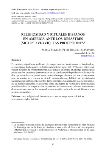
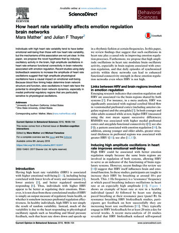
Available online at www.sciencedirect.comScienceDirectHow heart rate variability affects emotion regulationbrain networksMara Mather1 and Julian F Thayer2Individuals with high heart rate variability tend to have betteremotional well-being than those with low heart rate variability,but the mechanisms of this association are not yet clear. In thispaper, we propose the novel hypothesis that by inducingoscillatory activity in the brain, high amplitude oscillations inheart rate enhance functional connectivity in brain networksassociated with emotion regulation. Recent studies using dailybiofeedback sessions to increase the amplitude of heart rateoscillations suggest that high amplitude physiologicaloscillations have a causal impact on emotional well-being.Because blood flow timing helps determine brain networkstructure and function, slow oscillations in heart rate have thepotential to strengthen brain network dynamics, especially inmedial prefrontal regulatory regions that are particularlysensitive to physiological oscillations.Addresses1University of Southern California, United States2Ohio State University, United StatesCorresponding author: Mather, Mara (mara.mather@usc.edu)Current Opinion in Behavioral Sciences 2018, 19:98–104This review comes from a themed issue on Emotion-cognitioninteractionsEdited by Mara Mather and Michael FanselowFor a complete overview see the Issue and the Editorialin a rhythmic fashion at certain frequencies. In this paper,we review findings that suggest that such oscillations inheart rate play a causal role in improving emotion regulation processes. Furthermore, we propose that high amplitude oscillations in heart rate modulate brain oscillatoryactivity, especially in brain regions associated with emotion regulation, and that daily episodes of synchronizedactivity within these networks can lead to enhancedfunctional connectivity strength in these emotion regulation networks even when HRV is not high.Links between HRV and brain regions involvedin emotion regulationEmerging research indicates that emotion regulation andHRV are associated via the brain regions shared by bothsystems [7]. For instance, in a meta analysis, HRV wassignificantly associated with regional cerebral blood flowin ventromedial prefrontal cortex (including anterior cingulate regions) and the amygdala [7]. In both younger andolder adults scanned while at rest, higher HRV (measuredusing the root mean square successive differences;RMSSD) was associated with higher medial prefrontalcortex and amygdala functional connectivity ([8]; see also[9]), a pattern associated with emotion regulation [10]. Inaddition, among younger and older adults, greater structural thickness in prefrontal regions was associated withgreater HRV ([11]; see also [12,13]).Available online 8th January 52-1546/ã 2017 Elsevier Ltd. All rights reserved.IntroductionHaving high heart rate variability (HRV) is associatedwith higher emotional well-being [1–3], including beingcorrelated with lower levels of worry and rumination [4],lower anxiety [5], and better regulated emotionalresponding [6]. Thus, individuals with higher HRVappear to be better at regulating their emotions. However, it is not clear from these correlational studies if HRVis simply an output measure of regulatory brain health, orwhether it somehow increases prefrontal regulation effectiveness. In healthy individuals, high HRV is not simplythe result of random variability. Instead, much of thevariability is due to the heart responding to physiologicaloscillatory signals such as breathing and blood pressurefeedback, such that heart rate slows down and speeds upCurrent Opinion in Behavioral Sciences 2018, 19:98–104Inducing high amplitude oscillations in heartrate improves emotional well-beingHigh HRV could be associated with better emotionregulation simply because the same brain regions areinvolved in regulation of both systems, allowing HRVto serve as an indicator of the functioning of brain regulatory systems. However, recent findings (for review see[14]) suggest that HRV itself influences brain and emotional function. In these studies, participants are taught toincrease their HRV by breathing at around 10 s perbreath. This .1 Hz frequency is a ‘resonance’ frequencyat which paced breathing induces oscillations in heartrate at an especially high amplitude [15]. Figure 1shows an example of heart rate at rest in a healthyindividual (panel A) followed by heart rate duringpaced breathing at their resonance pace (panel B). Inresonance breathing HRV biofeedback studies, participants get feedback on how successfully they areincreasing heart rate oscillations [15]. They typicallyengage in HRV biofeedback for at least 20 min a day forseveral weeks. A recent meta-analysis of 24 studiesrevealed that HRV biofeedback reduced self-reportedwww.sciencedirect.com
Heart rate oscillations and emotion regulation Mather and Thayer 99Figure 180HRV(a)Heart Rate at RestHeart Rate (Beats Per Minute)70605080HRV(b)Heart Rate During Resonance Breathing706050Time (Tick Mark 30 s)Current Opinion in Behavioral Sciences(a) An example of heart rate variability during about a 2.5 min time period during quiet rest. (b) The same person’s heart rate during resonancebreathing during another 2.5 min time period.stress and anxiety with a large effect size [16]. Forinstance, in one study, basketball players scoring highon anxiety were randomly assigned to HRV-biofeedbackduring resonance paced breathing, to an active control, ora no-contact control condition [17]. Participants completed sessions 10 days in a row for 20 min in each sessionand were tested before and after the 10-day interventionand again a month later. The intervention reduced stateanxiety, and increased performance on standardized testsof basketball dribbling, passing and shooting. HRV biofeedback also has other positive effects on emotions.Coronary artery disease patients randomly assigned toHRV biofeedback during resonance breathing instead ofa wait-list control showed decreased expressive and suppressive hostility and these effects were maintained amonth after the 6-week intervention ended [18]. Likewise, veterans with post-traumatic stress disorderwww.sciencedirect.comrandomly assigned to HRV-biofeedback during resonance breathing showed reduced symptoms after 8 weeksof HRV-biofeedback whereas those assigned to treatment as usual did not show significant reductions insymptoms [19]. In addition, patients with post-strokedepression randomly assigned to treatment-as-usual inaddition to HRV biofeedback during resonance breathing showed greater reductions in some indices associated with depression than those not assigned to HRVbiofeedback [20]. Thus, in these studies, a series ofHRV-biofeedback sessions using resonance pacedbreathing enhanced emotional outcomes (Figure 2).Why resonance breathing increases theamplitude of heart rate oscillationsThe studies reviewed above using HRV biofeedbackduring paced breathing take advantage of the fact thatCurrent Opinion in Behavioral Sciences 2018, 19:98–104
100 Emotion-cognition interactionsHigh amplitude heart rate oscillations shouldpromote functional connectivity, especially inbrain regions involved in emotion regulationFigure 2Breathing at the same frequency asthe baroreflex feedback loop(resonance breathing;around 10-s per breath)High amplitude oscillations inheart rateHOW?High amplitude physiologicaloscillations stimulateoscillatory activity in thebrain, especially within brainregions sensitive tointeroceptive information.This strengthens functionalconnectivity within theseregions.Improved emotional well-beingCurrent Opinion in Behavioral Sciences(Article summary figure). How resonance breathing could lead toimproved emotional well-being by stimulating functional connectivity ofemotion regulation networks within the brain.We propose that episodes of high amplitude oscillationsin heart rate (like those observed during meditativepractice or HRV biofeedback) promote functional connectivity between certain brain regions, in particularamong brain regions involved in emotion regulation.Why might this be the case?First of all, brain activity is fueled by oxygen transportedby blood, and so should be affected by oscillations inblood flow. Indeed, heart rate contributes to blood-oxygen level dependent (BOLD) fluctuations during functional magnetic resonance imaging (fMRI) [31–33].Strong phase coupling of heart rate interval and BOLDoscillations have been observed in the mid cingulate andposterior cingulate regions at around the .1 Hz frequency[34]. These hemodynamics are likely to impact neuralactivity. In particular, oscillations in blood flow may leadto oscillations in the sensitivity of local cortical circuits tosensory stimuli [35].two physiological rhythms that have a strong influenceover the heart rate can be coordinated to induce highamplitude heart rate oscillations. The first of these physiological rhythms is the baroreflex. The vascular branch ofthe baroreflex has a lag time of approximately 10 s [21].When vessels are stretching, baroreceptors signal via thebrainstem to the heart to slow down the pace of heartbeats. There is a few-second delay in this feedback loop(between 4 and 6.5 s [22]) that creates oscillations in heartrate that take twice as long as the delay (from 0.075 to0.12 Hz, depending on the individual) to complete a fulloscillatory cycle [14,22]. The second major influence overHRV is breathing. As we breathe in, heart rate tends toincrease and as we breathe out, heart rate tends todecrease [23], although with a phase delay [24]. Weusually breathe at a faster frequency (between .15 and.4 Hz) than the baroreflex. However, unlike the baroreflex, which has a fixed frequency, we can alter the pace ofour own breathing. When breathing is slowed down to thesame frequency as the baroreflex feedback loop, thiscreates resonance, a non-linear effect that is greater thanan additive effect of the two influences. Thus, at anindividual’s resonance frequency, there is the potentialfor high amplitude oscillations in heart rate.Different brain regions vary in how long it takes for bloodto reach them, with distant brain regions sometimeshaving similar vascular delays. Estimating vascular delaytimes for each voxel in an fMRI image and then runningindependent component analyses reveals componentsthat resemble commonly identified resting state networks[36]. Resting state networks reflect brain regions thatactivate in correlated fashion at slow frequencies( .1 Hz; [37]). It is intriguing that just knowing thevascular delays of different brain regions provides enoughinformation to partially reconstruct resting state networks[36]. Indeed, part of what may lead some brain regions todevelop coordinated network activity with each othermay be their similar timing of blood delivery. Increasingthe amplitude of blood flow oscillations via resonancebreathing increases the impact of these coordinated vascular activities and thereby further stimulates networksthat were shaped in part by blood flow patterns. Repeatedbrief episodes of coordinated activity within a networkcan strengthen its internal pathways, promoting greaterfunctional connectivity during rest (e.g. [38,39]).A fascinating relevant phenomenon is that, in manymeditative and religious chanting practices, breathingslows to around a 10-s (.1 Hz) rate and heart rate oscillatesat this frequency [25–29]. For instance, reciting either therosary Ave Maria prayer or a yoga mantra leads to breathing at a 10-s/breath rate and increased blood pressure andheart rate oscillations at that .1 Hz resonance frequency[25]. Hypotheses about the mechanisms of the positiveeffects of meditative practices typically focus on the roleof attentional training and body awareness [30]. In contrast, there has been little focus on the role of physiological rhythms induced by the meditative practice.In addition to timing, regional differences in blood flowvolume are also associated with functional connectivity[40]. Brain regions with high resting cerebral blood flowalso show high functional connectivity with other brainregions, and the correlation between functional connectivity strength and blood flow is higher for measures oflong-range than for short-range functional connectivity[40]. Brain regions showing strong cross-subject correlations between functional connectivity strength and bloodflow include medial prefrontal cortex, anterior and posterior cingulate, and insula [40]. Thus, these brain regionsassociated with emotion regulation are among those thatCurrent Opinion in Behavioral Sciences 2018, 19:98–104www.sciencedirect.com
Heart rate oscillations and emotion regulation Mather and Thayer 101experience high regional blood flow and serve as hubregions for brain functional connectivity. These characteristics make oscillations in blood flow especially likelyto impact these brain regions.Breathing also influences brain rhythms. Breathing volume and pace help determine arterial CO2, which is acerebral vasodilator and is expelled during breath exhalation. Brain regions differ in the timing and strength oftheir responses to these CO2 fluctuations, likely related totheir proximity to large vessels, with strong correlationsseen in insula and midline cingulate regions [41,42].Breathing causes respiration-synched oscillations acrossmuch of the neocortex and gamma power waxes and wansdepending on the respiratory oscillation phase [43].Breathing through one’s nose synchronizes oscillationsin olfactory cortex as well as the amygdala and hippocampus [44]. In these limbic regions, nostril breathingentrains higher frequency oscillations in the delta, theta andbeta ranges to the respiratory phase [44]. Thus, the breathing component of resonance breathing biofeedback practiceshould also contribute to neural oscillatory activity, especially in the limbic regions during nostril breathing.Heartbeats also cause EEG responses known as heartbeat-evoked potentials that are particularly prominent inbrain regions associated with interoceptive sensation andemotion, including medial prefrontal cortex, cingulatecortex, insula, and amygdala [45,46]. Thus, heartbeatsshould be especially likely to influence brain rhythms inthese brain regions. Consistent with this, BOLD activityin the ventromedial prefrontal cortex covaries with heartrate more than does activity other brain regions [47], andas already reviewed, in general, medial prefrontal/anteriorcingulate regions show activity associated with HRV [7].In summary, resonance breathing stimulates high amplitude oscillations that can influence brain rhythms viaseveral channels, including fluctuations in blood flow,CO2 levels, and sensory input from breathing and fromheartbeats. These channels each are especially likely tomodulate activity in brain regions associated with emotion regulation networks [48].Slow oscillations can modulate fasterfrequencies of neural activityIn the previous section, we laid out the case that resonance breathing is likely to lead to oscillations in brainactivity. Here we argue that, in addition to provokingoscillations at the same frequency, resonance breathingshould also modulate faster oscillatory activity. Thepower density of EEG is inversely proportional to frequency, such that more powerful and widespread slowoscillations can modulate weaker but faster local oscillations [49,50]. Slow oscillations are also critical for brainnetworks, because the limited number and speed ofneuronal connections connecting distant regions meanwww.sciencedirect.comthat large-scale brain networks can only oscillate in tandem during slow oscillations [50].The ability of lower frequency oscillations to modulatethe phase of higher frequency oscillations leads to ahierarchical structure for EEG. For instance, in awakemacaque monkeys, delta phase (1–4 Hz) modulates thetaphase (4–10 Hz) and theta modulates gamma phase (20–50 Hz) amplitude, with these oscillations controllingbaseline excitability and leading to phasic oscillationsin responsiveness to stimuli [51]. Activity at one frequency is especially likely to modulate activity at otherfrequencies that are multiples of that frequency, a phenomenon known as harmonic frequency. Indeed, onespeculative proposal is that the heart rate is the basicfrequency and scaling factor for EEG frequency domains[52]. EEG is categorized into a set of different frequencybands (delta, theta, alpha, beta, gamma). The centerfrequency of each of these frequency bands (estimatedat 2.5, 5, 10, 20 and 40 Hz, respectively) is twice as highthan the previous lower frequency [52]. If the harmonicsequence of EEG frequency bands is extended downfrom delta to slower oscillations, the next lower one is1.25 Hz, which, at 75 beats per minute, is close to theaverage resting heart rate (e.g. [53]), suggesting that heartrate may be a basic frequency that serves as a scalingfactor (depending on individual differences in averageheart rate) for the EEG frequency domains [52]. Furthermore, if one continues going down to the subharmonicfrequencies, one of the frequencies overlaps with highfrequency HRV range influenced by breathing andanother overlaps with the low frequency HRV rangeinfluenced by baroreflex feedback [52]. Consistent withthese harmonic relationships, during sleep high frequencyHRV shows synchronization with each of the different EEGfrequency bands [54]. Furthermore, during wakefulness, theEEG spectral peak frequency (i.e., the alpha band peakfrequency) is correlated with heart rate and this correlationdecreases as sleep depth increases [55].Thus, low frequency oscillations induced by resonancebreathing should be able to promote functional connectivity between non-adjacent brain regions while alsomodulating oscillations at higher EEG frequency bands.Studies examining the oscillatory properties of brainactivity are consistent with this notion that oscillationsin the resonance frequency range help organize andmodulate activity at higher frequencies. For instance,oscillations in prefrontal oxyhemoglobin in the frequencyband between .07 and .13 Hz were coupled with EEGalpha and/or beta power oscillations [56]. Intracranialrecordings from human posteromedial cortex revealedthat the magnitude of cross frequency theta-to-gammamodulation fluctuated at around a 0.1 Hz frequency [57].Thus, oscillations in brain activity at around the resonance frequency can modulate interactions among fasterfrequency signals (see also [58]).Current Opinion in Behavioral Sciences 2018, 19:98–104
102 Emotion-cognition interactionsAnother potential pathway of action ofresonance frequency heart rate oscillationson the brainStimulation of the baroreflex with resonance pacedbreathing is also likely to modulate brainstem arousalpathways. As already touched upon, as part of theirfeedback loop, baroreceptors project to the nucleus ofthe solitary tract and stimulate both sympathoinhibitoryand vagal cardioinhibitory pathways that decrease heartrate [59]. In addition to its effects on the heart, thebaroreflex pathway also interacts bidirectionally withbrainstem and forebrain regions that regulate arousal[59]. Breathing also stimulates brainstem arousal centersand breathing and blood pressure signals interact inaffecting sympathetic activity (e.g. [60]). Thus, resonancebreathing should modulate arousal. Most likely, theseeffects will involve oscillatory influences. Consistent withthis possibility, during slow breathing, muscle sympathetic nerve activity decreases in a phasic fashion, reaching the same peak level as in control conditions butshowing phasic segments of suppression [61–63]. Ifinducing high amplitude oscillations in heart rate phasically suppresses sympathetic action while stimulatingparasympathetic action, this could help explain thestress-reducing and anxiety-reducing effects of resonancebreathing [16].ConclusionsPast research has focused on heart rate variability as adownstream measure, rather than something that itselfaffects emotion regulation. For instance, the Neurovisceral Integration Model proposed that the medial PFCalong with a core set of neural structures integratesinformation from different system to regulate the heart,and that HRV provides an index of the effectiveness ofthis ‘core integration’ system [7]. Furthermore, previousresearch has not distinguished whether it is random noiseor increases in the amplitude of oscillatory activity that isthe key component of HRV that is associated with betteremotional outcomes. The findings we outlined in thispaper suggest that heart rate oscillations can enhanceemotion by entraining brain rhythms in ways thatenhance regulatory brain networks.Conflict of interest statementNothing declared.AcknowledgementsWork on this review was supported by NIH grant R01AG057184.2.Kemp AH, Quintana DS: The relationship between mental andphysical health: insights from the study of heart ratevariability. Int J Psychophysiol 2013, 89:288-296.3.Shaffer F, McCraty R, Zerr CL: A healthy heart is not ametronome: an integrative review of the heart’s anatomy andheart rate variability. Front Psychol 2014:5.4.Ottaviani C, Thayer JF, Verkuil B, Lonigro A, Medea B,Couyoumdjian A, Brosschot JF: Physiological concomitants ofperseverative cognition: a systematic review and metaanalysis. Psychol Bull 2016, 142:231-259.5.Chalmers JA, Quintana DS, Abbott MJ, Kemp AH: Anxietydisorders are associated with reduced heart rate variability: ameta-analysis. Front Psychiatry 2014, 5:80.6.Appelhans BM, Luecken LJ: Heart rate variability as an index ofregulated emotional responding. Rev Gener Psychol 2006,10:229.7.Thayer JF, Åhs F, Fredrikson M, Sollers JJ, Wager TD: A metaanalysis of heart rate variability and neuroimaging studies:implications for heart rate variability as a marker of stress andhealth. Neurosci Biobehav Rev 2012, 36:747-756.Sakaki M, Yoo HJ, Nga L, Lee T-H, Thayer JF, Mather M: Heartrate variability is associated with amygdala functionalconnectivity with MPFC across younger and older adults.Neuroimage 2016, 139:44-52.For both younger and older adults, those with higher heart rate variabilityhad stronger amygdala-medial prefrontal cortex functional connectivity,indicating that functional connectivity within an emotion regulation circuitis related to heart rate variability.8.9.Jennings JR, Sheu LK, Kuan DCH, Manuck SB, Gianaros PJ:Resting state connectivity of the medial prefrontal cortexcovaries with individual differences in high-frequency heartrate variability. Psychophysiology 2016, 53:444-454.10. Etkin A, Büchel C, Gross JJ: The neural bases of emotionregulation. Nat Rev Neurosci 2015, 16:693.11. Yoo HJ, Thayer JF, Greening SG, Lee TH, Ponzio A, Min J,Sakaki M, Nga L, Mather M, Koenig J: Brain structuralconcomitants of resting state heart rate variability in theyoung and old: evidence from two independent samples. BrainStruct Funct 2017 http://dx.doi.org/10.1007/s00429-017-1519-7.(in press).12. Winkelmann T, Thayer JF, Pohlack S, Nees F, Grimm O, Flor H:Structural brain correlates of heart rate variability in a healthyyoung adult population. Brain Struct Funct 2016:1-8.13. Woodward SH, Kaloupek DG, Schaer M, Martinez C, Eliez S: Rightanterior cingulate cortical volume covaries with respiratorysinus arrhythmia magnitude in combat veterans. J Rehabil ResDev 2008, 45:451-464.14. Lehrer PM, Gevirtz R: Heart rate variability biofeedback: howand why does it work? Front Psychol 2014, 5:756.This review is an excellent source for further information on the mechanisms of how breathing rate interacts with heart rate and blood pressure.15. Lehrer P, Vaschillo B, Zucker T, Graves J, Katsamanis M, Aviles M,Wamboldt F: Protocol for heart rate variability biofeedbacktraining. Biofeedback 2013, 41:98-109.16. Goessl V, Curtiss J, Hofmann S: The effect of heart ratevariability biofeedback training on stress and anxiety: a metaanalysis. Psychol Med 2017:1-9.This meta-analysis of 24 studies found that HRV biofeedback trainingreduced self-reported stress and anxiety with a large effect size. Treatment efficacy was not moderated by the presence of an anxiety disorder,suggesting it had benefits even for those without clinical levels of anxiety.17. Paul M, Garg K: The effect of heart rate variability biofeedbackon performance psychology of basketball players. ApplPsychophysiol Biofeedback 2012, 37:131-144.References1.Beauchaine TP, Thayer JF: Heart rate variability as atransdiagnostic biomarker of psychopathology. Int JPsychophysiol 2015, 98:338-350.Current Opinion in Behavioral Sciences 2018, 19:98–10418. Lin IM, Fan S-Y, Lu H-C, Lin T-H, Chu C-S, Kuo H-F, Lee CS, Lu YH: Randomized controlled trial of heart rate variabilitybiofeedback in cardiac autonomic and hostility amongpatients with coronary artery disease. Behav Res Therapy 2015,70:38-46.www.sciencedirect.com
Heart rate oscillations and emotion regulation Mather and Thayer 10319. Tan G, Dao TK, Farmer L, Sutherland RJ, Gevirtz R: Heart ratevariability (HRV) and posttraumatic stress disorder (PTSD): apilot study. Appl Psychophysiol Biofeedback 2011, 36:27-35.20. Li X, Zhang T, Song L-P, Zhang Y, Zhang G-G, Xing C-X, Chen H:Effects of heart rate variability biofeedback therapy onpatients with poststroke depression: a case study. Chin Med J2015, 128:2542.21. Rosenbaum M, Race D: Frequency-response characteristics ofvascular resistance vessels. Am J Physiol-Legacy Content 1968,215:1397-1402.22. Vaschillo EG, Vaschillo B, Lehrer PM: Characteristics ofresonance in heart rate variability stimulated by biofeedback.Appl Psychophysiol Biofeedback 2006, 31:129-142.23. Yasuma F, Hayano J-I: Respiratory sinus arrhythmia: why doesthe heartbeat synchronize with respiratory rhythm? Chest2004, 125:683-690.24. Angelone A, Coulter NA: Respiratory sinus arrhythmia: afrequency dependent phenomenon. J Appl Physiol 1964,19:479-482.25. Bernardi L, Sleight P, Bandinelli G, Cencetti S, Fattorini L,Wdowczyc-Szulc J, Lagi A: Effect of rosary prayer and yogamantras on autonomic cardiovascular rhythms: comparativestudy. BMJ 2001, 323:1446-1449.This study demonstrates that the classic rosary prayer (the Ave Maria inLatin) and a typical yoga mantra (‘om-mani-padme-om’) both take about10 s to chant out loud and result in .1 Hz (10-s cycle) oscillations in heartrate and blood pressure in the person chanting. This suggests that theseclassic chants may have endured as calming practices because theystabilize the respiratory rate at the .1 Hz frequency that stimulatesparticularly high oscillations in heart rate and blood pressure.26. Friedman EH, Coats AJ: Neurobiology of exaggerated heart rateoscillations during two meditative techniques. Int J Cardiol2000, 73:199.27. Lehrer P, Sasaki Y, Saito Y: Zazen and cardiac variability.Psychosom Med 1999, 61:812-821.28. Peng CK, Mietus JE, Liu Y, Khalsa G, Douglas PS, Benson H,Goldberger AL: Exaggerated heart rate oscillations during twomeditation techniques. Int J Cardiol 1999, 70:101-107.oscillations that can be detected in peripheral regions such as fingertipsand toes. These systemic low frequency oscillations reach different brainregions at different time delays depending on local cerebral vasculature.The authors computed the delay time for these signals to reach eachvoxel in an functional magnetic resonance scan. Simulation data usingjust these voxel delay times in an independent components analysisrevealed a number of the well-known resting-state network patterns inbrain activity, indicating that such networks are related to systemicvascular fluctuations.37. Biswal BB, Mennes M, Zuo XN, Gohel S, Kelly C, Smith SM,Beckmann CF, Adelstein JS, Buckner RL, Colcombe S,Dogonowski AM, Ernst M, Fair D, Hampson M, Hoptman MJ,Hyde JS, Kiviniemi VJ, Kötter R, Li SJ, Lin CP, Lowe MJ, Mackay C,Madden DJ, Madsen KH, Margulies DS, Mayberg HS,McMahon K, Monk CS, Mostofsky SH, Milham MP: Towarddiscovery science of human brain function. Proc Natl Acad SciU S A 2010, 107:4734-4739.38. Ma L, Narayana S, Robin DA, Fox PT, Xiong J: Changes occur inresting state network of motor system during 4 weeks ofmotor skill learning. Neuroimage 2011, 58:226-233.39. Martı́nez K, Solana AB, Burgaleta M, Hernández-Tamames JA,Álvarez-Linera J, Román FJ, Alfayate E, Privado J, Escorial S,Quiroga MA, Karama S, Bellec P, Colom R: Changes in restingstate functionally connected parietofrontal networks aftervideogame practice. Hum Brain Map 2013, 34:3143-3157.40. Liang X, Zou Q, He Y, Yang Y: Coupling of functionalconnectivity and regional cerebral blood flow reveals aphysiological basis for network hubs of the human brain. ProcNatl Acad Sci U S A 2013, 110:1929-1934.41. Chang C, Glover GH: Relationship between respiration, endtidal CO2, and BOLD signals in resting-state fMRI. Neuroimage2009, 47:1381-1393.42. Di X, Kannurpatti SS, Rypma B, Biswal BB: Calibrating BOLDfMRI activations with neurovascular and anatomicalconstraints. Cereb Cortex 2013, 23:255-263.43. Heck DH, McAfee SS, Liu Y, Babajani-Feremi A, Rezaie R,Freeman WJ, Wheless JW, Papanicolaou AC, Ruszinkó M,Sokolov Y, Kozma R: Breathing as a fundamental rhythm ofbrain function. Front Neural Circ 2017, 10.29. Peng CK, Henry IC, Mietus JE, Hausdorff JM, Khalsa G, Benson H,Goldberger AL: Heart rate dynamics during three forms ofmeditation. Int J Cardiol 2004, 95:19-27.44. Zelano C, Jiang H, Zhou G, Arora N, Schuele S, Rosenow J,Gottfried JA: Nasal respiration entrains human limbicoscillations and modulates cognitive function. J Neurosci 2016,36:12448-12467.30. Hölzel BK, Lazar SW, Gard T, Schuman-Olivier Z, Vago DR, Ott U:How does mindfulness meditation work? Proposingmechanisms of action from a conceptual and neuralperspective. Perspect Psychol Sci 2011, 6:537-559.45. Babo-Rebelo M, Richter CG, Tallon-Baudry C: Neural responsesto heartbeats in the default network encode the self inspontaneous thoughts. J Neurosci 2016, 36:7829-7840.
[40]. Brain regions with high resting cerebral blood flow also show high functional connectivity with other brain regions, and the correlation between functional connec-tivity strength and blood flow is higher for measures of long-range than for short-range functional connectivity [40 Bra