Transcription
Guo et al. Diagnostic Pathology(2019) ARCHOpen AccessClinicopathological categorization of hydroavacciniforme-like lymphoproliferativedisorder: an analysis of prognosticimplications and treatment based on19 casesGuo Na1,2†, Chen Yueqiong1,3†, Wang Yu1,4, Huang Yuhua1,2, Feng Yanfen1,2, Li Min1,2 and Rao Huilan1,2*AbstractBackground: Hydroa vacciniforme-like lymphoproliferative disorder (HV-LPD) is a cutaneous form of chronic activeEpstein-Barr virus (EBV) infection, which occurs mainly in children in Latin America and Asia. It can progress tosystemic lymphoma. However, prognostic factors and treatment remain unclear.Methods: This retrospective study reviewed the clinical, morphologic, immunophenotypical features, and clinicaltreatment of 19 patients with HV-LPD.Results: All 19 patients had skin lesions in the face, extremities, or areas unexposed to the sun, including edema,blistering, ulceration, and scarring. The course was slowly progressive and relapsing. Histopathology showed anatypical lymphocytic infiltrate in the dermis and/or subcutaneous tissue. The lesions had a cytotoxic T/NK-cellimmunophenotype. Among 19 patients, 7 (37%) exhibited CD4 T cells, 5 (26%) exhibited CD8 T cells, and 7(37%) exhibited CD56 cells. Of 12 cases with a T-cell phenotype, molecular analyses demonstrated that 7 hadmonoclonal rearrangements in the T-cell receptor genes. Three cases had an NK-cell phenotype and had polyclonalrearrangements in the TCR genes. All cases were associated with EBV infections. Among 19 patients, 9 (47.4%)received chemotherapy. Only one patient received allogeneic transplantation and EBV-specific cytotoxic T lymphocytetreatment after chemotherapy. That patient was the only one alive without disease at the latest follow up. Ninepatients died of systemic lymphoma with disease progression, indicating irreversible process.Conclusions: This study confirmed that HV-LPD is a broad-spectrum EBV lymphoproliferative disorder. It progressedto EBV systemic T/NK lymphoma, although some patients had a more indolent, chronic course. Cytopenia, elevatedlactate dehydrogenase, destructive-multiorgan involvement, and older age were poor prognostic factors. Onlyallogeneic transplantation was curative.Keywords: Epstein-Barr virus-positive, Hydroa vacciniforme-like lymphoproliferative disorder, Clinicopathologic features,Prognostic factors, Treatment* Correspondence: raohl@sysucc.org.cn†Na Guo and Yueqiong Chen contributed equally to this work.1State Key Laboratory of Oncology in South China, Collaborative InnovationCenter for Cancer Medicine, Guangzhou, Guangdong 510060, People’sRepublic of China2Department of Pathology, Sun Yat-sen University Cancer Center, No. 651,Dongfeng Road East, Guangzhou, Guangdong 510060, People’s Republic of ChinaFull list of author information is available at the end of the article The Author(s). 2019 Open Access This article is distributed under the terms of the Creative Commons Attribution 4.0International License (http://creativecommons.org/licenses/by/4.0/), which permits unrestricted use, distribution, andreproduction in any medium, provided you give appropriate credit to the original author(s) and the source, provide a link tothe Creative Commons license, and indicate if changes were made. The Creative Commons Public Domain Dedication o/1.0/) applies to the data made available in this article, unless otherwise stated.
Guo et al. Diagnostic Pathology(2019) 14:82IntroductionHydroa vacciniforme-like lymphoproliferative disorder(HV-LPD) was nominated and introduced into the 4th revised edition of the WHO classification of Tumors ofHematopoietic and Lymphoid Tissues [1]. HV-LPD is a cutaneous form of Epstein-Barr Virus-positive (EBV ) T/NKlymphoproliferative disease, which occurs in childhood andcarries the risk of systemic lymphoma [2, 3]. Other termshave been used previously to describe HV-LPD, includinghydroa vacciniforme-like eruption, edematous scarring vasculitic panniculitis, classic and severe hydroa vacciniforme,hydroa-like cutaneous T-cell lymphoma, and hydroavacciniforme-like lymphoma (HVLL) [3]. The term HVLLwas included the 2008 WHO classification of peripheralmature T-cell lymphoma [4]. The nomenclature and definition changed from HVLL to lymphoproliferative disorder,due to its relationship with chronic active EBV (CAEBV)infections and its clinical course.HV-LPD is extremely rare. It manifests as recurrent skinlesions infiltrated with T cells or NK cells. It is mostly foundin children and young adults from Asian and Latin Americancountries. It is associated with CAEBV infections. It mightpersist only in genetically predisposed individuals [5–7].The clinical course of HV-LPD is variable; it lasts up to10–15 years [8, 9]. In the early stage, skin lesions tend tooccur on sun-exposed areas. Lesions comprise recurrentpapulovesicles, crusts, and eventually, vacciniform (pox-like)scars, after several weeks. In the late stage, this disorder involves areas unexposed to the sun and becomes more aggressive. Deep ulcerations, necrosis, and facial edema arecommon. Severe systemic manifestations might be present,such as intermittent fever, lymph node enlargement, andhepatosplenomegaly. When systemic dissemination occurs(hepatosplenomegaly, lymphadenopathy, and bone marrowinfiltration), it is best classified as systemic EBV T/NK-celllymphoma [2, 3].However, it is not always possible to distinguish betweenHV-LPD and systemic EBV T/NK-cell lymphoma, becausethese disorders often have overlapping clinical and pathological features at the time of presentation and/or duringdisease progression [10, 11]. Moreover, patients with HVLPD exhibit variable clinical courses, but often have fataloutcomes. There are no guidelines or consensus opinions toguide clinicians in making a HV-LPD diagnosis, evaluatingdisease progression, or selecting treatment. Therefore, weretrospectively reviewed 19 cases of HV-LPD, first tocharacterize further the clinicopathological and molecularfeatures, and second, to propose clinicopathological prognostic factors and potentially effective treatment.Materials and methodsPatient selectionAll patients were examined at the Department of Pathology, Sun Yat-sen University Cancer Center, fromPage 2 of 11September 2008 to May 2017. The HV-LPD diagnosiswas based on the 4th revised edition of the WHO classification of Tumors of Hematopoietic and LymphoidTissues [1]. The HV-LPD diagnosis was also based onstandard published clinical and histopathological criteria,immunohistochemistry results, and findings of T-cell receptor (TCR) rearrangements. Tissue specimens werefixed in neutral buffered formalin and embedded in paraffin for processing. Hematoxylin-eosin stained sections,immunohistochemical staining, in situ hybridization fordetecting EBV-encoded RNAs (EBERs), and molecularstudies were performed at the Department of Pathology.Patient follow-upOf the 19 patients identified, 9 patients had at least onetreatment record in the Sun Yat-sen University CancerCenter. Another five patients were treated at other hospitals. Clinical data and follow-up information were obtainedfrom the hospital and from private medical office records.All patients were followed up until May 2017. Diseaseprogression and recurrence were diagnosed based onclinical examinations, imaging assessments, and pathologicexaminations. Overall survival (OS) was measured from theinitiation of treatment to either the last follow-up or deathfrom disease. Progression-free survival (PFS) was measuredfrom the initiation of treatment to the first indications ofdisease progression or relapse, death from disease, or thelast follow-up.Hematoxylin-eosin stainingHV-LPD tissues were formalin-fixed, paraffin-embedded,and stained with hematoxylin and eosin. The histopathological features of the 19 cases were evaluated independentlyby two pathologists (Y-H H and H-L R).Immunohistochemical staining and EBV in situhybridizationThe tissue blocks were cut into 4-μm sections andprocessed for immunohistochemistry. The lineage ofEBV cells was determined by staining with antibodiesagainst CD3, CD5, CD4, CD8, CD56, CD30, TIA1, andKi67. Immunohistochemical stains were performed withan automated immunostainer (Ventana Medical Systems,Tucson, AZ), according to the manufacturer’s protocol.EBV was detected with the EBV Probe In SituHybridization Kit (TRIPLEX INTERNATIONAL BIOSCIENCES, CHINA, CO. LTD).T-cell clonality studiesPolymerase chain reaction (PCR) was performed toanalyze the clonality of T cells. We use the standardizedBIOMED-2 clonality assays-ABI Fluorescence Detection(IdentiClone, InVivo Scribe Technologies, San Diego, CA,USA) according to the manufacturer’s instructions. PCR
Guo et al. Diagnostic Pathology(2019) 14:82products were analysed by capillary electrophoresis usingan ABI 3500XL genetic analyser (Applied Biosystems,Foster City, CA, USA). DNA quality was assessed throughamplification of a control gene size ladder.First, after deparaffinization and proteinase K digestion,DNA was extracted from 10-mm paraffin sections,according to the standard procedure. Then, T-cell clonalexpansion was detected by analyzing rearrangements inthe TCR beta chain (Vb-Jb and Db-J-b) and TCR genes.Positive and negative controls were set appropriately in allexperiments. Clonality was assessed according to wellestablished recommendations.EBV DNA blood load measurementTotal plasma cell-free DNA was isolated with theQIAamp Blood Mini Kit (QIAgen, Inc., Valencia, CA,USA), according to the manufacturer’s protocol. Plasmalevels of EBV DNA toward the BamHI-W region weredetermined with the quantitative PCR method. Theprimers were designed as previously reported [12]. DNAfrom EBV-negative healthy volunteers comprised onenegative control, and another negative control was runwith no template. Controls were included on each plate.Results are expressed as the number of EBV copies/mlplasma. Zero copies/ml was selected for the cut-off valuein evaluating EBV DNA levels.There was a positive control used for EBV DNA detection each time. To estimate the sensitivity of the PCRassay, gradient dilutions of the synthetic EBV fragmentplasmid were prepared and amplified by the PCR assay.Using absolute quantification, a standard curve was madeby 103, 104, 105 and 106. And 103 and 105 were used aspositive controls .Statistical analysisData are described as the mean standard deviation(SD), range, frequency (number of cases), and percentages, when appropriate. All statistical calculations wereperformed with the Statistical Package for Social Sciences software, version 20 (SPSS, Inc. Chicago, IL, USA).ResultsClinical characteristicsNineteen cases were diagnosed in our center from September 2008 to May 2017. The incidence of HV-LPD was1.9% among patients with mature peripheral T-cell lymphoma in our center [13]. The cohort comprised 9 malesand 10 females, aged 2 to 21 years (median 8 years). Allpatients were ethnically Chinese. The clinicopathologicalfindings of our cases are summarized in Table 1.The main clinical features were papules (n 19/19,100%,) and fever (n 18/19, 94.7%). Only one patient (case8) had no B symptom at the diagnosis. All lesions were located in the skin, and they included papulovesicles,Page 3 of 11blisters, ulceration, and scars. Eruptions were seasonal;they typically occurred in spring or summer. Eighteen patients had recurrent and slowly progressive, relapsing clinical courses. One patient (case 19) presented with a severeHV. In the early stage, most skin lesions initially occurredafter sun exposure and subsided after sun protection. Thelesions typically appeared on the face and extremities. Theskin lesions typically healed within 1–2 weeks, withvacciniform scarring, and they recurred in the samedistribution. Rarely, cases presented with fever at the beginning. As the disease progressed, 15 patients (88%) presented with severe systemic manifestations, includingintermittent fever, wasting, lymphadenopathy, and hepatosplenomegaly. Severe skin lesions presented as deep ulcers, necrosis, plaques, or face edema in these patients(Fig. 1). The ulcerations took a longer time to heal, andthey left scars.Six cases had a history of mosquito-bite hypersensitivity. Among these, four had a CD4 phenotype, and theother two had a CD8 phenotype. No patient was hypersensitive to light.The available routine laboratory tests revealed that allpatients (11/11) had elevated lactate dehydrogenase(LDH) levels (range 312 to 1202 IU/L), when they wereinitially diagnosed with HV-LPD. Available data for sevenpatients showed positive EBV DNA blood loads in allpatients (n 7/7). In the 9 patients who had the treatmentrecord in Sun Yat-sen University Cancer Center, the rangeof time-to progression were from 0.2 to 7 years (median,0.63 yrs). Both the LDH and EBV DNA values at diseaseprogression or the last follow-up were shown in Table 2.Both of the LDH and EBV DNA values, the importantindex for lymphoma, reflected the effect of the treatment.During the treatments, both of the LDH and EBV DNAloads declined. However, both of the LDH and DNA loadsrose to high levels when the disease progressed (Table 2).HistopathologyHistologically, all the biopsies featured infiltration ofatypical lymphoid cells surrounding blood vessels and skinappendages in the dermis. In 12 cases (63%), atypical lymphocytes had infiltrated deep into fatty tissue or subcutaneous tissue. They were small to intermediate in size, andthey had enlarged, irregular nuclei with nucleoli, admixedwith an inflammatory background composed of smalllymphocytes, plasma cells, histocytes, and eosinophils(Fig. 2). Interestingly, nine cases developed into systemiclymphoma; these displayed dense and diffuse, atypical,medium- or large-sized lymphocytes (Fig. 3). Mitoses werebrisk. Tumor necrosis was observed. However, we observed no angio-invasion or vascular destruction in any ofthe nine cases analyzed.Seven patients had lymph node biopsies. Four biopsiesshowed preserved architecture with open sinuses and
everFeverPUCS/face, extremities,trunkPUCS, nodules/face,trunk, and extremitiesPUCS/face, extremities,trunk, and oral cavityFLFHSLFLPUCS, nodules/face,FHSLextremities, trunk; HMBPUCS/face, extremities,PUCS/areas exposedFSHLNPand unexposed to sun; nodules, HPSHMBPUCS, nodules/face,extremitiesPUCS/face, extremities,HMBPUCS/face, arms, areasnot exposed to sunPUCS, nodules/face,extremitiesPUCS/ areas exposedto sun; HMBPUCS, nodules/face,extremitiesErythema, PUCS/face,extremities, and areasnot exposed to sunMedium/dermis andsubcutaneous tissueMedium/dermis and fattytissueMedium/dermisMedium/dermisMedium/dermis and fattytissueMedium and large/dermisand subcutaneous tissueMedium/dermis andsubcutaneous tissueSmall and medium/dermisand subcutaneous tissueMedium and large/dermisSmall and medium/dermisSmall and medium/dermisand subcutaneous tissueMedium/dermisMedium/dermis and fattytissue/Fatty tissue /face, scalp,extremitiesSystemicSize of atypical cells/manifestations infiltration depth/necrosisCase Age(y)/ Sex/ Skin lesions/locationHistory (y)Table 1 Clinicopathologic features of patients with HV-LPD624324BM-EBV cells diffuselyinfiltrated LN andBMBM-EBV cells ininterfollicular areaof LNNDEBV cells diffuselyinfiltrated LN andBMNDNDNDNDNDCD56CD8CD4CD56CD56CD4CD8334.6 CD4358.9 CD56482318NA1202 CD4NA298.4 CD8NANANACD56CD56CD8CRNAPDPDNAPRNAChemotherapy (First Line:GEM for 6 cycles, SecondLine: VP-16 VCR for 1cycle, Third Line: VLB PDN for 1 cycle)Chemotherapy (GEM for6 cycles) and antivirustreatment (Ganciclovir)PRCRChemotherapy (PPDASP GEM L-OHP)Maintained herbal remedyChemotherapy (NHL-BFM- CR95) for 6 cycles; SiblingHSCT; and EBV-CTL treatment [15]NAChemotherapy(GEM VP16 MTX PASP thalidomide for 6cycles)NAChemotherapy (NHL-BFM- PR95) for 6 cycles [15]NAChemotherapy (IMR) andRadiotherapy. MaintainedHerbal remedyHerbal remedyChemotherapy (MTX VP- PR16) and ODDODDODAWDAWDDODAWDBest response Currentto treatment statusChemotherapy (NHL-BFM- PD90) for 3 cycles [14]Chemotherapy (IMR) andRadiotherapy. Maintainedherbal remedyLDH Phenotype Therapy(IU/L)EBV cells present450in interfollicular areaof LNEBV cells diffuselyinfiltrated LNEBV cells ininterfollicular areaof LNLN, BM 1Y8.7YSurvival afterDiagnosisGuo et al. Diagnostic Pathology(2019) 14:82Page 4 of 11
11/M/98/M/2F/5/3171819FeverFeverFHSLMedium/dermis andsubcutaneous tissueMedium/dermis andsubcutaneous tissueMedium/dermis andsubcutaneous tissueMedium/dermis and fattytissueNDBM-EBV cells diffuselyinfiltrated LN andBMBM-BM-LN, BM biopsyNACD8CD4CD4585312CD4NA323.2 ycin, acitretin,and cyclosporine)NAPRLFUAWDDODDODDODBest response Currentto treatment statusChemotherapy (First Line: PDNHL-BFM-95, Second Line:GEM for 2 cycles) [15]NAChemotherapy (GEM for6 cycles)LDH Phenotype Therapy(IU/L)LFU2.2Y2.4Y0.7Y1.3YSurvival afterDiagnosisPUCS papulovesicles, ulcers, crust, scars, HMB Hypersensitivity to mosquito bites, FHSL fever, hepatosplenomegaly, lymphadnopathy, FHS fever, hepatosplenomegaly, FL fever, lymphadnopathy, NP nodules nasopharynxnodules, HPS hemophagocytic syndrome, LN lymph node, BM bone marrow, BM- BM not involved, AWD Alive with disease, DOD dead of disease, NA not applicable, ND not done, LFU lost to follow-up, IMR interferon,mechlorethamine, and retinoic acid, NHL Non Hodgkin lymphoma, BFM Berlin-Frankfurt-Münster, MTX methotrexate, VP-16 etoposide, P-ASP pegaspargase, HSCT hematopoietic stem cell transplantation, GEMgemcitabine, L-OHP oxaliplatin, VCR vincristine, VLB vinblastine, PDN prednisone, CR complete remission, PD progressive disease, PR partial remissionPUCS/face, legs, andtrunkPUCS/face, arms, legs,and trunkPUCS, nodules/face,arms, legs, and areasnot exposed to sun;HMBPUCS/face, trunk, armsand legs; HMBFHSL12/F/416Medium/dermisFL7/F/415PUCS/face, extremitiesSystemicSize of atypical cells/manifestations infiltration depth/necrosisCase Age(y)/ Sex/ Skin lesions/locationHistory (y)Table 1 Clinicopathologic features of patients with HV-LPD (Continued)Guo et al. Diagnostic Pathology(2019) 14:82Page 5 of 11
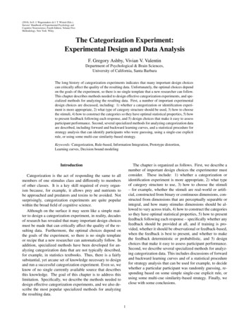
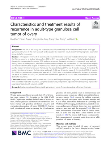
(2019) 14:82Guo et al. Diagnostic PathologyPage 6 of 11all cases displayed the presence of EBERs. Cells that carried EBERs were concentrated mainly around blood vessels and adnexa in the dermis (Figs. 2 and 3), but also insubcutaneous tissue.Of 19 patients, five biopsies (26%) had CD8 T cells, butno CD4 or CD56 cells (Fig. 2c). Seven (37%) had initialbiopsies with CD4 T cells, but no CD8 T cells orCD56 cells (Fig. 3c). Seven biopsies (37%) had CD56 cells, but no CD4 or CD8 T cells, which indicated anNK-cell phenotype.Molecular studiesFig. 1 Case 9 shows an ulcerated lesion and vacciniform scar on theface. Note the periorbital edemaparacortical hyperplasia. Cells that had infiltrated thelymph node were small- and medium-sized, with irregular nuclei and inconspicuous nucleoli. Cases 2, 9, and 17progressed to systemic NK/T cell lymphoma. They exhibited diffuse effacement of the normal nodal architecture, due to the infiltration of homogeneous mediumand large mixed lymphocytes with hyperchromatic nuclei and irregular nuclear contours. Cases 9, 13, and 17harbored bone marrow that had been infiltrated by atypical lymphocytes.Immunohistochemical studies and EBERsThe immunohistochemical findings are summarized inTable 3. The lymphoid cells that had infiltrated underthe epidermis were 100% positive for CD3 and TIA1,and partially expressed CD30. With in situ hybridization,Molecular analyses demonstrated that 7 of the 12 biopsies with a T-cell phenotype had undergone monoclonal rearrangements in the TCR genes. These sevencases were originally classified as HVLL, based on themonoclonality of the TCR gene rearrangements andfulfillment of the 2008 WHO classification criteria.The three biopsies with an NK-cell phenotype lackeddetectable clonal rearrangement.Treatment and outcomeDue to the long clinical course, the patients received avariety of treatments. Most patients were primarilytreated with immunomodulating or immunosuppressivetherapies, such as steroid cream or interferon, mechlorethamine, retinoic acid, etc. In the early stage, skin lesions typically improved with these treatments, but later,these treatments lost some efficacy.Among the 19 patients, nine (47.4%) received chemotherapy, one patient (5.3%) received an herbal remedy,one patient (5.3%) received a combination of chemotherapy and radiotherapy, one patient (5.3%) received a combination of chemotherapy and herbal remedy, and twopatients (10.5%) received sequential chemotherapy,radiotherapy, and herbal remedy. The other five patients(26.3%) received no anti-tumor treatment. The mainTable 2 EBV DNA loads and LDH level in the 9 patients who had the treatment record in Sun Yat-sen University Cancer CenterCaseEBV DNA (copy/ml)LDH 15.70.2yDODBeforetreatmentThe minimum levelduring he minimum levelduring treatmentProgression/last follow-upCase1NDNDND324NDCase2NDNDND6242156Case 7851.88 106.63 101.33 10298.4206.28880.8yDODCase 95.62 1062.0 1016.83 1041202248.711660.5yDODCase 1165.68 106.93 105.95 10318179.6259.8NAAlive withoutdiseaseCase 131.70 1056.0 104Case 1453.95 105442.69 103358.9238.6248.92.1yAWD34.53 104334.6203.7276.30.3yAWD346.59 10Case 156.94 101.20 107.56 10323.2251.7624.40.75yDODCase 171.58 1052.25 1043.09 105585119.4363.90.25yDODAWD Alive with disease, DOD dead of disease, NA not applicable, ND not done
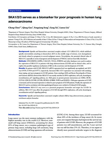
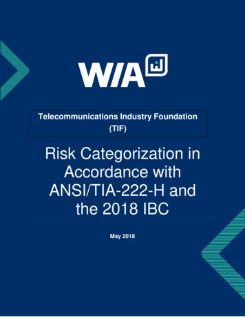
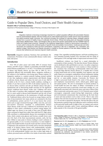
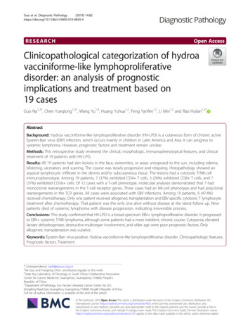
Guo et al. Diagnostic Pathology(2019) 14:82Page 7 of 11Fig. 2 HV-LPD morphology and immunophenotype of case 10. a Skin biopsy shows edema and vesicles in the epidermis (hematoxylin and eosin[H&E], 40 ). b Skin biopsy shows an infiltration of atypical lymphocytes surrounding adnexa and blood vessels (H&E, 200 ). c ImmunohistochemicalCD8 stain shows that the cells surrounding the adnexa and blood vessels are strongly CD8-positive (200 ). d In situ hybridization for EBV-encodedRNA showed positive signals in infiltrating lymphocytes (200 )chemotherapy regimens in our study were BFM-90/95,for children with non-Hodgkin lymphoma (NHL), andgemcitabine for adults with extranodal NK/T-celllymphoma, nasal type. Only one patient (case 11) in ourstudy received an allogeneic hematopoietic stem celltransplantation and EBV-specific cytotoxic T lymphocyte(EBV-CTL) treatment after chemotherapy.Among the 14 patients that received treatment, fourpatients (28.6%) achieved complete remission (CR), fivepatients (35.7%) achieved partial remission (PR), and fivepatients (35.7%) had progressive disease. Among the latter, four patients developed systemic lymphoma and diedof disease during the anti-tumor treatment. Three patients died of disease during the follow-up with the besteffect of CR or PR. The 2-year, 3-year and 5-year OSwas78.6, 56.3 and 45.0%, respectively.Among all of the19 patients in our study, the 2-year,3-year and 5-year OS were 75.0, 47.6 and 38.1%, respectively, excluding 3 patients who lost to the follow up.Nine patients developed systemic lymphoma and died ofdisease with or without treatment.Differences between patients with T-cell and NK-cellphenotypesWe compared the clinical features between patients withT-cell and NK-cell phenotypes. The male-to-female ratioswere quite different (T cell: 2:1 vs. NK cell: 1:6), showingfemale dominance in NK phenotype. But the median ageswere similar between groups (7.5 vs. 8 years). The twogroups had similar skin lesions, and both had systemicsymptoms at presentation. HMB was documented onlyfor the T-cell phenotype, including five patients with aCD4 T-cell phenotype and one patient with a CD8 Tcell phenotype. Progression to T/NK-cell lymphoma inthe nasal region occurred in one patient (case 9) with aCD4 T-cell phenotype. The two groups had similarincidences of systemic lymphoma, and the clinicaloutcomes were similar in all patients with T-cell lineagedisease (CD4 and CD8 ) and those with NK-cell lineagedisease (p 0.578 for T-cell vs. NK-cell, Fig. 4).DiscussionHV-LPD is extremely rare in Western countries. Moststudies described recurrent skin lesions with T-cell or NKcell phenotypes in children and adolescents from Asianand Latin American countries [11, 16, 17]. South Chinahas a high-incidence of EBV-associated nasopharyngealcarcinoma and extranodal NK/T-cell lymphoma, nasaltype [13]. In our previous study, HV-LPD comprised 8.1%of peripheral T/NK-cell neoplasms in children and adolescents [18], indicating the prevalence of specific EBVstrains and genetic background or genetic predisposition,a hypothesis that requires further study for confirmation.In our study, all cases of HV-LPD had undergone aninitial smoldering stage that lasted from 1 to 10 years.The early stage clinical features fit the criteria for hydroavacciniforme. All patients had self-limited vesiculopapular eruptions on exposed areas, including the face, dorsalsurfaces of the hands, and lower limbs, with no manifestation or only mild fever. As the disease progressed,
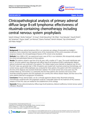
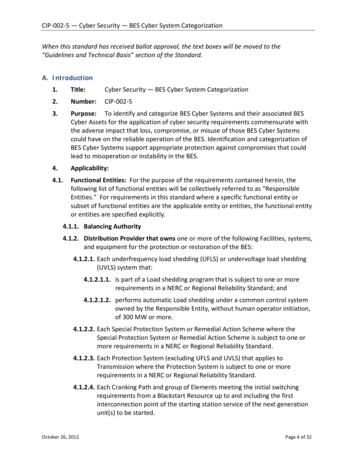
Guo et al. Diagnostic Pathology(2019) 14:82Page 8 of 11Fig. 3 Morphology and immunophenotype of case 9, which progressed to systemic T-cell lymphoma. a Skin biopsy shows a diffuse, atypicallymphocyte infiltrate, with a marked epidermis and subcutaneous distribution (hematoxylin and eosin stain [H&E], 40 ). b Skin biopsy showsmedium- to large-sized lymphocytes, with enlarged, oval and pleomorphic nuclei (H&E, 200 ). c Strong staining of CD4 cells (IHC, 200 ). dGranB Positive cells (IHC 100 ). e Ki67 expression is high in 70% of tumor cells (IHC 100 ). f In situ hybridization shows that neoplastic cells arepositive for EBV-encoded RNA (100 )most patients exhibited severe, extensive skin lesionswith systemic manifestations, including fever, hepatosplenomegaly, and lymphadenopathy, consistent withother reports [2, 19, 20]. The term HV-LPD changedfrom hydroa vacciniforme-like lymphoma to lymphoproliferative disorder by the 4th revised edition of WHOclassifications would be a more appropriate classificationand a more descriptive term [1, 21, 22].The histopathology of biopsies from 11 patients showedthat atypical lymphocytes had infiltrated, and subcutaneous tissues were involved. Nine patients died of systemicT/NK lymphoma. All had elevated LDH levels (range: 318to 1202 IU/L) and higher EBV DNA blood loads. Ourstudy confirmed previous results that indicated that HVLPD and HVLL groups comprised a spectrum of cutaneous CAEBV forms. HV-LPD is an EBV-associated lymphoproliferative disorder of T or NK-cell phenotype withrisk for progression to HVLL.There is no method in the literature for distinguishingbetween HV-LPD and progression to systemic NK/T celllymphoma. This lack of distinction often confuses pathologists and chemotherapy physicians. The challenge remainsto identify morphological or clinical markers for predictingwhich patients are at high risk of progressing to systemiclymphoma. We found that the presence of a TCR gene rearrangement was not useful for making a diagnosis oflymphoma, because, in our study, case 14 and 18 hadmonoclonalities in the early stage. Four patients developedsystemic lymphoma and died of the disease progressionduring chemotherapy treatment. As shown in Table 1, allthose patients displayed atypical, medium- to large-sizedcells with deep skin ulcers, highly elevated LDH, longlasting high EBV DNA loads (typically 1 105 copies), severe systemic manifestations, diffuse effacement of normalnodal architecture by atypical cell infiltration, and bonemarrow involvement. Older patients tended to developlymphoma, which was probably associated with anirreversible process [23].HV-LPD was originally thought to be derived from skinhoming cytotoxic T-cells or NK cells. The majority of
Guo et al. Diagnostic Pathology(2019) 14:82Page 9 of 11Table 3 Immunophenotype and molecular analysis of biopsies from 19 patients with HV-LPDCaseCD3CD8CD4CD56TIA1CD30Ki67EBERsTCR rearrangement1 –– –30% –2 –– –60% –3 –– –30% ND4 – – P 80% –5 –– ND30% –6 –– ND30% –7 –– –30% –8 –– P 50% ND9 – – 70% 10 –– ND20% 11 –– –30% 12 –– –30% ND13 –– 70% F14 – – ND40% 15 –– –60% –16 – – P 70% 17 – – 40% 18 – – –10% 19 – – –60% –EBERs EBV-encoded RNAs, TCR T-cell receptor, P partial, ND not done, F failure to extract sample DNAFig. 4 Kaplan-Meier survival curves for patients with HV-LPD, categorized by a clinicopathological feature. Patients with T-cell lineage disease(CD4 and CD8) had similar clinical outcomes with those of NK-cell lineage disease for all the patients with or without the treatment (n 19,p 0.578 for T-cell vs. NK-cell). Survival-time differences were analyzed with the log-rank test
Guo et al. Diagnostic Pathology(2019) 14:82studies on HV-LPD reported a T-cell phenotype withdivergent immunoexpression of either CD4 or CD8 [24,25]. However, EBV-infected cells might be CD4 T cells,CD8 T cells, or NK cells. EBV-infected cellular lineageshave varied in different studies and populations. Doeden etal. described patients with a NK-cell phenotype [6].Interestingly, our cases exhibited the CD4 phenotype (7/19), the CD8 phenotype (5/19), and the NK-cell phenotype (7/19). The clinical course of the CD56 phenotypewas not more aggressive than that of the T-cell phenotype;only two cases died of systemic NK lymphoma. The othertwo patients with the CD56 phenotype had longer clinicalcourses, and were alive with disease after 3 or 7 years offollow up. This finding was consistent with
Six cases had a history of mosquito-bite hypersensitiv-ity. Among these, four had a CD4 phenotype, and the other two had a CD8 phenotype. No patient was hyper-sensitive to light. The available routine laboratory tests revealed that all patients (11/11) had elevated lactate dehydrogenase (LDH) levels (range 312 to 1202IU/L), when they were