Transcription
Ichikawa et al. Experimental Hematology & Oncology 2013, HExperimentalHematology & OncologyOpen AccessClinicopathological analysis of primary adrenaldiffuse large B-cell lymphoma: effectiveness ofrituximab-containing chemotherapy includingcentral nervous system prophylaxisSatoshi Ichikawa1, Noriko Fukuhara1*, Ai Inoue1, Hiroki Katsushima2, Rie Ohba1, Yuna Katsuoka1, Yasushi Onishi1,Joji Yamamoto3, Osamu Sasaki4, Jun Nomura5, Osamu Fukuhara6, Kenichi Ishizawa1, Ryo Ichinohasama3and Hideo Harigae1,7AbstractBackground: Primary adrenal lymphoma (PAL) is an extremely rare subtype of extranodal non-Hodgkin’slymphoma. Some researchers have reported some of the characteristics of PAL and its association with poorprognosis; however, the clinicopathological features of PAL remain to be elucidated.Methods: From 2008 to 2011 we experienced seven cases of PAL in our institutions. We retrospectively analyzedthe clinical and pathological features of these patients.Results: The patients ranged in age from 50 to 85 years, with a median of 71 years. The overall male:female ratiowas 6:1. All seven patients were diagnosed with diffuse large B-cell lymphoma (DLBCL) pathologically. Bilateraladrenal involvement was confirmed in five patients. The median largest tumor diameter at diagnosis was 58 mm.The Ki-67 index was generally high ( 70%). All patients were treated with rituximab-containing chemotherapy, andcentral nervous system (CNS) prophylaxis was conducted for three patients. One patient with CNS involvement atthe time of the diagnosis also received whole-brain radiation. The overall survival rate at two years was 57%(median follow-up; 24.8 months). It is noteworthy that the three patients who received a full course of therituximab-containing regimen and CNS prophylaxis are currently alive without disease relapse, and that none of theseven patients died due to progression of lymphoma.Conclusions: Primary adrenal DLBCL can be a clinically aggressive disease entity. Rituximab-containingchemotherapy combined with CNS prophylaxis could be a reasonable option for the treatment of PAL; however,analyses of more PAL cases are needed for the establishment of this strategy.Keywords: Primary adrenal lymphoma, Diffuse large B-cell lymphoma, Adrenal insufficiency, Central nervous systeminfiltration, Rituximab* Correspondence: nfukuhara@med.tohoku.ac.jp1Department of Hematology and Rheumatology, Tohoku University GraduateSchool of Medicine, 1-1 Seiryo-cho, Sendai 980-8574, JapanFull list of author information is available at the end of the article 2013 Ichikawa et al.; licensee BioMed Central Ltd. This is an Open Access article distributed under the terms of the CreativeCommons Attribution License (http://creativecommons.org/licenses/by/2.0), which permits unrestricted use, distribution, andreproduction in any medium, provided the original work is properly cited.
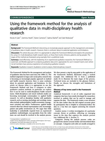
Ichikawa et al. Experimental Hematology & Oncology 2013, ctionMalignant lymphoma primarily arising in the adrenalgland, i.e., primary adrenal lymphoma (PAL), is an extremely rare disease and accounts for less than 1% of allnon-Hodgkin’s lymphoma. Around 100 cases of PAL havebeen reported in the English literature to date; with theexception of a few investigational reports based on severalcases, most have been single case reports and literature reviews [1-5]. These reports suggested the following clinicaland pathological characteristics of PAL: a predominancein elderly males, diffuse large B-cell lymphoma (DLBCL)as the most common histology, frequent bilateral adrenalinvolvement with insufficiency of adrenal function and atendency for patients to develop central nervous system(CNS) infiltration. Some researchers have argued thatthere may be a correlation between PAL and Epstein–Barrvirus infection [1,6], while others have reported thattumor cells of primary adrenal DLBCL usually expressBCL-2 with a non-germinal center B-cell phenotype [7].However, the accumulation of more information is neededto further elucidate the clinicopathological features ofPAL. PAL has also been reported to generally have a poorprognosis with conventional chemotherapy, such as theCHOP regimen, and the optimal therapy for PAL remainsto be established. However, a substantial proportion ofcases reported in the literature were treated before the development of rituximab, and it is possible that rituximabwould improve the prognosis of primary adrenal DLBCL,as it does of typical DLBCL.We herein analyzed the clinicopathological features andclinical courses of seven cases of PAL encountered at ourinstitutions in recent years; all were pathologically diagnosed as DLBCL and treated with rituximab-containingchemotherapy.ResultsClinical characteristicsFrom 2008 to 2011 in our institutions, seven cases werenewly diagnosed as primary adrenal DLBCL. We thoroughly reviewed their clinical and pathological findingsretrospetctively. The clinical features of the patients areshown in Table 1. The patients ranged in age from 50 to85 years, with a median of 71 years. The overall male:female ratio was 6:1. Only one patient (Case 5) presentedwith symptoms of adrenal insufficiency and fever. The adrenal tumor of three cases (Cases 1, 2 and 3) was incidentally found by a CT that was performed to evaluate otherunderlying diseases, such as a renal tumor, chronic hepatitis and pulmonary fibrosis. The remaining three patientshad back pain. B-symptoms (fever, weight loss of morethan 10% within six months, or night sweating) were observed in only one patient (Case 5).The mean maximum diameter of the tumor at diagnosiswas 58 mm, with a range from 40 to 80 mm. Two patientsPage 2 of 8with unilateral adrenal involvement underwent surgical resection due to an initial suspicion of adrenal carcinoma. Onthe other hand, the remaining five patients with bilateraladrenal involvement were diagnosed by needle biopsy. Fourbilateral cases (Cases 3, 5, 6 and 7) had paraaortic lymphnode lesions; all of which were smaller than the adrenal lesions. These patients were stratified to be in a high-riskgroup by the International Prognosis Index (IPI) [8]. Noadditional lesions, including mediastinal and CNS lesions,were detected in any cases on CT.Positron emission tomography (PET) with 2-[18F] fluoro2-deoxy-D-glucose (FDG) was performed in all cases. Theuptake of FDG in the adrenal tumor was confirmed in allcases, and in four cases, uptake was also detected in adjacent paraaortic lymph nodes. No other lesions with significant FDG uptake were detected in any of the patients.Uptake foci at the adrenal tumors were revealed in all cases,and the maximum standardized uptake values (SUVmax)in each case were generally high (8.0 – 26.0, median, 21.5).The SUVmax seemed to be higher in the cases with bilateraladrenal infiltration (n 5; median, 22.4) than in the caseswith unilateral infiltration (n 2; median, 15.5). In addition,the values of the SUVmax in adjacent lymph nodes were alllower than those in the adrenal tumors. Bone marrow infiltration was not detected in any of the patients.Apparent CNS infiltration was not confirmed at the timeof diagnosis in any of the cases except in Case 4, in whicha brain tumor and bilateral adrenal tumors were detectedsimultaneously. Both the cerebral and adrenal lesions werebiopsied and concluded to have the same histology asDLBCL. In Case 5, multiple cerebral infarctions emerged amonth before the patient’s presentation to our institutions.This may have been caused by the infiltration of lymphomacells toward cerebral vessels, although magnetic resonanceimaging of the brain and cytology of the cerebrospinal fluiddid not detect any infiltration of lymphoma cells.The serum lactic dehydrogenase (LDH) level was abovethe normal limits in all cases. The serum level of solubleinterleukin-2 receptor (sIL-2R) was also elevated in all sixcases in which it was measured. The rapid adrenocorticotropic hormone (ACTH) stimulation test, in which acortisol level less than 18 – 20 μg/dL at 60 minutes afterACTH challenge is regarded to be a poor response [9],revealed partial adrenal insufficiency in two cases withbilateral adrenal involvement without clinically apparent adrenal insufficiency.Pathological analysesThe pathological and immunohistochemical findings of allseven cases are shown in Table 2, and representative pathological findings are shown in Figure 1. There were nounique morphological characteristics of the PAL cases. Allcases were diagnosed as DLBCL. The tumor cells expressedCD20 (7 of 7), CD79a (6 of 6), BCL2 (5 of 7), BCL6 (4 of 7)
Case/ Initial presentationAge/SexTumor BiopsytypestrategyClinical PS IPI CT findingsstageLaterality 1/81/ IncidentalomaMDLBCL LaparoscopicadrenalectomyIE-A0L-I Unilateral2/69/ IncidentalomaMDLBCL OpenadrenalectomyIE-A03/71/ IncidentalomaMDLBCL CT-guided needlebiopsyIIE-A4/62/ HemiparesisMDLBCL CT-guided needlebiopsy5/50/ Fever, shock CerebralMinfarctionPET findingsLaboratory dataOtherlymphomalesions[Maximaldiameter(mm)]Serum cortisolSUVmaxSUVmaxLDH sIL-2R Serum cortisolin adrenal in adjacent (IU/L) (U/mL) level at baseline level after(μg/dL)ACTHtumorLNsstimulation*(μg/dL)40 [L]None11.8No lesions372†565†13.724.8L-I Unilateral80 [L]None19.2No lesions376†1374†7.221.92H-I Bilateral52 [L]paraaorticLNs [17]8.04.3319†1367†NDNDIV-A2HBilateral60 [L]Brain24.5No lesions381†NDNDNDDLBCL CT-guided needlebiopsyIIE-B4L-I Bilateral57 [L]paraaorticLNs [17]22.43.5415†10468† 7.815.7‡6/73/ Back painMDLBCL CT-guided needlebiopsyIIE-A3H-I Bilateral57 [L]paraaorticLNs [39]26.014.6345†7303†8.811.1‡7/85/ Back painFDLBCL CT-guided needlebiopsyIIE-A2H-I Bilateral67 [L]paraaorticLNs [19]21.58.6529†7777†10.813.0‡33 [R]Ichikawa et al. Experimental Hematology & Oncology 2013, 2:19http://www.ehoonline.org/content/2/1/19Table 1 Summary of clinical findings67 [R]58 [R]22 [R]39 [R]ACTH adrenocorticotropic hormone, CT computed tomography, DLBCL diffuse large B-cell lymphoma, F female, H high; H-I high-intermediate, IPI international prognosis index, L left, LDH serum lactate dehydrogenase,L-I low-intermediate, LNs lymph nodes, M male, ND not done, PET positron emission tomography, PS performance status, R right, sIL-2R serum soluble interleukin-2 receptor, SUVmax maximumstandardized uptake value.*Each serum cortisol level was measured at 60 min after ACTH challenge.†The value exceeds upper limit of normal. ‡The value is below lower limit of normal.Page 3 of 8
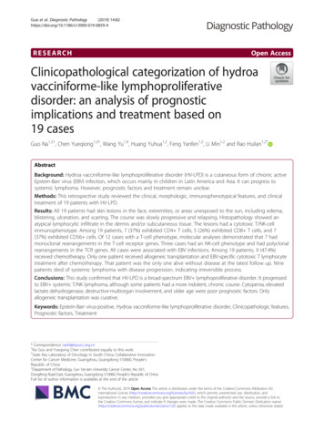
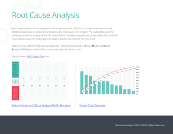
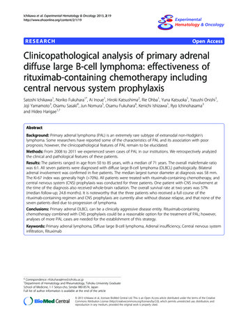
Ichikawa et al. Experimental Hematology & Oncology 2013, 2:19http://www.ehoonline.org/content/2/1/19Page 4 of 8Table 2 Summary of pathological and immunohistochemical findingsCasePathological diagnosisCD5CD10CD20CD79aBCL2BCL6EBERMUM1Ki-67 indexPhenotype1DLBCL, NOS 70-80%Non-GCB2DLBCL, NOS 90%GCB3DLBCL, NOS 80%Non-GCB4DLBCL, NOS ND 75-90%Non-GCB5DLBCL, NOS 90-99%Non-GCB6DLBCL, NOS 75-90%Non-GCB7DLBCL, NOS 90-99%Non-GCBDLBCL diffuse large B-cell lymphoma, GCB germinal center B-cell phenotype, ND not done, NOS not otherwise specified.and MUM1 (5 of 7). CD10 was negative in all cases. CD5was positive in two cases. The Ki-67 index was generallyhigh ( 70%). A flow cytometric analysis of the lymphomacells (data not shown) was available for Cases 6 and 7, anddid not conflict with the immunohistochemical data described above. According to Hans’ criteria [10], most of thecases (6 of 7) were classified as the non-germinal center Bcell subtype.Treatment and outcomesAll patients were treated with rituximab-containing chemotherapy, such as cyclophosphamide, doxorubicin, vincristine,prednisolone and rituximab (R-CHOP), with a successfulclinical outcome (Table 3). The courses of chemotherapyand the doses of agents were modified at the discretion ofthe attending physicians according to the clinical conditionof the patients. In Case 4, because of the apparent coexistence of CNS lymphoma, combined immunochemotherapyplus whole-brain radiotherapy (WBRT), high-dose methotrexate (HD-MTX), high-dose cytarabine and rituximab,was performed according to a previous report [11]. Case 5,in which progression toward the CNS could not be excluded, received systemic intravenous administration ofhigh-dose methotrexate. Two additional cases (Cases 2Figure 1 Representative pathological findings of primary adrenal diffuse large B-cell lymphoma (Case 1). The black lines in the lowerright corner of each figure indicate 100 μm. (A, B) The results of hematoxylin and eosin staining. On low-power magnification (A), the lymphomawas located in the zona reticularis (represented by r) of the adrenal cortex. f and g represent the zona fasiculata and zona glomerulosa,respectively. High-power magnification (B) showed medium- to large-sized atypical lymphoid cells with dispersed chromatin. (C)Immunohistochemical staining for CD20 showed that the large lymphoma cells were positive for CD20. (D) Immunohistochemical staining forCD3 showed reactivity in small lymphocytes, but not in large lymphoma cells.
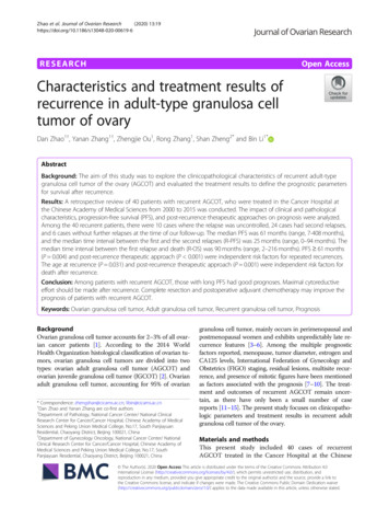
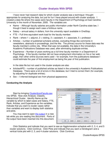
Ichikawa et al. Experimental Hematology & Oncology 2013, 2:19http://www.ehoonline.org/content/2/1/19Page 5 of 8Table 3 Summary of clinical coursesCaseTreatmentResponse to treatmentOverall survival duration (months)RelapseOutcomeCause of death1R-CHOP 5PR45.0NoDeadCholangiocarcinoma2R-CHOP 6PR7.9NoDeadPneumonia3R-CHOP 2NA4.0 DeadExacerbation of pulmonary fibrosis4R-MPV 4CR40.1NoAlive CR28.0NoAlive CR24.8NoAlive NA2.6 DeadPneumonia(75% dose)IT*(70% dose)IT*HD-Ara-C 2WBRT5R-CHOP 6HD-MTX 2IT*6R-CHOP 8IT*7R-COP 1(60% dose)*IT represents prophylactic administration of methotrexate and/or cytarabine.CR complete response, CRu unconfirmed complete response, HD-Ara-C high-dose cytarabine, HD-MTX high-dose methotrexate, IT intrathecal themotherapy, NAnot assessable, R-CHOP rituximab, cyclophosphamide, doxorubicin, vincristine, and prednisolone, R-COP rituximab, cyclophosphamide, vincristine, andprednisolone, R-MPV rituximab, high-dose methotrexate, procarbazine, and vincristine, WBRT Whole-brain radiotherapy.and 6) received prophylactic intrathecal injection ofmethotrexate.After the completion of chemotherapy, we assessed theresponse in each case. CT scans were performed in allcases, and FDG-PET was also performed in some cases(Cases 4, 5 and 6). In the five patients who received a fullcourse of rituximab-containing chemotherapy and theadditional therapy mentioned above, a favorable clinicaloutcome (complete response in Cases 4, 5 and 6, and partial responses in Cases 1 and 2) was achieved, and therehave been no cases of relapse as of the writing of thismanuscript. The other two patients died of complicationsduring the course of chemotherapy (Case 3, exacerbationof pulmonary fibrosis; Case 7, pneumonia). In both cases,no signs of lymphoma progression, such as further elevation of the serum LDH level, were detected within theirclinical course after the initiation of chemotherapy.The median survival time of all patients from diagnosis until death or the date of the last follow-up was 24.8months (range, 2.6 – 45.0 months). No patients experienced a relapse of lymphoma, and none died due to theprogression of lymphoma. Both the OS and FFS at twoyears were 57.1% (95% CI 17.2 – 83.7, Figure 2).DiscussionPAL is an extremely rare disease entity and only about 100cases have been reported in the English literature to date.The characteristics of our series of were largely consistentwith the previous reports; all were pathologically diagnosed as DLBCL, and the patients were predominantlyelderly males [1-4]. It might be unique that two cases werepositive for CD5 in our case series; for CD5-positive primary adrenal DLBCL cases have scarcely been reportedand only one case was reported recently [12]. However,CD5 expression has not been evaluated even in some recent studies which included a relatively large number ofprimary adrenal DLBCL cases, and the significance ofCD5 expression in primary adrenal DLBCL remains to beknown. Three patients were asymptomatic at presentation,and they were incidentally suspected to have adrenalFigure 2 The overall survival in all seven cases of PAL.
Ichikawa et al. Experimental Hematology & Oncology 2013, 2:19http://www.ehoonline.org/content/2/1/19tumors based on CT scans performed as a screening foranother disease (i.e., incidentalomas). These observationssuggested that PAL might be more prevalent than hasbeen reported.The definition of primary adrenal lymphoma has not beenmade clear in the literature, unlike other rare extranodallymphomas, such as primary bone lymphoma [13] or primary breast lymphoma [14]. A common feature of the definition of the two diseases mentioned above is that thelymphoma arises in a specific extranodal organ, with orwithout adjacent lymph node swelling. It is thought to bereasonable that this definition is also applicable to primaryadrenal lymphoma (PAL). In our cases which had paraaorticlymph node lesions (Cases 3, 5, 6 and 7), the size of the adrenal tumors was larger than that of the lymph node lesionson CT (Table 1). Images of PET-CT scans showed that theSUVmax of the adjacent lymph node lesions was also lowerthan that of the adrenal lesions (Table 1). These findingssuggested that the adrenal lesion was the primary tumor ineach case. In Case 4, cerebral and adrenal lesions weredetected simultaneously. It is reported that DLBCL with adrenal involvement could have a tendency to develop CNSinfiltration [15], so we think that simultaneous involvementof CNS and adrenal glands by lymphoma would be considered as PAL with secondary CNS involvement, as Rashidi Aet al. recently stated [16].The most common symptoms of PAL at presentationwere reported to be abdominal or back pain, fever of unknown origin and signs of adrenal insufficiency [1-4]. Bilateral adrenal involvement of PAL is frequently accompaniedby clinical adrenal insufficiency [3,4,17-22]. In our series,clinically definite adrenal insufficiency was seen in onlyone of the five cases in whom the adrenal function was examined. The serum cortisol levels at baseline were not reduced in any of these cases. Partial adrenal insufficiency,which was asymptomatic and detected by the rapid ACTHtest, was established in two cases of bilateral adrenal involvement. Lymphoma cells show diffuse infiltration intothe adrenal parenchyma, and it is reasonable that bilateralinvolvement of lymphoma would lead to a condition of adrenal insufficiency, even if it is not clinically obvious.Gene expression studies of DLBCL have identifiedtwo subtypes; the germinal-center B-cell-like (GCB)subtype and the non-GCB subtype (combining the activated B-cell-like subtype and the type 3 subtype) [23];the latter are associated with a significantly lower survival than the former. Moreover, Li et al. recentlyshowed that non-GCB DLBCL cases with high Ki-67 expression, which is an index of proliferation, received alimited survival benefit from standard R-CHOP therapy[24]. Following the Hans’ algorithm [10], we confirmedthat most of our cases were classified as the non-GCBsubtype, and all had high Ki-67 expression (over 70%).In other previous reports [1,7], most PAL cases werePage 6 of 8subclassified as the non-GCB type; these findings maybe associated with the poor prognosis of PAL.In addition to the pathological factors mentioned above,some clinical factors for predicting the prognosis ofDLBCL have recently been reported. For example, a highsIL-2R level was reported to be associated with a poorprognosis among patients with DLBCL who were treatedwith R-CHOP [25]. It has also been reported that a highSUVmax on PET scans is associated with a shorter survival in patients with DLBCL [26]. Moreover, Shou et al.recently reported that lymphomas showing intense FDGuptake have an increased capacity for proliferation andrapid growth, which was confirmed by the Ki-67 expression [27]. In the present study, the bilateral cases had highlevels of sIL-2R and a high SUVmax on PET/CT compared with unilateral cases. The Ki-67 index was also generally high in all cases. These points suggest that bilateraladrenal DLBCL should be a clinically aggressive disease.We successfully treated PAL patients in a tolerable general condition with rituximab-containing systemic chemotherapy and CNS prophylaxis. PAL is frequently treatedwith cyclophosphamide, doxorubicin hydrochloride, vincristine and prednisolone (CHOP), or a CHOP-like regimen; however, the prognosis is generally poor [1,2]. Inrecent years, rituximab has been added to the regimen,and there have been a few reports about successful outcomes with rituximab-containing regimens for PAL[5,28,29]. However, CNS relapses of PAL occur frequently,and most patients with CNS relapse are reported to havedied early [3,4,19,30,31]. We treated most of the patients inthe present study by R-CHOP, with modifications as necessary, and added HD-MTX and/or intrathecal chemotherapyfor the patients who were considered to potentially haveCNS infiltration at presentation. It is noteworthy that all ofthe patients who received a full course of rituximabcontaining chemotherapy and CNS-oriented treatment arecurrently alive without disease progression or relapse. Nopatients died of the progression of lymphoma, and therewere no cases of CNS relapse. This is despite the non-GCBDLBCL subtype, elevated sIL-2R level and Ki-67 indexnoted in these patients. Therefore, in younger PAL patientsin good general condition, an aggressive chemotherapeuticstrategy consisting of a rituximab-containing chemotherapy regimen and CNS-oriented treatment is thought to bepromising, and could improve the prognosis of PAL.ConclusionsPAL with DLBCL histology is a clinically aggressive disease. Rituximab-containing chemotherapy combinedwith CNS prophylaxis could be a reasonable option forthe treatment of PAL; however, further accumulation ofclinical experiences, as well as additional research, isnecessary to establish the optimal therapeutic strategyfor PAL.
Ichikawa et al. Experimental Hematology & Oncology 2013, Case selection and clinical dataFrom 2008 to 2011 in our institutions, a total of 563 patients were diagnosed with DLBCL. We reviewed the records of the patients with newly diagnosed primary adrenalDLBCL. Cases with secondary adrenal involvement andthose with human immunodeficiency virus infection wereexcluded. A total of seven cases were included in this study.All had unilateral or bilateral adrenal tumors, which werethe predominant tumors considered to be primary lesions(and were biopsied), while some other lesions, such asparaaortic lymph node swelling or central nervous systemlesions, were also detected in several cases. The therapeutic strategy was chosen at the discretion of the attending physicians.All available data, including the clinical presentations, laboratory findings, radiological findings, therapeutic regimens and follow-up information, were evaluated. Thestaging procedures included a complete physical examination, computed tomography (CT) scans of the neck, chestand abdomen, positron emission tomography (PET) scansand bone marrow biopsy and/or aspiration. This study wasapproved by the Ethics Committee of each hospital. Theprocedures performed for the present study were done inaccordance with the Declaration of Helsinki.Pathological evaluationTissue specimens were fixed in 10% formalin solution,routinely processed and embedded in paraffin. The sections were stained with hematoxylin-eosin for microscopic examination. Immunohistochemical studies wereperformed using antibodies against CD3, CD5, CD10,CD20, CD45RA, CD79a, BCL2, BCL6, MUM1, Ki-67and Epstein–Barr virus-encoded small RNA (EBER).For two cases with sufficient materials available, flowcytometry was performed using various monoclonalantibodies against CD2, CD3, CD4, CD5, CD7, CD8,CD10, CD13, CD19, CD20, CD22, CD30, CD45, CD56,TCRαβ, TCRγδ, Igκ and Igλ. Markers that wereexpressed on more than 20% of the cells were judged tobe positive.Statistical analysisAll statistical analyses were performed using the GraphPadPrism software program (version 5.04 for Windows;GraphPad Software Inc., San Diego, CA). The treatmentoutcomes were measured by the failure-free survival (FFS)and overall survival (OS). The FFS was defined as the timefrom initial diagnosis to the first occurrence of progression,relapse after a response or death from any cause. Thefollow-up of patients not experiencing one of these eventswas censored at the date of last contact. The OS was measured from the time of the initial diagnosis until deathfrom any cause, with surviving patient follow-up censoredPage 7 of 8at the last contact date. Estimates of the FFS and OS weredetermined using the Kaplan–Meier method.Competing interestsThe authors declare no competing interests.Authors’ contributionsSI, the first author, designed the research project, analyzed the data,reviewed the literature, drafted the manuscript and revised the finalmanuscript. NF, the corresponding author, designed the research project,contributed to discussions and reviewed the manuscript. AI, RO, YK, YO, JY,OS, JN and OF were in charge of the treatment and follow-up of thepatients and collected the patient information. HK interpreted thepathological slides. KI contributed to the discussion and review of themanuscript. RI interpreted the pathological slides and contributed to thediscussion and review of the manuscript. HH, the last author, supervised theoverall project and contributed to the discussion and review of themanuscript. All authors reviewed and approved the final version of themanuscript.Author details1Department of Hematology and Rheumatology, Tohoku University GraduateSchool of Medicine, 1-1 Seiryo-cho, Sendai 980-8574, Japan. 2Department ofHematopathology, Tohoku University Graduate School of Medicine, Sendai,Japan. 3Department of Internal Medicine, Sendai City Hospital, Sendai, Japan.4Department of Hematology, Miyagi Prefectural Cancer Center, Sendai,Japan. 5Department of Internal Medicine, NTT East Tohoku Hospital, Sendai,Japan. 6Department of Internal Medicine, Sendai Red Cross Hospital, Sendai,Japan. 7Department of Molecular Hematology/Oncology, Tohoku UniversityGraduate School of Medicine, Sendai, Japan.Received: 20 June 2013 Accepted: 31 July 2013Published: 2 August 2013References1. Mozos A, Ye H, Chuang WY, Chu JS, Huang WT, Chen HK, Hsu YH, BaconCM, Du MQ, Campo E, Chuang SS: Most primary adrenal lymphomas arediffuse large B-cell lymphomas with non-germinal center B-cellphenotype, BCL6 gene rearrangement and poor prognosis. ModernPathol 2009, 22:1210–1217.2. Singh D, Kumar L, Sharma A, Vijayaraghavan M, Thulkar S, Tandon N:Adrenal involvement in non-Hodgkin’s lymphoma: four cases and reviewof literature. Leukemia Lymphoma 2004, 45:789–794.3. Mantzios G, Tsirigotis P, Veliou F, Boutsikakis I, Petraki L, Kolovos J,Papageorgiou S, Robos Y: Primary adrenal lymphoma presenting asAddison’s disease: case report and review of the literature. Ann Hematol2004, 83:460–463.4. Grigg AP, Connors JM: Primary adrenal lymphoma. Clin Lymphoma 2003,4:154–160.5. Kim YR, Kim JS, Min YH, Yoon DH, Shin HJ, Mun YC, Park Y, Do YR, JeongSH, Park JS, et al: Prognostic factors in primary diffuse large B-celllymphoma of adrenal gland treated with rituximab- CHOPchemotherapy from the Consortium for Improving Survival ofLymphoma (CISL). J Hematol Oncol 2012, 5:49.6. Ohsawa M, Tomita Y, Hashimoto M, Yasunaga Y, Kanno H, Aozasa K:Malignant lymphoma of the adrenal gland: its possible correlation withthe Epstein-Barr virus. Modern Pathol 1996, 9:534–543.7. Ide M, Fukushima N, Hisatomi T, Tsuneyoshi N, Tanaka M, Yokoo M,Tomimasu R, Funai N, Sueoka E: Non-germinal cell phenotype and bcl-2expression in primary adrenal diffuse large B-cell lymphoma. LeukemiaLymphoma 2007, 48:2244–2246.8. The International Non-Hodgkin's Lymphoma Prognostic Factors Project: Apredictive model for aggressive non-Hodgkin’s lymphoma. New Engl JMed 1993, 329:987–994.9. May ME, Carey RM: Rapid adrenocorticotropic hormone test in practice.Retrospective review. Am J Med 1985, 79:679–684.10. Hans CP, Weisenburger DD, Greiner TC, Gascoyne RD, Delabie J, Ott G,Muller-Hermelink HK, Campo E, Braziel RM, Jaffe ES, et al: Confirmationof the molecular classification of diffuse large B-cell lymphoma byimmunohistochemistry using a tissue microarray. Blood 2004,103:275–282.
Ichikawa et al. Experimental Hematology & Oncology 2013, 2:19http://www.ehoonline.org/content/2/1/1911. Shah GD, Yahalom J, Correa DD, Lai RK, Raizer JJ, Schiff D, LaRocca R, GrantB, DeAngelis LM, Abrey LE: Combined immunochemotherapy withreduced whole-brain radiotherapy for newly diagnosed primary CNSlymphoma. J Clin Oncol 2007, 25:4730–4735.12. Rashidi A, Bergeron CW, Fisher SI, Chen IA: Primary adrenal de novo CD5positive diffuse large B cell lymphoma. Ann Hematol 2013. doi:10.1007/s00277-013-1690-8 [Epub ahead of print].13. Ostrowski ML, Unni KK, Banks PM, Shives TC, Evans RG, O’Connell MJ, TaylorWF: Malignant lymphoma of bone. Cancer 1986, 58:2646–2655.14. Wiseman C, Liao KT: Primary lymphoma of the breast. Cancer 1972,29:1705–1712.15. Tomita N, Yokoyama M, Yamamoto W, Watanabe R, Shimazu Y, Masaki Y,Tsunoda S, Hashimoto C, Murayama K, Yano T, et al: Central nervoussystem event in patients with diffuse large B-cell lymphoma in therituximab era. Cancer Sci 2012, 103:245–251.16. Rashidi A, Fisher SI: Primary adrenal lymphoma: a systematic review.Ann Hematol 2013. doi:10.1007/s00277-013-1812-3 [Epub ahead ofprint].17. Kuyama A, Takeuchi M, Munemasa M, Tsutsui K, Aga N, Goda Y, KanetadaK: Successful treatment of primary adrenal non-Hodgkin’s lymphomaassociated with adrenal insufficiency. Leukemia Lymphoma 2000,38:203–205.18. Hamid Zargar A, Ahmad Laway B, Alam Bhat K, Shah A, Ahmad M, AejazAziz S, Iftikhar Bashir M, Iqbal Wani A, Hayat
It is noteworthy that the three patients who received a full course of the rituximab-containing regimen and CNS prophylaxis are currently alive without disease relapse, and that none of the . or night sweating) were ob-served in only one patient (Case 5). . adrenal infiltration (n 5; median, 22.4) than in the cases with unilateral .