Transcription
Iranian Journal of Medical PhysicsVol. 14, No. 1, March 2017, 1-7Received: September 16, 2016; Accepted: December 10, 2016Original ArticleQuality Control Assessment of Conventional Radiology Devices in IranMohsen Asadinezhad1, Mohammad Taghi Bahreyni Toossi2*, Ali Ebrahiminia3, Masoumeh Giahi1AbstractIntroductionQuality control (QC) techniques are used in monitoring and maintenance of the components of an x-raysystem. QC of radiology devices plays a significant role in reduction of medication dose and optimization ofimage quality. This study aimed to conduct QC tests on randomly selected radiology devices, installed indiagnostic imaging departments of Iran.Materials and MethodsIn total, quality control tests were conducted on 51 conventional radiology devices installed in 20 cities ofIran in order to assess the accuracy of peak kilovoltage (kVp), exposure time, exposure linearity andreciprocity, reproducibility of exposure and determination of half-value layer (HVL) using a calibrated MultO-Meter.ResultsIn this study, 38.6% of devices had intolerable variance of kVp accuracy. The results of 34.5% of deviceswere out of the acceptable limits in exposure time accuracy test. In 46.7% and 53.1% of devices, variancewas greater than the acceptable range for exposure linearity and exposure reciprocity, respectively. In termsof reproducibility of exposure test, the reproducibility variance and percentage of tube output variations in19.4% of devices exceeded the limits. Moreover, the thickness of first HVL was lower than the acceptablelimit in 14.7% of devices.ConclusionAccording to the results of this study, there were wide variations in QC test results, perhaps mainly due tothe fact that it is not an obligation to implement QC programs in Iran. The most important problems werenon-reciprocity of exposure, nonlinearity of exposure with milliampere-second (mAs), kVp and timerinaccuracy. Involvement of medical physicists, radiologists and radiographers in the implementation of QCprograms at various stages of development, installation and use of equipment should enable the gradualimprovement in equipment performance.Keywords: Quality control, Diagnostic X-ray, Radiology1- Department of Radiology Technology, School of Paramedical Sciences, Mashhad University of Medical Sciences,Mashhad, Iran2- Medical Physics Research Center, Mashhad University of Medical Sciences, Mashhad, Iran3- Department of Biochemistry & Biophysics, Faculty of Medicine, Guilan University of Medical Sciences, Rasht,Iran*Corresponding author: Tel: 98 51 38002317; Fax: 98 511 8002320; Email: bahreynimt@mums.ac.ir1Iran J Med Phys., Vol. 14, No. 1, March 2017
Mohsen Asadinezhad, et al.1. IntroductionAccording to the classification published by theunited nations scientific committee on the effectsof atomic radiation (UNSCEAR) and dataprovided by the World Health Organization(WHO), Iran is a country of level two healthcare [1, 2]. Annually, more than 20 million Xray examinations are performed in radiologycenters of Iran [3]. One of the fastest and easiestways for a physician to view the internal organsand structures of the human body is X-rayimaging, which has no proper alternative.According to the safety protocol, it isrecommended that the radiation exposure bemaintained as low as reasonably achievable(ALARA) in order to keep exposures to ionizingradiation as far below the dose limits aspractical, and at the same time, be able toprovide valuable images of high usability [4]. Inorder to achieve this goal, quality assuranceprograms have been implemented in thediagnostic radiology and medical imagingdepartments [5, 6].The purpose of quality control (QC) program isto ensure that there is optimal performancerelated to all imaging components [5]. The QCprograms can give rise to the highest qualityimagining with the lowest possible radiationdose to patients and to radiation workers throughmaintaining high diagnostic quality [5]. Asstated by the American Association of Physicistsin Medicine (AAPM), designing and supervisinga quality assurance program is the primaryresponsibility of medical physicist [7]. In 1997,the European commission of protection againstionizing radiation published guidance for QC ofdiagnostic units [8]. The main components ofQC programs have been described in a report byAAPM in 2002 [9]. A wide variety of studieshave been implemented on QC of diagnosticradiographic units and some guidelines havebeen established for QC tests [10-18]. In a studyby Ortiz et al. the results were indicative ofminimized exposure dose to patients duringradiological procedures through the evaluationand revision of the QC parameters [16]. Inanother study by Godechal et al. (1995), qualityassurance program was used for X-ray devicesto assess the efficiency of their specificationsthrough a systematic measurement. According totheir results, the main limitation for X-raydevices was inadequate filtration [11].Many studies have been performed on the QC ofdiagnosticradiographicequipmentinChaharmahal and Bakhtiari, Zanjan, Khorasan,Lorestan, Golestan, Khuzestan, Hormozgan andKerman provinces of Iran [19-27]. Saghatchi(1999) performed QC assessment ofradiographic equipment in Zanjan province andmarked that the status of 57%, 42%, 14%, and7% of the units was not acceptable in terms ofkVp accuracy, exposure linearity, timeraccuracy, and timer reproducibility, respectively[20]. In 2004, Shahbazi conducted an assessmenton medical equipment in order to measure theentrance dose and compare the results before andafter the QC [19]. The results demonstrated thatQC conduction led to 40% decrease in the meandose required for chest examination. Resultsobtained by Khoshbin Khoshnazar et al. (2013)indicated that timer accuracy was a commonproblem of X-ray units in Golestan province[23]. In another study, Gholamhosseinian-Najjaret al. (2014) observed that the status of 27% and45% of apparatuses in Khorasan province wereunacceptable regarding kVp accuracy and timeraccuracy, respectively [21]. Moreover, Rasuli etal. (2014) and Gholami et al. (2015) evaluatedthe performance of radiographic X-rayequipment in Khuzestan and Lorestan provinces,respectively [22, 24]. Jomehzadeh et al. (2016)conducted a study in Kerman province andaffirmed that kVp accuracy, kVp reproducibility,timer accuracy, timer reproducibility, exposurereproducibility, mA/timer linearity and halfvalue layer (HVL) were not within theacceptable limits in 25%, 4%, 29%, 18%, 11%,12%, and 7% of the evaluated units, respectively[26]. Before this study, no comprehensivenational program for quality assurance ofradiology devices has been developed in Iran. Tothe best of our knowledge, no comprehensivenational QC program has been developed yet inIran. With this background in mind, this studywas conducted to perform QC tests on randomlyselected radiology devices installed in diagnosticimaging departments of Iran.Iran J Med Phys., Vol. 14, No. 1, March 20172
Quality Control of Radiology Devices in Iran2. Materials and MethodsIn total, 51 conventional radiology devicesfrom 31 radiology centers in 20 cities of Iran(Arak, Isfahan, Ahvaz, Amol, Mahshahr,Bushehr, Tabriz, Tehran, Rasht, Zahedan,Sanandaj, Shahriar, Shiraz, Qazvin, Karaj,Lahijan, Mashhad, Mamasani, Hashtrood andHashtgerd) were selected using systematicrandom sampling. kVp accuracy, exposuretime accuracy, exposure linearity, exposurereciprocity, reproducibility of exposure anddetermination of HVL were the evaluated QCtests, performed to assess the devices. Toevaluate these tests, a calibrated Mult-O-MeterModel 303 (Unfors, Sweden) was placed onthe radiographic tabletop on top of a leadapron, 100 cm from the focal spot and in thecenter of the field. The lead apron can absorbbackscatter from the table top material;therefore, it can prevent the reduction of anyreadings inaccuracy. Inaccuracy of kVp andexposure rate measurements was 2%, whereasit was 0.5% for time measurements. All QCtests were performed according to standardsset forth in the “quality management in theimaging science” [28].Data are presented as mean standard deviation(SD). Data analysis was performed in SPSSversion 17.2.1. kVP AccuracyAt SSD 100 cm, we measured kVp from 50100 (50, 60, 70, 80, 90 and 100) in two mA(100, 300 or 320) and identified the differencebetween the selected and measured values.This difference should be within 5% [28].2.2. Exposure Time AccuracyVariable times of ( 10, 20,80,100 and 200mSec) were selected at a fixed condition(kVp 60, mA 100). The average of threeexposes were used in order to measureexposure time. Exposure time accuracy wasdetermined using the equation 1. In this test, 5% variation was acceptable for exposuretimes 10 mSec and 20% for exposure time 10 mSec [28].𝑚𝑒𝑎𝑠𝑢𝑟𝑒𝑑 𝑡𝑖𝑚𝑒 𝑛𝑜𝑚𝑖𝑛𝑎𝑙 𝑡𝑖𝑚𝑒 100(1)𝑛𝑜𝑚𝑖𝑛𝑎𝑙 𝑡𝑖𝑚𝑒32.3. Exposure LinearityThis test was performed in stationaryconditions (kVp 70, exposure time 100mSec) and various mA, including 50, 100, 200and 400. Using the equation (2), exposurelinearity variance was obtained:𝑚𝑅𝑚𝑅 )𝑚𝐴𝑠𝑚𝑎𝑥 𝑚𝐴𝑠𝑚𝑖𝑛𝑚𝑅2 𝑚𝐴𝑠𝑎𝑣𝑒𝑟𝑎𝑔𝑒(Linearity variance (2)This variance should be 0.1 (or 10%) [28].2.4. Exposure ReciprocityThis test was performed in kVp 80 using fivedifferent combinations of mA and time, in allof which mAs was equal to 20 (mAs 20). Theamount of reciprocity variance obtained fromequation (3) should be 0.1 (or 10%) [28].𝑚𝑅𝑚𝑅 )𝑚𝐴𝑠𝑚𝑎𝑥 𝑚𝐴𝑠𝑚𝑖𝑛𝑚𝑅2 ty variance (3)2.5. Reproducibility of ExposureFive exposures were measured at 80 kVp, 100mA and 100 mSec. Reproducibility variancewas obtained from the equation (4):𝑚𝑅𝑚𝑎𝑥 𝑚𝑅𝑚𝑖𝑛Reproducibility variance (4)𝑚𝑅𝑚𝑎𝑥 𝑚𝑅𝑚𝑖𝑛This variance should be 0.05 [28].The variation in tube output can be obtainedfrom equation (5):𝑚𝑅𝑚𝑅 )𝑚𝐴𝑠𝑚𝑎𝑥 ��𝑥(Output variation (5)The maximum allowable current quantity is10% [28].2.6. Determination of HVLThe aluminum HVL attenuator set was used todetermine the HVL of X-ray beams. The firstHVL for three phase devices at kVp 80 andmAs 50 must be at least equal to 2.3 mm ofaluminum [28]. All of the devices in ourresearch included three phases of radiation,and the HVL was determined in kVp 80.3. ResultsAmong 51 devices, 19 were Shimadzu, 10Siemens, five Varian, five General Electric,three Parspad and the rest were manufactured byother companies. Mean age of the equipmentwas 11.90 9.79 years. The maximum rates ofkVp and mA were 165 and 1000, respectively.Iran J Med Phys., Vol. 14, No. 1, March 2017
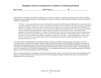
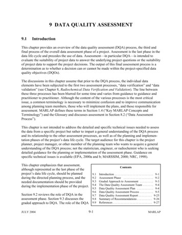
Mohsen Asadinezhad, et al.Table 1. Kilovolt peak accuracy test resultsmA100300or320Kilovolt peakSetMeasured kilovoltpeak ( SD)57.08 (8.00)50607080901005060708090100relationship between exposure time accuracyand age of equipment. Results of Pearson’scorrelation test revealed a correlation coefficientat level of α 0.01between exposure timeaccuracy and age of equipment.3.3. Exposure LinearityIn 46.7% of the devices, variance was greaterthan the acceptable range. Moreover, meanvariance was 14.84 12.04.3.4. Exposure ReciprocityThe results demonstrated that variance was outof range in 53.1% of equipment. Mean value forexposure reciprocity was 15.06 20.22.3.5. Reproducibility of ExposureOur findings indicated that 19.4% of devicesfailed to achieve acceptable results. Meanvariance was 1.98 3.01.3.6. Determination of HVLIn 14.7% of the devices, thickness of the firstHVL was lower than the limited range. Meanvalue of HVL was 3.02 0.58 mm 5.535.533.360.59 (5.04)68.45 (5.24)78.33 (5.98)87.88 (6.82)96.97 (6.56)56.39 (5.55)59.91 (4.64)68.69 (5.25)77.80 (5.67)87.29 (6.12)97.04 (7.23)Table 2. Exposure time accuracy test resultsExposure time set(mSec)6 to 10 (mean 7.75)2080100200Measured exposuretime ( SD) (mSec)8.68 (2.39)21.00 (6.73)80.24 (14.04)105.77 (23.26)205.07 (29.39)Failure(%)37.5%64.5%35.5%20.0%14.8%4. DiscussionThe results related to all of QC tests on the 51radiology devices are presented in Figure 2. Itis evident that a relatively high percentage ofdevices failed to successfully pass the tests. Inthis regard, the most common problems werereciprocity of exposure, linearity of exposurewith mAs, kVp and timer accuracy. In Table 3,the QC test results of the present study werecompared with some other Iranian studies.3.1. kVP AccuracyResults related to kVp accuracy are presented inTable 1. According to this table, 38.6% ofdevices had intolerable variance of kVpaccuracy. Mean kVp accuracy was 6.10 4.47.3.2. Exposure Time AccuracyAccording to the results, 34.5% of the deviceswere out of the acceptable limits, as provided inTable 2. In addition, Figure 1 illustrated theTable 3. Comparison of the quality control test results of the present study with other Iranian studiesQC testPresentstudyEsmaeilli[25]Kilovolt peak accuracyExposure time accuracyExposure linearityReciprocity of exposureReproducibilityofexposureDetermination of halfvalue 274554---19.4303014.7------Failure ---Iran J Med Phys., Vol. 14, No. 1, March 20174
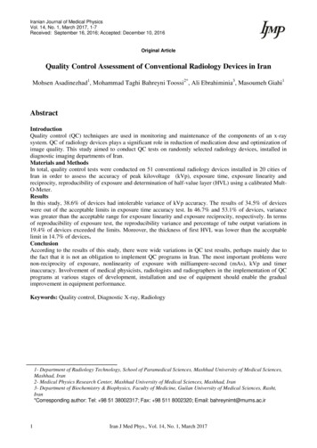
Exposure Time Accuracy (%)Quality Control of Radiology Devices in Iran1614121086420123456791012Age of Equipment (year)1416233031Figure 1. Relationship between exposure time accuracy and age of equipment6050403020100kVp Accuracy Exposure Time Linearity of Reciprocity of Reproducibility DeterminationAccuracyExposure withExposureof Exposureof HVLmAsFigure 2. Failure percentage in all QC test resultsIn kVp accuracy test, 38.6% of devices failedto pass the test. In previous studies in Iran, 6.757% of devices failed this test [20, 21, 23, 25,26]. Variations in kVp may be caused byvariations in the line voltage supplying the xray generator by faulty high voltage cables orproblems with the autotransformer/kVpselection circuitry. Given the significantsimilarity between our findings and previousstudies in this regard, it is important toevaluate and correct any defects in thesedevices.Differences between measured kVp andnominal kVp at different mA stations may bemore important than across-the-board errors.Unfortunately, there is no similar Iranian studyto compare the results. Kilovoltage settings5tend to drift over time, primarily as a result oftubing aging. Results of the t-test s between measured kVp andnominal kVp at different mA settings(P 0.15).Furthermore, in the exposure time accuracytest, 34.5% of devices did not meet thestandards. This amount was 14-45% in otherIranian studies [20, 21, 23, 25, 26]. However,Rasuli et al. affirmed that the results met thestandard criteria in all devices [24]. Age ofequipment is an effective factor for timercircuit function. As a result, correction of thisvariable is vital in order to have an appropriateradiograph and avoid repeating the radiographand increasing patient exposure.Iran J Med Phys., Vol. 14, No. 1, March 2017
Mohsen Asadinezhad, et al.Exposure linearity test demonstrated that46.7% of devices were out of the standardrange. This amount was 11-54% in some ofthe previously conducted studies in Iran [20,21, 23, 25, 26]. In a study by Rasuli et al. [24],the results were in line with the definedstandards. Given the use of mAs selector forsetting the cathode filament temperature in Xray generator in order to determine thequantity of X-ray tube output along withexposure time, the selected mA as theaccuracy of exposure time is important. As aresult, it is necessary to amend this rate offailure.In the exposure reciprocity test, 53.7% of thedevices failed to pass the test. The result ofthis test is in congruence with the resultsobtained by Khoshbin khoshnazar et al.( 30%) [23]. In these studies, the maximumfailure was observed in QC test, whichconsisted more than half of the devices. Giventhe lack of research in this area, it is suggestedthat more studies be conducted in the future inorder to reach more accurate results. Thepercentage of failure in the reproducibility ofexposure in observed devices was 19.4%,which was 7-39% in the other Iranian studies.Meanwhile, the results by khoshbinkhoshnazar et al. and Rasuli et al. were in theallowed range and had pass the defined limits[20, 21, 23-26]. In this test, no difference wasobserved between the results of observation,which is needed to correct the devices for thepurpose of ALARA goals.In 14.7% of the evaluated devices, the tubeoutput beam had HVL less than the allowedlimit. In a study by Jomeh zadeh et al., theresults indicated that HVL was in anacceptable level, with the exception of twodevices (13% less than the limit) [26]. Resultsobtained by Khoshbin khoshnazar et al.demonstrated that the measured amount ofHVL was more than the acceptable limit [23].The minimum filtration is needed to omit thelow energy beam but the extra filtration causesthe elimination of suitable beams. As a result,more exposure is needed to reach the desirabledose, which leads to consequent increase ofpatient dose.In the united states of America and Europeancountries, QC has been an important issue fora long time. In many developed countries, QCis an obligatory activity. In Iran, many peoplehave emphasized the importance of this issue;nevertheless, it seems that this subject has notcome to practice yet.5. ConclusionAccording to the results of this study, widevariations in QA test results might be due tothe fact that it is not mandatory to implementQA programs in Iran. Non-reciprocity ofexposure, nonlinearity of exposure with mAs,kVp and timer inaccuracy were reported to bethe most important problems. Involvement ofmedicalphysicists,radiologistsandradiographers in the implementation of QAprograms at various stages of development,installation and use of equipment shouldenable the gradual improvement in equipmentperformance.AcknowledgementsHereby, we extend our gratitude to all theradiologists and the radiographers whocooperated in this study.References1.2.3.4.United Nations. Scientific Committee on the Effects of Atomic Radiation. Sources and effects of ionizingradiation: sources. United Nations Publications; 2000.World Health Organization. World health statistics 2006. World Health Organization; 2006.Asadinezhad M, Toossi MT. Doses to patients in some routine diagnostic X-ray examinations in Iran:proposed the first Iranian diagnostic reference levels. Radiation protection dosimetry. 2008 Dec1;132(4):409-14. DOI: 10.1093/rpd/ncn308.Pernicka F, McLean ID. Dosimetry in diagnostic radiology: an international code of practice. InternationalAtomic Energy Agency. 2007.Iran J Med Phys., Vol. 14, No. 1, March 20176
Quality Control of Radiology Devices in 22.23.24.25.26.27.28.7National Council on Radiation Protection and Measurements. Quality assurance for diagnostic imaging,NCRP Report 99, Bethesda, Md; 1990.Protection R. ICRP publication 103. Ann. ICRP. 2007;37(2.4):2.Gray JE, Barnes GT, Bronskill MJ. The role of the clinical medical physicist in diagnostic radiology. AAPMReport; 1994.Council of the European Commission. Council directive 97/43/EURATOM of 30 June 1997 on healthprotection of individuals against the dangers of ionizing radiation in relation to medical exposure. OfficialJournal of the European Communities. 1997; 180:22-7.Shepard SJ, Lin PJ, Boone JM. Quality control in diagnostic radiology. AAPM: Report. 2002(74).Bosnjak J, O. Ciraj-Bjelac, and B. Strbac. Implementation of quality assurance in diagnostic radiology inBosnia and Herzegovina (Republic of Srpska). Radiation Protection Dosimetry. 2008; 129(1-3): 249-52.DOI: 10.1093/rpd/ncn011.Godechal D, Delhove J, Mambour C, Coomans J, Wambersie A. A quality assurance programme for medicalX ray diagnostic units carried out in Belgium. Radiation Protection Dosimetry. 1995 Jan 1;57(1-4):309-13.Gori C, Belli G, Calvagno S, Capaccioli L, Guasti A, Spano G, et al. Quality control in the radiologicaldepartments of the Florence General Hospital. Radiation Protection Dosimetry. 1995 Jan 1;57(1-4):315-6.DOI: 10.1093/oxfordjournals.rpd.a082550Jankowski J, Staniszewska MA. Methodology for the set-up of a quality control system for diagnostic X rayunits in Poland. Radiation protection dosimetry. 2000 Aug 1;90(1-2):259-62. DOI:10.1093/oxfordjournals.rpd.a033133Kharita MH, Khedr MS, Wannus KM. A comparative study of quality control in diagnostic radiology.Radiation protection dosimetry. 2008 Jul 1;130(4):447-51. DOI: 10.1093/rpd/ncn096.Neofotistou V, Molfetas M, Panagiotakis N. Quality Control in Conventional Diagnostic Radiology rtiz P, Maccia C, Padovani R, Vano E, Carlsson GA, Schibilla H. Results of the IAEA-CEC co-ordinatedresearch programme on radiation doses in diagnostic radiology and methods for reduction. RadiationProtection Dosimetry. 1995 Jan 1;57(1-4):95-9. DOI: 10.1093/oxfordjournals.rpd.a082503Servomaa A, Rannikko S, Parviainen T, Holmberg P, Kuss E, Müürsepp T, et al. Quality control and patientdose from x ray examinations in some hospitals in Estonia. Radiation Protection Dosimetry. 1995 Jan 1;57(14):297-300. DOI: 10.1093/oxfordjournals.rpd.a082546Zoetelief J. Quality Control in Diagnostic Radiology in the Netherlands. Radiation Protection Dosimetry.1998; 77(4): 257-66. DOI: 10.1093/oxfordjournals.rpd.a032321Shabazi GD. Quality control of the radiological equipment in Chaharmahal & Bakhtiari Hospitals. Journal ofShahrekord University of Medical Sciences. 2004; 5(4): 11-8.Saghatchi F. Quality control of diagnostic X-ray units in hospitals of Zanjan University of Medical Sciences(Doctoral dissertation, Master Thesis of Medical Physics, Mashhad University of Medical Sciences).Gholamhosseinian-Najjar H, Bahreyni-Toosi MT, Zare MH, Sadeghi HR, Sadoughi HR. Quality ControlStatus of Radiology Centers of Hospitals Associated with Mashhad University of Medical Sciences. IranianJournal of Medical Physics. 2014 Apr 1;11(1):182-7. DOI: 10.22038/ijmp.2014.2625.Gholami M, Nemati F, and Karami V. The Evaluation of Conventional X-ray Exposure Parameters IncludingTube Voltage and Exposure Time in Private and Governmental Hospitals of Lorestan Province, Iran. IranianJournal of Medical Physics. 2015; 12(2): 85-92. DOI: 10.22038/ijmp.2015.4770.Khoshbin Khoshnazar A, Hejazi P, Mokhtarian M, Nooshi S. Quality control of radiography equipments inGolestan Province of Iran. Iranian Journal of Medical Physics. 2013 Jan 1;10(1):37-44. DOI:10.22038/ijmp.2013.917.Rasuli B, Pashazadeh AM, Tahmasebi Birgani MJ, Ghorbani M, Naserpour M, Fatahi-Asl J. quality controlof conventional radiology devices in selected hospitals of Khuzestan province, Iran. Iranian Journal ofMedical Physics. 2015 Jul 1;12(2):101-8. DOI: 10.22038/ijmp.2015.4773.Esmaeili, S. Measurement of patient skin dose of common techniques in diagnostic radiology in 15 radiologycenters and quality control of those units in Mashhad, in Medical Physics. 2006, Mashhad University ofMedical Sciences.Jomehzadeh Z, Jomehzadeh A, Tavakoli MB. Quality Control Assessment of Radiology Devices in KermanProvince, Iran. Iranian Journal of Medical Physics. 2016 Mar 1;13(1):25-35. DOI: 10.22038/ijmp.2016.7142.Haghparast M, Afzali Pour R, Ahmadi S, Golverdi Yazdi MS, Dindarloo Inaloo K, Saanei M. Quality controlof radiology devices in Health Centers Affiliated with Hormozgan University of Medical Sciences.Hormozgan Medical Journal. 2015; 19 (1) :51-57.Papp J. Quality management in the imaging sciences. 4th. ed. 2011: Mosby.Iran J Med Phys., Vol. 14, No. 1, March 2017
Keywords: Quality control, Diagnostic X-ray, Radiology 1- Department of Radiology Technology, School of Paramedical Sciences, Mashhad University of Medical Sciences, . set forth in the "quality management in the imaging science" [28]. Data are presented as mean standard deviation (SD). Data analysis was performed in SPSS