Transcription
Clinical practiceguideline on diagnosisand treatmentof hyponatraemia1
Disclaimer:This document is written on behalf of ERBP which is an official body of theERA-EDTA (European Renal Association – European Dialysis and TransplantAssociation) and is based on the official Publication in Nephrology, Dialysisand Transplantation. ERBP only takes full responsibility for the original fullguideline in English as published inhttp://ndt.oxfordjournals.org/content/29/suppl 2/i1.full.pdf html2
CONTENTSGuideline Development Group41. Introduction and methodology52. Diagnosis of Hyponatraemia62.1. Classification of hyponatraemia62.1.1. Definition of hyponatraemia based on biochemical severity62.1.2. Definition of hyponatraemia based on time of development62.1.3. Definition of hyponatraemia based on symptoms62.2. Confirming hypotonic and excluding non-hypotonic hyponatraemia72.3. Which parameters to use for differentiating causes of hypotonichyponatraemia?83. Treatment of Hypotonic Hyponatraemia3.1. Hyponatraemia with severe symptoms3.1.1. First hour management, regardless of whether hyponatraemia isacute or chronic1010103.1.2. Follow up management in case of improvement of symptoms after a5 mmol/L increase in serum sodium concentration in the first hour, regardlessof whether hyponatraemia is acute or chronic113.1.3. Follow up management in case of no improvement of symptoms aftera 5 mmol/L increase in serum sodium concentration in the first hour,regardless of whether the hyponatraemia is acute or chronic113.2. Hyponatraemia with moderately severe symptoms123.3. Acute hyponatraemia without severe or moderately severe symptoms133.4. Chronic hyponatraemia without severe or moderately severe symptoms133.4.1. General management133.4.2. Patients with expanded extracellular fluid143.4.3. Patients with syndrome of inappropriate antidiuresis143.4.4. Patients with contracted circulating volume143.5. What to do in case hyponatraemia is corrected too rapidly?15Tables16Figures213
Guideline development group co-chairsGoce SpasovskiConsultant Nephrologist, State University Hospital Skopje, Skopje,Macedonia.Raymond VanholderConsultant Nephrologist, Ghent University Hospital, Ghent, Belgium.Work GroupBruno AllolioConsultant Endocrinologist, Würzburg University Hospital, Würzburg,Germany.Djillali AnnaneConsultant Intensivist, Raymond Poincaré Hospital, University of VersaillesSaint Quentin, Paris, France.Steve BallConsultant Endocrinologist, Newcastle Hospitals and Newcastle University,Newcastle, UK.Daniel BichetConsultant Nephrologist, Hospital, Montreal, Canada.Guy DecauxConsultant Internal Medicine, Erasmus University Hospital, Brussels,Belgium.Wiebke FenskeConsultant Endocrinologist, Würzburg University Hospital, Würzburg,Germany.Ewout HoornConsultant Nephrologist, Erasmus Medical Centre, Rotterdam,The Netherlands.Carole IchaiConsultant Intensivist, Nice University Hospital, Nice, France.Michael JoannidisConsultant Intensivist, Innsbruck University Hospital, Innsbruck, Austria.Alain SoupartConsultant Internal Medicine, Erasmus University Hospital, Brussels,Belgium.Robert ZietseConsultant Nephrologist, Erasmus Medical Centre, Rotterdam,The Netherlands.ERBP methods support teamMaria HallerSpecialist Registrar Nephrology, KH Elisabethinen Linz, Linz, Austria.Evi NaglerSpecialist Registrar Nephrology, Ghent University Hospital, Ghent, Belgium.Wim Van BiesenConsultant Nephrologist, Chair of ERBP, Ghent University Hospital, Ghent,Belgium.Sabine van der VeerImplementation Specialist, Amsterdam Medical Centre, Amsterdam,TheNetherlands.4
1. Introduction and MethodologyHyponatraemia, defined as a serum sodium concentration 135 mmol/L, isthe most common disorder of body fluid and electrolyte balance encountered in clinical practice. Hyponatraemia is present in 15–20% of emergencyadmissions to hospital and occurs in up to 20% of critically ill patients. Itcan lead to a wide spectrum of clinical symptoms, from subtle to severe oreven life threatening and is associated with increased mortality, morbidityand length of hospital stay in patients presenting with a range of conditions.Despite this, the management of patients remains problematic. The prevalence of hyponatraemia in widely different conditions and the fact that hyponatraemia is managed by clinicians with a broad variety of backgrounds, hasfostered diverse institution- and speciality-based approaches to diagnosisand treatment.Against this background, the European Society of Intensive Care Medicine(ESICM), the European Society of Endocrinology (ESE) and the EuropeanRenal Association - European Dialysis and Transplant Association (ERA-EDTA),represented by European Renal Best Practice (ERBP) have developed thisClinical Practice Guideline on the diagnostic approach and treatment ofhyponatraemia as a joint venture of three societies representing specialistswith a natural interest in hyponatraemia. In addition to a rigorous approachto methodology and evaluation, we were keen to ensure the documentfocused on patient-important outcomes and had utility for clinicians involvedin every-day practice.This condensed and translated version of the Clinical Practice Guideline onDiagnosis and Treatment of Hyponatraemia focuses on recommendations ondiagnosis and treatment of hyponatraemia. For aspects of conflict ofinterest, purpose and scope, methods of guideline development andpathophysiology of hyponatraemia, we refer to the full version of theguideline, which is free available onhttp://ndt.oxfordjournals.org/content/29/suppl 2/i1.full.pdf htmlDisclaimer: this guideline was translated with approval of ERBP, the officialguideline body of ERA-EDTA. However, ERBP only takes full responsibility forthe original full guideline in English as published in Nephrol. Dial. Transplant.(2014) 29 (suppl 2): i1-i39. doi: 10.1093/ndt/gfu040 - First publishedonline: February 25, 2014http://ndt.oxfordjournals.org/content/29/suppl 2/i1.full.pdf htmlhttp://european-renal-best-practice.org/5
2. Diagnosis of Hyponatraemia2.1. Classification of hyponatraemia2.1.1 Definition of hyponatraemia based on biochemicalseverityWe define ‘mild’ hyponatraemia as a biochemical finding of a serum sodiumconcentration between 130 and 135 mmol/L as measured by ion specificelectrode.We define ‘moderate’ hyponatraemia as a biochemical finding of a serumsodium concentration between 125 and 129 mmol/L as measured by ionspecific electrode.We define ‘profound’ hyponatraemia as a biochemical finding of a serumsodium concentration 125 mmol/L as measured by ion specific electrode.2.1.2. Definition of hyponatraemia based on time ofdevelopmentWe define ‘acute’ hyponatraemia as hyponatraemia that is documented toexist 48 hours. We define ‘chronic’ hyponatraemia as hyponatraemia thatis documented to exist for at least 48 hours.If the hyponatraemia cannot be classified, we consider it being chronic,unless there is clinical or anamnestic evidence of the contrary (Table 1, 2).2.1.3. Definition of hyponatraemia based on symptomsWe define ‘moderately symptomatic’ hyponatraemia as any biochemicaldegree of hyponatraemia in the presence of moderately severe symptoms ofhyponatraemia (Table 1).We define ‘severely symptomatic’ hyponatraemia as any biochemical degreeof hyponatraemia in the presence of severe symptoms of hyponatraemia(Table 1).Hyponatraemia can be classified based on different parameters, such asserum sodium concentration, rate of development, symptom severity,serum osmolality, and volume status. We intended to make the classificationdirectly relevant for patient management. However, treatment strategiescannot be adequately classified with reference to a single criterion. Hence,treatment strategies have been classified according to combinations of thesecriteria.Published research suggests using a threshold of 48 hours to distinguish"acute" from "chronic" hyponatraemia, as brain oedema seems to occurmore frequently when hyponatraemia develops in less than 48 hours.Experimental studies also suggest that the brain needs approximately 48hours to adapt to a hypotonic environment. Before adaptation, there is a riskof brain oedema, because the lower extracellular osmolality promotes a shift6
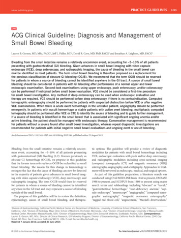
of water into the cells. However, once adaptation is completed, brain cellscan again sustain damage if the serum sodium concentration increases toorapidly. Breakdown of the myelin sheath insulating individual neurons canresult in what is called the osmotic demyelination syndrome. It is thusimportant to distinguish between acute and chronic hyponatraemia toassess whether someone is at greater risk of immediate brain oedema thanof osmotic demyelination. In clinical practice, the distinction between acuteand chronic hyponatraemia is often unclear, particularly for patientspresenting to the emergency room. If classification as acute or chronic is notpossible or when there is doubt, it should be considered chronic, unlessthere are reasons to assume it is acute (see Table 10 of original document).The classification based on symptoms aims to reflect the degree of brainoedema and the extent of immediate danger. It allows matching treatmentto the immediate risk, with more aggressive treatment for symptoms thatare more severe. Nevertheless, a classification based only on symptomseverity has several shortcomings, as patients may porgress from moderatelysevere to severe symptoms within hours. In addition, symptoms ofhyponatraemia are nonspecific and clinicians need to assess thepossibility that symptoms can be caused by conditions other thanhyponatraemia on itself. In general, one should be particularly carefulwhen attributing moderately severe to severe symptoms to hyponatraemiawhen the biochemical degree of hyponatraemia is only mild.Patients with hyponatraemia may be hypovolaemic, euvolaemic, orhypervolaemic, and many traditional diagnostic algorithms start with aclinical assessment of volume status [33] .The sensitivity and specificity ofclinical assessments of volume status are low, potentially leading tomisclassification early in the diagnostic tree. In addition, there might beconfusion regarding the compartment the fluid is in (circulating orextracellular). Therefore, we have used the terms ‘effective circulatingvolume’ and ‘extracellular fluid volume’ throughout the text to reduceambiguity.2.2. Confirming hypotonic and excluding non-hypotonichyponatraemiaWe recommend excluding hyperglycaemic hyponatraemia by measuring theserum glucose concentration and correcting the measured serum sodiumconcentration for the serum glucose concentration if the latter is increased.(1D)Hyponatraemia with a measured osmolality 275 mOsm/kg always reflectshypotonic hyponatraemia. (Not Graded)Accept as ‘hypotonic hyponatraemia’ a hyponatraemia without evidence forcauses of non-hypotonic hyponatraemia as listed in Table 3. (Not Graded)Estimates of the serum sodium concentration corrected for the presence of7
European JCorrection ons:On page G20, onlyunitsshould frombe usedthe equationfor estimates of the serum sodfor the presence of hyperglycaemia and not as published. The correct equation is as followCorrected serum ðNaCÞ Z measured ðNaCÞ C 2:4 !Corrected ðNaCÞ Z measured ðNaCÞ C 2:4 !ðglucose ðmmol lÞK100 ðmmol lÞ100 mmol lðglucose ðmmol lÞK5:5 ðmmol lÞ5:5 mmol l† [Na ], serum sodium concentration; [Glucose], serum glucose concentration.This translates into adding 2.4 mmol/L to the measured serum sodiumconcentration for every 5.5 mmol/L (100 mg/dL) incremental rise in serumglucose concentration above a standard serum glucose concentration of 5.5mmol/L (100 mg/dL).2.3. Which parameters to use for differentiating causes of hypotonic hyponatraemia? (figure 1)We recommend interpreting urine osmolality of a spot urine sample as afirst step. (1D)If www.eje-online.orgurine osmolality 100 mOsm/kg,weSocietyrecommendacceptingrelative excess2014 Europeanof Endocrinology,Published by Bioscientifica Ltd.Society of Intensive Care Medicine,DOI: 10.1530/EJE-13-1020ePrinted in Great Britainwaterintake as a causeEuropeanof tion-EuropeanDialysis andTransplant AssociationIf urine osmolality 100 mOsm/kg, we recommend interpreting the urinesodium concentration on a spot urine sample taken simultaneously with ablood sample. (1D)If urine sodium concentration 30 mmol/L, we suggest accepting low effective arterial volume as a cause of the hypotonic hyponatraemia. (2D)If urine sodium concentration 30 mmol/L, we suggest assessing extracellular fluid status and use of diuretics to further differentiate likely causes ofthe hyponatraemia. (2D)We suggest against measuring vasopressin for confirming the diagnosis ofSIADH. (2D)Advice for clinical practice Correct interpretation of laboratory measurements requires contemporaneous collection of blood and urine specimens. For practical reasons, urine osmolality and sodium concentration arebest determined in the same urine sample. If clinical assessment indicates the volume of extracellular fluid is notoverly increased and the urine sodium concentration 30 mmol/L,exclude other causes of hypotonic hyponatraemia before implicatingSIAD. Consider using the diagnostic criteria listed in Table 4 and lookingfor known causes of SIAD (Table 5 and 6). Consider primary or secondary adrenal insufficiency as an underlyingcause of the hypotonic hyponatraemia.8
Kidney disease complicates differential diagnosis of hyponatraemia.Besides possibly contributing to the hyponatraemia, the ability of thekidneys to regulate urine osmolality and urine sodium is often diminished, much like with the use of diuretics. As urine osmolality andsodium may no longer reflect the effects of the regular hormonal axesregulating water and sodium homeostasis, any diagnostic algorithmfor hyponatraemia must be used with caution in patients with kidneydisease. The water-loading test is generally not helpful for differential diagnosisof hypotonic hyponatraemia and may be dangerous in this setting.9
3. Treatment of Hypotonic HyponatraemiaHow to use the treatment recommendationsIndividual recommendations and statements on management of hyponatraemia can only be correctly interpreted and implemented if consideredwithin the structure illustrated in figure 2.The guideline development group felt that with severe or moderately severesymptoms, the acute risk of brain oedema outweighs the risk of osmotic demyelination syndrome. They felt it justifies urgent treatment in these conditions, irrespective of biochemical degree or timing (acute versus chronic) ofhyponatraemia. Conversely, the guideline development group believed thatin the absence of severe or moderately severe symptoms, there is time fordiagnostic assessment, and cause-specific treatment is the most reasonableapproach.It is crucial to understand that for correctly classifying symptoms as ‘severe’ or ‘moderately severe’, there must be sufficient confidence that thesymptoms are caused by hyponatraemia itself. If hyponatraemia is mild andsymptoms are severe or moderately severe, the guideline developmentgroup advises to only accept causality in exceptional cases. Consequently,generally, chapters 3.1, 3.2, and 3.3 are not applicable when hyponatraemiais mild (see chapters 7.1, 7.2 and 7.3 full guideline publication). It is alsoessential to understand that the guideline development group distinguishesbetween targets and limits. A target is a goal one is aiming for; it is the change in serum sodium concentration that one wishes and expects to achievewith a particular treatment. In contrast, a limit is a change in serum sodiumconcentration one does not want to exceed and if surpassed, requiresprompt counter-regulating intervention. In addition, the reader should bearin mind that the absolute numbers provided as ‘targets’ or ‘limits’ shouldalways be interpreted in the clinical context of the individual patient.3.1. Hyponatraemia with severe symptoms3.1.1. First hour management, regardless of whetherhyponatraemia is acute or chronicWe recommend prompt intravenous infusion of 150 mL 3% hypertonic salineor equivalent over 20 minutes. (1D)We suggest checking the serum sodium concentration after 20 minuteswhile repeating an infusion of 150 mL 3% hypertonic saline or equivalentover the next 20 minutes. (2D)We suggest repeating the two therapeutic recommendations above twiceor until a target of 5 mmol/L increase in serum sodium concentration isachieved. (2D)Manage patients with severely symptomatic hyponatraemia in an environment where close biochemical and clinical monitoring can be provided. (NotGraded)10
3.1.2. Follow up management in case of improvement ofsymptoms after a 5 mmol/L increase in serum sodiumconcentration in the first hour, regardless of whether hyponatraemia is acute or chronicWe recommend stopping the infusion of hypertonic saline. (1D)We recommend keeping the intravenous line open by infusing the smallestfeasible volume of 0.9% saline until cause-specific treatment is started. (1D)We recommend starting a diagnosis specific treatment if available, aiming atleast to stabilize sodium concentration. (1D)We recommend limiting the increase in serum sodium concentration to atotal of 10 mmol/L during the first 24 hours and an additional 8 mmol/Lduring every 24 hours thereafter until the serum sodium concentrationreaches 130 mmol/L. (1D)We suggest checking the serum sodium concentration after 6 and 12 hours,and daily afterwards until the serum sodium concentration has stabilisedunder stable treatment. (2D)3.1.3 Follow up management in case of no improvement ofsymptoms after a 5 mmol/L increase in serum sodiumconcentration in the first hour, regardless of whether thehyponatraemia is acute or chronicWe recommend continuing an intravenous infusion of 3% hypertonic salineor equivalent aiming for an additional 1 mmol/L/h increase in serum sodiumconcentration. (1D)We recommend stopping the infusion of 3% hypertonic saline or equivalentwhen the symptoms improve, the serum sodium concentration increases 10mmol/L in total or the serum sodium concentration reaches 130 mmol/L,whichever occurs first. (1D)We recommend additional diagnostic exploration for other causes of thesymptoms than hyponatraemia. (1D)We suggest checking the serum sodium concentration every 4 hours as longas an intravenous infusion of 3% hypertonic saline or equivalent iscontinued. (2D)Advice for clinical practice Prompt infusion of hypertonic saline may save lives. However, preparinga 3% hypertonic saline infusion takes time and errors may occur incalculating the required amount of sodium chloride. Therefore, it maybe wise for the pharmacy to store pre-prepared 150 mL bags of 3% hypertonic saline. It ensures that solutions are prepared under sterile conditions, by either the pharmacist or the manufacturer, and are availablefor immediate infusion without having to prepare them on the spot.11
Consider using weight based (2mL/kg) rather than the fixed 150 mLinfusion volumes of 3% hypertonic saline in case of obviously deviantbody composition. Do not expect patients with severe symptoms to completely recoverimmediately, as it may take some time for the brain to fully recover. Beaware that sometimes it may not be possible to assess an improvementin symptoms, e.g. because the patient is intubated and sedated. Inthese cases, we advise to follow guidance as described under 3.1.2. (seechapter (7.1.2. full guideline publication). Keep in mind that if hypokalaemia is present, correction of the hypokalaemia will contribute to an increase in serum sodium concentration. To achieve the 1 mmol/L/h increase advised in 3.1.3. (see chapter 7.1.3.full guideline publication)., the formula of Adrogué-Madias may beused, but keep in mind that the actual increase may exceed the calculated increase :𝐶𝐶ℎ𝑎𝑎𝑎𝑎𝑎𝑎𝑎𝑎 𝑖𝑖𝑖𝑖 𝑠𝑠𝑠𝑠𝑠𝑠𝑠𝑠𝑠𝑠 [𝑁𝑁𝑁𝑁 ] 𝐶𝐶ℎ𝑎𝑎𝑎𝑎𝑎𝑎𝑎𝑎 𝑖𝑖𝑖𝑖 𝑠𝑠𝑠𝑠𝑠𝑠𝑠𝑠𝑠𝑠 [𝑁𝑁𝑁𝑁 ] ��𝑖𝑖𝑖 [𝑁𝑁𝑁𝑁 ] 𝑠𝑠𝑠𝑠𝑠𝑠𝑠𝑠𝑠𝑠 [𝑁𝑁𝑁𝑁 ]𝑡𝑡𝑡𝑡𝑡𝑡𝑡𝑡𝑡𝑡 𝑏𝑏𝑏𝑏𝑏𝑏𝑏𝑏 𝑤𝑤𝑤𝑤𝑤𝑤𝑤𝑤𝑤𝑤 𝑖𝑖𝑖𝑖 [𝑁𝑁𝑁𝑁 ] ��𝑖𝑖𝑖 [𝐾𝐾 ]) 𝑠𝑠𝑠𝑠𝑠𝑠𝑠𝑠𝑠𝑠 [𝑁𝑁𝑁𝑁 ]𝑡𝑡𝑡𝑡𝑡𝑡𝑡𝑡𝑡𝑡 𝑏𝑏𝑏𝑏𝑏𝑏𝑏𝑏 𝑤𝑤𝑤𝑤𝑤𝑤𝑤𝑤𝑤𝑤 1† [Na ], sodium concentration in mmol/L; [K ], potassium concentration inmmol/L§ The numerator in formula 1 is a simplification of the expression in formula2, with the value yielded by the equation in mmol/L. The estimated totalbody water (in litres) is calculated as a fraction of body weight. The fractionis 0.6 in nonelderly men and 0.5 in nonelderly women; and 0.5 and 0.45 inelderly men and women respectively. Normally, extracellular andintracellular fluids account for 40% and 60% of total body water respectively.The formula of Adrogue Madias estimates the effect of 1L infusion of asolution with the specified sodium concentration on serum Na .3.2. Hyponatraemia with moderately severe symptomsWe recommend starting prompt diagnostic assessment. (1D)Stop, if possible, medications and other factors that can contribute to orprovoke the hyponatraemia. (Not Graded)We recommend cause-specific treatment. (1D)We suggest immediate treatment with a single intravenous infusion of 150mL 3% hypertonic saline or equivalent over 20 minutes. (2D)We suggest aiming for a 5 mmol/L/24 h increase in serum sodium concentration. (2D)We suggest limiting the increase in serum sodium concentration to 10mmol/L in the first 24 hours and 8 mmol/L during every 24 hours thereafter,until a serum sodium concentration of 130 mmol/L is reached. (2D)12
We suggest checking the serum sodium concentration after one, 6 and 12hours. (2D)We suggest additional diagnostic exploration for other causes of thesymptoms if the symptoms do not improve with an increase in serumsodium concentration. (2D)We suggest considering to manage the patient as in severely symptomatichyponatraemia if the serum sodium concentration further decreases despitetreating the underlying diagnosis. (2D)3.3. Acute hyponatraemia without severe or moderatelysevere symptomsMake sure that the serum sodium concentration has been measured usingthe same technique as used for the previous measurement and that noadministrative errors in sample handling have occurred. (Not Graded)If possible, stop fluids, medications and other factors that can contribute toor provoke the hyponatraemia. (Not Graded)We recommend starting prompt diagnostic assessment. (1D)We recommend cause-specific treatment. (1D)If the acute decrease in serum sodium concentration exceeds 10 mmol/L, wesuggest a single intravenous infusion of 150 mL 3% hypertonic saline orequivalent over 20 minutes. (2D)We suggest checking the serum sodium concentration after four hours, usingthe same technique as used for the previous measurement. (2D)3.4. Chronic hyponatraemia without severe or moderatelysevere symptoms3.4.1. General managementStop non-essential fluids, medications and other factors that can contributeto or provoke the hyponatraemia. (Not Graded)We recommend cause-specific treatment. (1D)In mild hyponatraemia, we suggest against treatment with the sole aim ofincreasing the serum sodium concentration. (2C)In moderate or profound hyponatraemia, we recommend avoiding an increase in serum sodium concentration of 10 mmol/L during the first 24 hoursand 8 mmol/L during every 24 hours thereafter. (1D)In moderate or profound hyponatraemia, we suggest checking the serumsodium concentration every six hours until the serum sodium concentrationhas stabilised under stable treatment. (2D)13
In case of unresolved hyponatraemia, reconsider the diagnostic algorithmand ask for expert advice. (Not Graded)3.4.2. Patients with expanded extracellular fluidWe recommend against a treatment with the sole aim of increasing theserum sodium concentration in mild or moderate hyponatraemia. (1C)We suggest fluid restriction to prevent further fluid overload. (2D)We recommend against vasopressin receptor antagonists. (1C)We recommend against demeclocycline. (1D)3.4.3. Patients with syndrome of inappropriate antidiuresisIn moderate or profound hyponatraemia, we suggest restricting fluid intakeas first- line treatment. (2D)In moderate or profound hyponatraemia, we suggest the following can beconsidered equal second line treatments: increasing solute intake with 0.25to 0.50 g/kg/day of urea or a combination of low dose loop diuretics andoral sodium chloride. (2D)In moderate or profound hyponatraemia, we recommend against lithium ordemeclocycline. (1D)In moderate hyponatraemia, we do not recommend vasopressin receptorantagonists. (1C)In profound hyponatraemia, we recommend against vasopressin receptorantagonists. (1C)3.4.4. Patients with contracted circulating volumeWe recommend restoring extracellular volume with intravenous infusion of0.9 % saline or a balanced crystalloid solution at 0.5 to 1.0 mL/kg/h. (1B)Manage patients with haemodynamic instability in an environment whereclose biochemical and clinical monitoring can be provided. (Not Graded)In case of haemodynamic instability, the need for rapid fluid resuscitationoverrides the risk of an overly rapid increase in serum sodium concentration.(Not Graded)Advice for clinical practice A sudden increase in urine output to 100 mL/h signals increased risk ofoverly rapid rise in serum sodium concentration. If vasopressin activityis suddenly suppressed, as happens when intravascular volume is restored in hypovolaemia, free water clearance can dramatically increase,resulting in serum sodium concentrations rising more rapidly than expected. If urine output suddenly increases, we would advise measuring14
the serum sodium concentration every two hours until it has stabilisedunder stable treatment. The implicit advice to monitor urine outputdoes not imply we advise a bladder catheter solely for this purpose.Most patients will be able to void spontaneously and collect urine foroutput monitoring. As a means of increasing solute intake, we suggest daily intake of 0.25to 0.50 g/kg urea can be used. The bitter taste can be reduced by combining it with sweet tasting substances. The pharmacist may be askedto prepare the following as sachets: urea 10 g NaHCO3 2 g citric acid1.5 g sucrose 200 mg, to be dissolved in 50 to 100 mL water. This willresult in a more palatable, slightly sparkling solution.3.5. What to do in case hyponatraemia is corrected toorapidly?We recommend prompt intervention for relowering the serum sodiumconcentration if it increases 10 mmol/L during the first 24 hours or 8 mmol/L in any 24 hours thereafter. (1D)We recommend discontinuing the ongoing active treatment. (1D)We recommend consulting an expert to discuss if it is appropriate to startan infusion of 10 mL/kg body weight of electrolyte-free water (e.g. glucosesolutions) over one hour under strict monitoring of urine output and fluidbalance. (1D)We recommend consulting an expert to discuss if it is appropriate to addintravenous desmopressin 2 µg, with the understanding that this should notbe repeated more frequently than every 8 hours. (1D)15
TablesTable 1 (Table 5 of the online full document): Classification ofsymptoms of hyponatraemiaSeverityModerately severeSevereSymptomNausea without y distressAbnormal and deep somnolenceSeizuresComa (Glasgow Coma Scale 8)The guideline development group wants to underscore that these symptomscan also be induced by other conditions. Clinical and anamnestic data shouldbe taken into account when assessing the causal relation between the hyponatraemia and a certain symptom (i.e. to assess whether the symptom hasbeen caused by the hyponatraemia or the hyponatraemia by the underlying condition/symptom). The less pronounced (e.g. mild) the biochemicaldegree of hyponatraemia, the more caution should be taken when considering that the hyponatraemia is the cause of the symptoms. This list is notexhaustive, and all symptoms that can be signs of cerebral oedema shouldbe considered as severe or moderate symptoms that can be caused by hyponatraemia.Table 2 (Table 8 of the online full document): Drugs andconditions associated with acute hyponatraemia ( 48 hours)hyponatraemia ( 48 hours)Postoperative phasePost-resection of the prostate, post-resection of endoscopic uterine surgeryPolydipsiaExerciseRecent thiazides prescription3,4-methyleendioxymethamfetamine (MDMA, XTC)Colonoscopy preparationCyclophosphamide (intravenous)OxytocinRecently started desmopressin therapyRecently started terlipressin, vasopressin16
Table 3 (Table 10 of the online full document): Causes of nonhypotonic hyponatraemiaSettingSerum osmolalityExamplesPresence of “effective” osmoles that raiseserum osmolality and can causehyponatraemiaIsotonic phane-ketoglutarateHyperosmolar radiocontrast mediaMaltosePresence of “ineffective” osmoles thatraise serum osmolality but do not causehyponatraemiaIsotonic orhyperosmolarUreaAlcoholsEthylene-glycolPresence of endogenous solutes thatcause pseudohyponatraemia (laboratoryartifact)IsotonicTriglycerides, cholesterol , proteinIntravenous immunoglobulinsMonoclonal gammapathiesTable 4 (Table 6 of the full online document): Diagnosticcriteria for the syndrome of inappropriate antidiuresisEssential criteria Effective serum osmolality 275 mOsm/kg Urine osmolality 100 mOsm/kg at some level of decreased effective osmolality Clinical euvolaemia Urine sodium concentration 30 mmol/L with normal dietary salt and water intake Absence of adrenal, thyroid, pituitary or renal insufficiency No recent use of diuretic agentsSupplemental criteria Serum uric acid 0.24 mmol/L ( 4 mg/dL) Serum urea 3.6 mmol/L ( 21.6 mg/dL) Failure to correct hyponatraemia after 0.9% saline infusion Fractional sodium excretion 0.5% Fractional urea excretion 55% Fractional uric acid excretion 12% Correction of hyponatraemia through fluid restrictionAdapted from Schwartz WB et al. Am J Med 1957; 23: 529-543. and Ja
Consultant Endocrinologist, Würzburg University Hospital, Würzburg, Germany. Djillali Annane Consultant Intensivist, Raymond Poincaré Hospital, University of Versailles Saint Quentin, Paris, France. Steve Ball Consultant Endocrinologist, Newcastle Hospitals and Newcastle University, Newcastle, UK. Daniel Bichet