Transcription
PRACTICE GUIDELINESnature publishing groupCMEACG Clinical Guideline: Diagnosis and Management ofSmall Bowel BleedingLauren B. Gerson, MD, MSc, FACG1, Jeff L. Fidler, MD2, David R. Cave, MD, PhD, FACG3 and Jonathan A. Leighton, MD, FACG4Bleeding from the small intestine remains a relatively uncommon event, accounting for 5–10% of all patientspresenting with gastrointestinal (GI) bleeding. Given advances in small bowel imaging with video capsuleendoscopy (VCE), deep enteroscopy, and radiographic imaging, the cause of bleeding in the small bowel cannow be identified in most patients. The term small bowel bleeding is therefore proposed as a replacement forthe previous classification of obscure GI bleeding (OGIB). We recommend that the term OGIB should be reservedfor patients in whom a source of bleeding cannot be identified anywhere in the GI tract. A source of small bowelbleeding should be considered in patients with GI bleeding after performance of a normal upper and lowerendoscopic examination. Second-look examinations using upper endoscopy, push enteroscopy, and/or colonoscopycan be performed if indicated before small bowel evaluation. VCE should be considered a first-line procedurefor small bowel investigation. Any method of deep enteroscopy can be used when endoscopic evaluation andtherapy are required. VCE should be performed before deep enteroscopy if there is no contraindication. Computedtomographic enterography should be performed in patients with suspected obstruction before VCE or after negativeVCE examinations. When there is acute overt hemorrhage in the unstable patient, angiography should be performedemergently. In patients with occult hemorrhage or stable patients with active overt bleeding, multiphasic computedtomography should be performed after VCE or CTE to identify the source of bleeding and to guide further management.If a source of bleeding is identified in the small bowel that is associated with significant ongoing anemia and/oractive bleeding, the patient should be managed with endoscopic therapy. Conservative management is recommendedfor patients without a source found after small bowel investigation, whereas repeat diagnostic investigations arerecommended for patients with initial negative small bowel evaluations and ongoing overt or occult bleeding.Am J Gastroenterol 2015; 110:1265–1287; doi:10.1038/ajg.2015.246; published online 25 August 2015Bleeding from the small intestine remains a relatively uncommon event, accounting for 5–10% of all patients presentingwith gastrointestinal (GI) bleeding (1,2). Known previously asobscure GI hemorrhage (OGIB), we propose in this guidelinethat the former term referred to as OGIB be reclassified as smallbowel bleeding. The reason for this change in terminology isowing to the fact that the cause of bleeding can now be detectedin the majority of patients given advances in small bowel imaging with video capsule endoscopy (VCE), deep enteroscopy, andradiographic imaging. The term OGIB would then be reservedfor patients in whom a source of bleeding cannot be identifiedanywhere in the GI tract and may represent a source of bleedingoutside of the small bowel.The purpose of this guideline will be to review the definition,epidemiology, causes of small bowel bleeding, and therapeu-tic options. The guideline will provide a review of diagnosticmodalities for patients with small bowel hemorrhage includingVCE, endoscopic evaluation with push and/or deep enteroscopy,and radiographic modalities including cross-sectional imaging(computed tomography (CT) and magnetic resonance (MR))enterography, angiography, and scintigraphy. Approaches to treatment will be reviewed as endoscopic, medical, and surgical options.As part of this guideline preparation, a literature search wasconducted using Ovid MEDLINE from 1946 to present, EMBASE1988 to present, and SCOPUS from 1980 to present using majorsearch terms and subheadings including “obscure” or “occult,”“gastrointestinal hemorrhage,” “iron-deficiency anemia,” “capsule endoscopy,” “enteroscopy” “angiography,” “computed tomographic enterography,” “magnetic resonance enterography,”“tagged red blood cell,” “angioectasia,” “Meckel’s diverticulum,”1Division of Gastroenterology, California Pacific Medical Center and Department of Medicine, University of California School of Medicine, San Francisco,California, USA; 2Division of Radiology, Mayo Clinic School of Medicine, Rochester, Minnesota, USA; 3Division of Gastroenterology, University of MassachusettsMedical Center, Worcester, Massachusetts, USA; 4Division of Gastroenterology, Mayo Clinic School of Medicine, Scottsdale, Arizona, USA. Correspondence:Lauren B. Gerson, MD, MSc, Director of Clinical Research, GI Fellowship Program, Division of Gastroenterology, California Pacific Medical Center, 2340 ClayStreet, 6th Floor, San Francisco, California 94115, USA. E-mail: GersonL@sutterhealth.orgReceived 7 January 2015; accepted 21 June 2015 2015 by the American College of GastroenterologyThe American Journal of GASTROENTEROLOGY1265
1266Gerson et al.Table 1. Recommendation statementsDiagnosis of small bowel bleeding1. Second-look upper endoscopy should be considered in cases of recurrent hematemesis, melena, or a previously incomplete exam (strong recommendation, low level of evidence).2. Second-look colonoscopy should be considered in the setting of recurrent hematochezia or if a lower source is suspected (conditional recommendation,very low level of evidence).3. If the second-look examinations are normal, the next step should be a small bowel evaluation (strong recommendation, moderate level of evidence).4. Push enteroscopy can be performed as a second-look examination in the evaluation of suspected small bowel bleeding (conditional recommendation,moderate level of evidence).5. Video capsule endoscopy (VCE) should be considered as a first-line procedure for SB evaluation after upper and lower GI sources have been excluded,including second-look endoscopy when indicated (strong recommendation, moderate level of evidence).6. Owing to the lower detection rate of lesions in the duodenum and proximal jejunum with VCE, push enteroscopy should be performed if proximal lesionsare suspected (strong recommendation, very low level of evidence).7. Total deep enteroscopy should be attempted if there is a strong suspicion of a small bowel lesion based on clinical presentation (strong recommendation,moderate level of evidence).8. Any method of deep enteroscopy can be used when endoscopic evaluation and therapy is required based on similar diagnostic yields (strong recommendation, high level of evidence).9. Intraoperative enteroscopy is a highly sensitive but invasive diagnostic and effective therapeutic procedure. Its usage should be limited to scenarios whereenteroscopy cannot be performed, such as patients with prior surgeries and intestinal adhesions (strong recommendation, low level of evidence).10. VCE should be performed before deep enteroscopy to increase diagnostic yield. Initial deep enteroscopy can be considered in cases of massive hemorrhage or when VCE is contraindicated (strong recommendation, high level of evidence).Usage of radiographic examinations11. Barium studies should not be performed in the evaluation of small bowel bleeding (strong recommendation, high level of evidence).12. Computed tomographic enterography (CTE) should be performed in patients with suspected small bowel bleeding and negative capsule endoscopybecause of higher sensitivity for the detection of mural-based small bowel masses, superior capability to locate small bowel masses, and ability to guidesubsequent deep enteroscopy (strong recommendation, low level of evidence).13. CT is preferred over magnetic resonance (MR) imaging for the evaluation of suspected small bowel bleeding. MR can be considered in patients withcontraindications for CT or to avoid radiation exposure in younger patients (conditional recommendation, very low level of evidence).14. CTE could be considered before VCE in the setting of established inflammatory bowel disease, prior radiation therapy, previously small bowel surgery,and/or suspected small bowel stenosis (strong recommendation, very low level of evidence).15. In patients with suspected small bowel bleeding and negative VCE examination, CTE should be performed if there is high clinical suspicion for a smallbowel source despite performance of a prior standard CT of the abdomen (conditional recommendation, very low level of evidence).16. In acute overt massive GI bleeding, conventional angiography should be performed emergently for hemodynamically unstable patients (strongrecommendation, low level of evidence).17. In hemodynamically stable patients with evidence of active bleeding, multiphasic CT (CTA) can be performed to identify the site of bleeding and guidefurther management (strong recommendation, low level of evidence).18. In patients with acute overt GI bleeding and slower rates of bleeding (0.1–0.2 ml/min), or uncertainty if actively bleeding, tagged red blood cell scintigraphy should be performed if deep enteroscopy or VCE are not performed to guide timing of angiography (strong recommendation, moderate level ofevidence).19. In brisk active overt bleeding, CT angiography (CTA) is preferred over CTE (conditional recommendation, very low level of evidence).20. Conventional angiography should not be performed as a diagnostic test in patients without overt bleeding (conditional recommendation, very low level ofevidence).21. Provocative angiography can be considered in the setting of ongoing overt bleeding and negative VCE, deep enteroscopy, and/or CT examination (conditional recommendation, very low level of evidence).22. In younger patients with ongoing overt bleeding and normal testing with capsule endoscopy and enterography examinations, a Meckel’s scan should beperformed (conditional recommendation, very low level of evidence).Treatment and outcomes23. If a source of bleeding is found by VCE and/or deep enteroscopy in the small intestine that is associated with significant ongoing anemia or active bleeding, then the patient should be managed with endoscopic therapy (strong recommendation, low level of evidence).24. If after appropriate small bowel investigation no source of bleeding is found, the patient should be managed conservatively with oral iron or by intravenous infusion as is dictated by the severity and persistence of the associated iron-deficiency anemia. In this context, a small vascular lesion found oncapsule endoscopy does not always need treatment (strong recommendation, very low level of evidence).Table 1 continued on following pageThe American Journal of GASTROENTEROLOGYVOLUME 110 SEPTEMBER 2015 www.amjgastro.com
Guidelines for Small Bowel BleedingTable 1. Recommendation statements25. If bleeding persists in either of the above situations with worsening anemia, a further diagnostic workup should include a repeated upper and lowerendoscopy, video capsule examination, deep enteroscopy, CT or MRI enterography as is appropriate for the clinical situation and availability ofinvestigative devices (strong recommendation, low level of evidence).26. If bleeding persists or recurs or a lesion cannot be localized consideration may be given to medical treatment with iron, somostatin analogs, orantiangiogenic therapy (strong recommendation, moderate level of evidence).27. Anticoagulation and/or antiplatelet therapy should be discontinued if possible in patients with small bowel hemorrhage (conditional recommendation,very low level of evidence).28. Surgical intervention in massive small bowel bleeding may be useful, but is greatly aided with presurgical localization of the site of bleeding by markingthe lesion with a tattoo (strong recommendation, low level of evidence).29. Intraoperative enteroscopy should be available at the time of the surgical procedure to provide assistance to localize the source of bleeding and toperform endoscopic therapy (conditional recommendation, low level of evidence).30. Patients with Heyde’s syndrome (aortic stenosis and angioectasia) and ongoing bleeding should undergo aortic valve replacement (conditionalrecommendation, moderate level of evidence).31. For patients with recurrence of small bowel bleeding, endoscopic management can be considered depending on the patient’s clinical course andresponse to prior therapy (conditional recommendation, moderate level of evidence).CTA, CT angiography; CTE, computed tomographic enterography; MRI, magnetic resonance imaging; VCE, video capsule endoscopy.and “telangiectasia.” The full literature search strategy is demonstrated in the Appendix.To evaluate the level of evidence and strength of recommendations, we used the Grading of Recommendations Assessment,Development, and Evaluation (GRADE) system (3). The level ofevidence could range from “high” (implying that further researchwas unlikely to change the authors’ confidence in the estimate ofthe effect) to “moderate” (further research would be likely to havean impact on the confidence in the estimate of effect), “low” (further research would be expected to have an important impact onthe confidence in the estimate of the effect and would be likely tochange the estimate), or “very low” (any estimate of effect is veryuncertain). The strength of a recommendation was graded as“strong” when the desirable effects of an intervention clearly outweigh the undesirable effects and as “conditional” when there isuncertainty about the trade-offs. We preferentially used meta-analyses or systematic reviews when available, followed by clinical trialsand retrospective cohort studies. To determine the level of evidence, we entered data from the papers of highest evidence into theGRADE program (accessible at http: //www.gradepro.org). The recommendation statements from this guideline are shown in Table 1.Summary statements, when listed, are designed to be descriptive innature without associated evidence-based ratings.Definition of overt or occult small bowel bleedingSummary statements1. A source of small bowel bleeding should be considered inpatients with overt or occult GI hemorrhage after performance of a normal upper and lower endoscopic examination.2. Patients should be classified as having small bowel bleedingif a source of bleeding is identified distal to the ampulla ofVater and/or proximal to the ileocecal valve.3. After normal upper and lower endoscopic examinations andbefore performance of capsule endoscopy, patients should beclassified as having “potential small bowel bleeding.” 2015 by the American College of Gastroenterology4. “Overt small bowel bleeding” refers to patients presentingwith either melena or hematochezia with a source ofbleeding identified in the small intestine. The term “occultsmall bowel bleeding” can be reserved for patients presentingwith iron-deficiency anemia with or without guaiac-positivestools who are found to have a small bowel source ofbleeding.5. The term “obscure GI bleeding” should be reserved forpatients not found to have a source of bleeding after performance of standard upper and lower endoscopic examinations, small bowel evaluation with VCE and/or enteroscopy,and radiographic testing.The traditional definition of “OGIB” before the introduction ofVCE and deep enteroscopy included patients with overt or occultGI bleeding who underwent normal upper and lower endoscopicexaminations in addition to a small bowel series that did notreveal a source of bleeding. Patients with overt obscure bleedingwere defined as patients presenting with either hematochezia ormelena, whereas patients with occult obscure bleeding were classified based on the presence of a positive fecal occult blood test withor without iron-deficiency anemia.With the introduction of VCE in the United States in 2001 anddeep enteroscopy in 2004, the majority ( 75%) of patients previously classified as having obscure bleeding were found to havesources of bleeding identified in the small intestine (4). The diagnostic yield included any causes of bleeding detected distal to theampulla of Vater or proximal to the ileocecal valve by any testingmodality including push enteroscopy, ileoscopy, deep enteroscopy,VCE, angiography, or an enterography examination. We wouldtherefore propose that patients with small bowel sources identifiedbe classified as having small bowel bleeding, reserving the priorterm of OGIB for patients without a source of bleeding identifiedafter comprehensive evaluation of the small bowel as described inthe sections below.The American Journal of GASTROENTEROLOGY1267
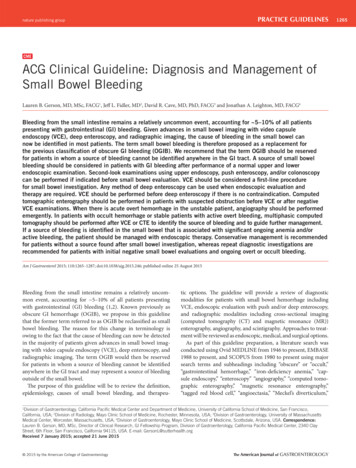
1268Gerson et al.Suspected small bowel bleedingOvertOccultPositiveTreataccordinglyRepeat endoscopy ifwarrantedNegativeProceed with smallbowel evaluationPossible obstructionNo obstructionNegativeNegative–no obstructionCTE/MREVCEPositiveNegativeNegative PositiveFurther evaluationwarrantedNoSpecific management:push or deep enteroscopysurgery intraoperativeenteroscopyYesConsider repeat endoscopy/VCE/Meckel’sscan/surgery intraoperative enteroscopyObservation/iron supplementsFigure 1. Algorithm for suspected small bowel bleeding. CTE, computed tomographic enterography; MRE, magnetic resonance enterography; VCE, videocapsule endoscopy.Brisk/massive suspectedsmall bowel bleedingUnstableStabilize patientRed cell scan or CTangiographyNegativePositiveAngiographySpecific managemententeroscopy vs surgery andintraoperative enteroscopyNegativePositiveEmbolizationFigure 2. Algorithm for brisk or massive suspected small bowel bleeding. CT, computed tomography.Sub-acute ongoingsmall bowel bleedingStabilize patientConsider VCE vs CTENegativeConsider RBC scan and orangiography or surgery intraoperative endoscopyPositiveProceed to deep endoscopyNegativePositiveTreat accordinglyFigure 3. Algorithm for sub-acute ongoing suspected small bowel bleeding. CTE, computed tomographic enterography; RBC, red blood cell; VCE, videocapsule endoscopy.The American Journal of GASTROENTEROLOGYVOLUME 110 SEPTEMBER 2015 www.amjgastro.com
Guidelines for Small Bowel BleedingTable 2. Causes of small bowel bleedingCommon causesRare causesUnder age 40 yearsOver age 40 yearsHenoch–Schoenlein purpuraInflammatory boweldiseaseAngioectasiaSmall bowel varices and/orportal hypertensive enteropathyDieulafoy’s lesionsDieulafoy’s lesionsAmyloidosisNeoplasiaNeoplasiaBlue rubber bleb nevussyndromeMeckel’s diverticulumNSAID ulcersPseudoxanthoma elasticumPolyposis syndromesOsler–Weber–Rendu syndromeKaposi’s sarcoma with AIDSPlummer–Vinson syndromeEhlers–Danlos syndromeInherited polyposis syndromes(FAP, Peutz–Jeghers)Malignant atrophic papulosisand double-balloon enteroscopy (DBE) (4), the prevalence of smallbowel vascular lesions based on 10 studies was 24% for both VCE(N 371) and DBE (N 364). For inflammatory findings, the yieldwas 18% for VCE (N 343) and 16% for DBE (N 336), and theyield was 11% for mass lesions (VCE, N 343 and DBE, N 336).An analysis comparing diagnostic yields from Western comparedto Asian countries demonstrated that patients undergoing DBEin Asian countries were more likely to have neoplastic findings,whereas angioectasia were more common in Western countries.Age has been known to be a determinant for the type of smallbowel pathology detected. Patients under the age of 40 years are morelikely to have inflammatory bowel disease or Meckel’s diverticulum.Small bowel neoplasms (e.g., GI stromal cell tumor, lymphoma,carcinoid, adenocarcinoma, or other polypoid lesions) and Dieulafoy’s lesions can occur in both younger and older patient cohorts(6–11). Angioectasia, other vascular lesions, and ulcers secondaryto anti-inflammatory agents are more likely in patients over theage of 40 years. Data regarding ethnicity and small bowel findingshas not been extensively published to date.HematobiliaAorto-enteric fistulaHemosuccus entericusFAP, familial adenomatous polyposis; NSAID, nonsteroidal anti-inflammatorydrug.Epidemiology and natural history of small bowel bleedingSummary statements1. The type of lesion responsible for small bowel bleeding isdependent on patient age but not gender or ethnicity.2. Small bowel angioectasia are the most common cause ofsmall bowel bleeding.3. Risk factors for angioectasia include advancing age, presenceof aortic stenosis, chronic renal failure, left ventricular assistdevices, and other hereditary disorders.4. Risk factors for recurrent small bowel bleeding from angioectasia include number of lesions, advanced age, presence ofcomorbid conditions, and anticoagulant therapy.Prevalence and etiology of small bowel bleeding. The prevalenceof small bowel lesions has been estimated to be 5–10% in patientspresenting with GI bleeding (1,2). Details pertaining to the clinicalpresentation are critically important in the determination of theetiology. A history of a bleeding diathesis as with von Willebranddisease and medication usage including aspirin, nonsteroidalanti-inflammatory drugs, anticoagulants, and/or other antiplatelet agents also can lend clues to the diagnosis. Knowledge of comorbidities such as valvular heart disease and prior procedures/surgeries such as liver biopsy, liver transplantation, abdominalaortic aneurysm repair, or bowel resection again can be very helpful. Common causes of small bowel bleeding are listed in Table2 and are found in 75% of patients with suspected small bowelbleeding (5). Based on a 2008 meta-analysis combining data fromWestern and Asian countries and reporting yields on both VCE 2015 by the American College of GastroenterologyDifferences in findings between patients with overt or occultsmall bowel bleeding. Studies using VCE and deep enteroscopyhave demonstrated higher diagnostic yields for patients withovert bleeding compared with patients with occult bleeding. Forpatients with prior overt bleeding, the diagnostic yield was lessthan that for current overt bleeders, and decreased substantiallywith time. In a 2004 study by Pennazio et al. (12) of 100 patientsundergoing VCE, the diagnostic yield was 92% for patients withovert bleeding, 44% for occult bleeders, 67% for patients with prior overt bleeding who were studied within 10–14 days, and 33%at 3–4 weeks postbleeding episode. In a 2010 study of 200 patientswith bleeding undergoing DBE, the diagnostic yield was 77% forovert bleeding, 67% for patients with occult hemorrhage, and 59%for patients with prior overt bleeding (13).In addition to higher diagnostic yields for patients with overtbleeding, recurrence rates may be higher in patients presentingwith overt bleeding. In a multicenter US study assessing long-termoutcomes post-DBE, recurrence of overt bleeding occurred in34% of patients presenting with overt hemorrhage compared with13% of patients with occult bleeding at 12 months postprocedure(P 0.06) (14). These recurrence rates, however, were not significant at 30 months of follow-up (27% vs. 20%, P NS).Rare causes and non-small bowel sources of bleeding. Rarecauses of small bowel bleeding are shown in Table 2. Patientswith disorders associated with portal hypertension and/or withendoscopic evidence of varices or portal hypertension have alsodemonstrated portal hypertensive changes in the small bowel onVCE or enteroscopy studies (15). Other rare causes of bleedingfrom the small bowel have included Kaposi’s sarcoma associatedwith acquired immunodeficiency syndrome, Plummer–Vinsonsyndrome, pseudoxanthoma elasticum, Ehlers–Danlos syndrome,Henoch–Schoenlein purpura, neurofibromatosis, malignantatrophic papulosis, and other inherited polyposis syndromes. Afamily history of polyposis syndromes may provide importantThe American Journal of GASTROENTEROLOGY1269
1270Gerson et al.clues to the underlying etiology of small bowel bleeding. Physicalexamination, including a detailed dermatological evaluation, mayalso be useful in the diagnosis of systemic syndromes, includinghereditary hemorrhagic telangiectasia and blue-rubber bleb nevus syndrome. Uncommon non-small bowel sources of obscureGI bleeding not shown in the table have included hematobilia,hemosuccus pancreatitis, and aortoenteric fistulae.Prior clinical guidelines have listed celiac disease as a cause ofsmall bowel bleeding (16), but there is emerging evidence thatceliac disease leads to iron-deficiency anemia because of malabsorption and not because of the presence of occult GI bleeding(17). Although complications associated with celiac disease such asulcerative jejunitis, lymphoma, and/or adenocarcinoma can causebleeding from the small intestine, the entity of celiac disease is nolonger listed as a cause of small bowel bleeding.Diagnosis of small bowel bleeding (Figure 1)Recommendations1. Second-look upper endoscopy should be considered in casesof recurrent hematemesis, melena, or a previously incompleteexam (strong recommendation, low level of evidence).2. Second-look colonoscopy should be considered in the settingof recurrent hematochezia or if a lower source is suspected(conditional recommendation, very low level of evidence).3. If the second-look examinations are normal, the next stepshould be a small bowel evaluation (strong recommendation,moderate level of evidence).4. Push enteroscopy can be performed as a second-look examination in the evaluation of suspected small bowel bleeding(conditional recommendation, moderate level of evidence).5. VCE should be considered a first-line procedure for smallbowel (SB) evaluation after upper and lower GI sources havebeen excluded, including second-look endoscopy when indicated (strong recommendation, moderate level of evidence).6. Owing to the lower detection rate of lesions in the duodenumand proximal jejunum with VCE, push enteroscopy shouldbe performed if proximal lesions are suspected (strong recommendation, very low level of evidence).7. Total deep enteroscopy should be attempted if there is astrong suspicion of a small bowel lesion based on clinicalpresentation or abnormal VCE study (strong recommendation, moderate level of evidence).8. Any method of deep enteroscopy can be used when endoscopicevaluation and therapy is required based on similar diagnosticyields (strong recommendation, high level of evidence).9. Intraoperative enteroscopy (IOE) is a highly sensitive but invasive diagnostic and effective therapeutic procedure. Its usageshould be limited to scenarios where enteroscopy cannot beperformed, such as patients with prior surgeries and intestinaladhesions (strong recommendation, low level of evidence).10. VCE should be performed before deep enteroscopy toincrease diagnostic yield. Initial deep enteroscopy can beconsidered in cases of massive hemorrhage or when VCEis contraindicated (strong recommendation, high level ofevidence).The American Journal of GASTROENTEROLOGYThe main limitations of SB evaluation in the past were related toits length ( 6 m) and limited intubation with conventional endoscopy; these shortcomings have been largely overcome by recentadvances in endoscopic technology, including VCE, deep enteroscopy (including DBE, SB enteroscopy, and spiral enteroscopy),and radiologic modalities including CT enterography (CTE) andMR enterography. These new advances, as well as the capacity tosuccessfully perform endoscopic therapeutic interventions, haveled to significant improvement in the management of patients withsmall bowel bleeding, and a decline in invasive surgical procedures(IOE, laparoscopy, and exploratory laparotomy) (18–21).Second-look endoscopyMost small intestinal bleeding is undramatic in presentation andeither presents as stable overt or occult bleeding. The prior literature demonstrated that a high percentage of patients designatedas having “potential small bowel bleeding” were found to havemissed bleeding sources within reach of conventional upper andlower endoscopy including diagnostic yields ranging from 2 to25% in patients undergoing repeat esophagogastroduodenoscopyand 6 to 23% on repeat colonoscopy (22–24). More recent studies using DBE and capsule endoscopy have also confirmed thesefindings (25–30).Most overt bleeding can be evaluated first with a second-lookprocedure to exclude upper and lower bleeding that can be readilyreached with a standard endoscope. Instead of repeating an upperendoscopy, a push enteroscopy may be performed to examine thedistal duodenum and proximal jejunum. During the colonoscopy,every effort should be made to intubate the terminal ileum to visualize the ileal mucosa and to inspect for blood coming from amore proximal location of the small intestine. For expediency ofwork up, it is sometimes appropriate to use VCE as the first-linetest after having had a negative upper endoscopy and colonoscopy.In fact, one study did not show that second-look endoscopy wascost effective (31). However, the distal duodenum and proximaljejunum would still need to be examined unless the VCE revealsthe source of the suspected small bowel bleeding.Push enteroscopyPush enteroscopy is an extended upper endoscopy performedwith a long endoscope such as a pediatric colonoscope (32) orwith a commercially available push enteroscope, which is typically 250 cm in length. Push enteroscopy allows only limited evaluation of the proximal SB, 70 cm distal to the ligament of Treitz.Push enteroscopy using a colonoscope typically can be passed45–60 cm beyond the ligament of Treitz (33). When push enteroscopy is carried out with the variable stiffness design, it reaches adeeper distance of nearly 90 cm (34). The diagnostic yield of pushenteroscopy is reported to range from 3 to 70%, with the majority of SB findings being vascular lesions (16,35–38). Interestingly,most of the lesions diagnosed on push enteroscopy have beenfound in locations accessible to standard esophagogastroduodenoscopy, emphasizing the importance of second-look endoscopy(22,39). When a dedicated push enteroscope is used, it may beperformed with an overtube designed to reduce looping in theVOLUME 110 SEPTEMBER 2015 www.amjgastro.com page
anywhere in the GI tract and may represent a source of bleeding outside of the small bowel. Th e purpose of this guideline will be to review the defi nition, epidemiology, causes of small bowel bleeding, and therapeu-tic options. Th e guideline will provide a review of diagnostic modalities for patients with small bowel hemorrhage including