Transcription
Clin Auton Res (2014) 24:15–23DOI 10.1007/s10286-013-0213-yRESEARCH ARTICLEAutonomic deficit not the cause of death in West Nile virusneurological diseaseHong Wang Venkatraman SiddharthanJeffery O. Hall John D. Morrey Received: 22 May 2013 / Accepted: 20 August 2013 / Published online: 25 October 2013Ó The Author(s) 2013. This article is published with open access at Springerlink.comAbstractIntroduction Some West Nile virus (WNV)-infectedpatients have been reported to manifest disease signsconsistent with autonomic dysfunction. Moreover, WNVinfection in hamsters causes reduced electromyographyamplitudes of the gastrointestinal tract and diaphragm, andthey have reduced heart rate variability (HRV), a read-outfor the parasympathetic autonomic function.Methods HRV was measured in both hamsters and miceusing radiotelemetry to identify autonomic deficits. Toidentify areas of WNV infection within the medullaoblongata mapping to the dorsal motor nucleus of vagus(DMNV) and the nucleus ambiguus (NA), fluorogold dyewas injected into the cervical trunk of the vagus nerve ofhamsters. As a measurement of the loss of parasympatheticfunction, tachycardia was monitored contiguously over thetime course of the disease.Results Decrease of HRV did not occur in all animals thatdied, which is not consistent with autonomic function beingthe mechanism of death. Fluorogold-stained cells in theDMNV were not stained for WNV envelope protein.Fourteen percent of WNV-stained cells were co-localizedwith fluorogold-stained cells in the NA. These data,H. Wang V. Siddharthan J. D. Morrey (&)Department of Animal, Dairy, and Veterinary Sciences,School of Veterinary Medicine, Institute for AntiviralResearch, Utah State University, 4700 Old Main Hill,Logan, UT 84322-4700, USAe-mail: john.morrey@usu.eduJ. O. HallDepartment of Animal, Dairy, and Veterinary Sciences,Utah Veterinary Diagnostic Laboratory,School of Veterinary Medicine, Utah State University,Logan, UT 84322-5700, USAhowever, did not suggest a fatal loss of autonomic functions because tachycardia was not observed in WNVinfected hamsters.Conclusion Parasympathetic autonomic function deficitwas not a likely mechanism of death in WNV-infectedrodents and possibly in human patients with fatal WNneurological disease.Keywords West Nile virus Autonomic Parasympathetic Heart rate variabilityIntroductionWe have previously reported that autonomic nervous system function deficits can be observed in hamsters infectedwith WNV [32]. This is consistent with human clinicalstudies and case reports documenting WNV disease signssuch as respiratory distress [29], diaphragmatic paralysis[2], gastrointestinal involvement, urinary retention [31],bladder dysfunction [29], fatigue [19], and cardiacarrhythmia [4]. West Nile virus infection is fatal in \1 %of human cases [20], but the fatality rate in experimentallyinfected mice and hamsters is typically reported to bebetween 20 and 80 % [23, 28, 35]. Therefore, rodentsprovided an opportunity to ask questions about the mechanism of death from WN neurological disease.In WNV-hamster studies, autonomic function was suppressed as measured by reduced heart rate variability(HRV), which is a well-accepted read-out for monitoringsympathetic and parasympathetic autonomic functions [11,14]. The major influence of the parasympathetic HRV iscardiopulmonary coupling, which is the neurological connectivity that causes the heart rate to increase duringresting respiratory inspiration and decrease during123
16expiration, which is physiologically referred to as respiratory sinus arrhythmia [12]. This autonomic process resultsin efficient cardiopulmonary function. Therefore, healthyautonomic parasympathetic function results in higherHRV. Conversely, unhealthy parasympathetic functionresults in reduced HRV. The parasympathetic input forcardiopulmonary coupling or sinus arrhythmia occursthrough the cervical trunk of the vagus nerve innervated byneurons in the DMNV and NA of the medulla [34]. IfDMNV or NA neurons were damaged or their numberswere reduced, the sympathetic response would be dominant. Reduced parasympathetic and increased sympatheticresponses typically result in tachyarrhythmia, and if severeenough, cardiac arrest [13]. Therefore, our approach tomeasure the effects of WNV on the parasympathetic cardiopulmonary coupling was to measure HRV, infection ofthe DMNV and NA neurons, and tachyarrhythmia ininfected rodents.We have recently determined that respiratory insufficiency is highly correlated with mortality in WNV-infectedhamsters, and that respiratory insufficiency was due toneurological disease [26, 33]. This respiratory insufficiencycould be due to the neurodegeneration of neurons controlling respiratory function including the raphe obscurus[8], retrotrapezoid nucleus and parafacial respiratory group[1, 10], rostral ventrolateral medulla [15], or pre-Bötzingercomplex [3] in the brainstem, phrenic motor neurons in thecervical cord, or in the posterior hypothalamus [18]. Eventhough WNV suppresses respiratory function, we did notknow specifically if autonomic dysfunction correlated torespiratory insufficiency or mortality, which was the subject of this study.Materials and methodsEthical statementThis study was conducted in accordance with the approvalof the Institutional Animal Care and Use Committee ofUtah State University (IACUC approval #1079 and #1488;WNV APHIS Permit # 47210). The work was done in theAAALAC-accredited Laboratory Animal Research Centerof Utah State University and in accordance with theNational Institutes of Health Guide for the Care and Use ofLaboratory Animals.Animals and virusesAdult female Syrian golden hamsters or C57BL/6 mice[7 weeks of age were used (Charles River Laboratories,Wilmington, MA). Animals were randomized to treatmentgroups. Rodents were judged as moribund if they did not step123Clin Auton Res (2014) 24:15–23forward if prodded, or if they did not right themselves whenplaced on their backs. WNV was diluted in minimal essentialmedium immediately prior to subcutaneous (s.c.) injection inthe groin area [24, 25, 27]. Hamsters were injected with avolume of 0.1 mL containing 5.7 9 107 pfu of a New YorkWNV isolate from crow brain [16, 17]. Mice were injectedwith a volume of 0.1 mL containing 2.5 9 106 pfu of aWN02 isolate designated as Kern 515 from Dr. Robert Tesh(Mosquito, 10/05/07, Kern County, CA, TVP 10799 BBRClot # WNVKERN515-01, University of Texas MedicalBranch Arbovirus Reference Collection). These two strainsof WNV were used because of suitable mortality in the tworodent strains used and to demonstrate that the findings ofthis report are not rodent- or virus strain specific.Heart rate variability (HRV) analysisRadio telemetry devices (TA10EEAT-F20, Data SciencesInternational, New Brighton, MN) were surgicallyimplanted in a subcutaneous pocket [21, 30, 32]. Tworecording-leads were subcutaneously tunneled toward theleft and right clavicular region where the tips of leads weresutured to the pectoral muscles. The DSI ETA-F20 transmitter transmitted ECG tracings with a sampling rate of1,000 Hz as an analog radio signal with a digital signature.The receivers, under the cages, were connected to a dataacquisition matrix hard-wired to a PC-based computerrunning Dataquest A.R.T. Silver System software (DataScience International, DSI). The duration of each readingwas 2 min and one reading was obtained every hour. Theacquisition software was IOX Base 8c software, and theECG analysis software was ECG auto with HRV ? module to generate HRV data including the Poincaré plot(emka Technologies, USA, Falls Church, VA). Theacquired data from the DSI software were converted to datasuitable for IOX software analysis by using ECG Auto 3.1(emka Technologies, USA, Falls Church, VA). The timesof the resting status of the hamsters were recorded multipletimes each day. Data from resting hamsters, as opposed toalert moving hamsters, were used to minimize backgroundnoise from movement of the animals.The Poncairé plot is constructed by plotting each R to R(RR) interval in milliseconds (ms) on the x-axis, and theprevious adjacent RR interval on the y-axis [5]. The morerapid the heart rate, the smaller is the RR interval. Conversely, the slower the heart rate, the larger is the RRinterval. Since the heart rate is neurologically coupled tothe inspiration and expiration of the lung, referred to ascardiopulmonary coupling, the RR intervals will be different than the previous RR intervals as the animalbreathes. This cardiopulmonary coupling is controlled byautonomic function. If the autonomic function is healthy,the individual dots on the Poincaré plot will be widely
Clin Auton Res (2014) 24:15–23distributed in a comet shape. As autonomic function isreduced, the comet shape will condense to a small shape ordot [5]. Data sample times were 2 min for hamsters and5 min for C57BL/6 mice. The Poincaré data were quantified as nonlinear measurements of HRV by measuring thestandard deviations in the perpendicular direction of thepattern (SD1) and the SD of the parallel direction of thedata pattern (SD2) using the Kubios HRV Analysis software (version 21.).Radiotelemetry for temperature, heart rate, and activityThe same radio telemetry chips and surgical proceduresused for HRV analysis were used. Data were obtainedcontinuously throughout the course of the experiment. Thedata were graphed using GraphPad Prism 5.0.Fluorogold and WNV envelope stainingDMNV and NA regulate considerable parasympatheticautonomic function to the heart through the cervical trunkof the vagus nerve [6]. To fluorescently locate the DMNVand NA, the cervical trunk of the vagus nerve was injectedwith 1 lL of 2 mg/mL of fluorogold. The tissues werefixed for histological analysis 3 days after fluorogoldinjection. The hamsters were injected with euthasol(100 mg/kg, i.p) and cardiac perfused (i.v.) with 0.01 MPBS followed by 4 % paraformaldehyde. The brainstemsmaintained in 30 % sucrose overnight were cut into coronal sections (10 lm) by a cryostat instrument (Leica CM1900). They were permeabilized in 0.5 % triton X-100,blocked with 5 % goat serum in 0.1 % BSA, incubatedwith 7H2 anti-WNV mouse mAb (1:450, BioReliance,Invitrogen Bioservices, Rockville, MD) and rabbit antifluorogold polyclonal antibody (1:3000, Fluorochrome,LLC, Denver, Colorado) at 4 C overnight. The sectionswere then incubated with Alexa-fluorÒ 568 goat antimouse IgG antibody and Alexa-fluorÒ 488 goat anti-rabbitIgG antibody (Life Technologies, Grand Island, NY).Slides were examined using a Carl Zeiss Axioskop-2microscope (Carl Zeiss Microscopy, LLC, Thornwood,NY) and X-Cite fluorescence illumination (Ontario, Canada), connected with CRI-Nuance (Grantham, UK) multispectral image system.ResultsAll six WNV-infected hamsters lost some autonomicfunction during the course of the disease as evidenced bythe Poincaré plots, wherein two representative WNVinfected hamsters compared to a sham-infected hamster areshown (Fig. 1a). As explained in ‘‘Materials and methods’’,17the autonomic HRV is healthy if the dots are widely distributed. If the function or numbers of parasympatheticautonomic neurons are diminished, the HRV is diminishedand the dots condense to a smaller shape. Hamsters displayed patterns of diminished autonomic nervous functionduring the course of infection as illustrated with hamsters#229 and #236, which became moribund on days 10 and25, respectively. The condensed data pattern of hamster#229 continued until the day of termination, whereas thecondensed patterns of hamster #236 disappeared later inthe course of disease as illustrated on day 18 (Fig. 1a). Onthe day of termination at day 25, the distribution of datapoints expanded even more. Overall, condensed data pointpatterns characteristic of autonomic dysfunction fromWNV-infected hamsters did not consistently correlate withthe time of mortality (data of all animals not shown). Noneof the sham-infected hamsters possessed condensed datapatterns, as illustrated with hamster #234 (Fig. 1a).The HRV of each hamster was quantified from the Poincaré data by measuring the standard deviation (SD) in theperpendicular direction of the pattern (SD1) and the SD ofthe parallel direction of the data pattern (SD2) (KubiosHRV Analysis, version 2.1). The smaller numbers of SD1and SD2 indicated smaller heart rate variability. Two SDbelow the mean of sham-infected animals was used as acutoff for normal values, i.e., values below 2SD wereconsidered abnormal (Table 2). The HRV from animals#227, 229, 236, and #238 appeared to be low on days 8 and12 after WNV challenge. Some of the values from thesehamsters were below the cutoff of 2SD. One hamster(#238) gained higher HRV values before succumbing to thedisease. The remainder of the hamsters (#239 and #240)did not develop low HRV according to this quantitativeanalysis, but still succumbed to the disease.Poincaré plots were generated for WNV- and shaminfected C57BL/6 mice (Fig. 1b). Unlike the hamsters,condensed data point patterns did not develop in nearly allof the mice with fatal WNV infection. Mouse #44 andmouse #50 developed values below 2SD of the shaminfected means, but the low HRV values were not associated with the time of mortality (Table 1). Conversely, thedistribution of data points often expanded during the courseof disease, which supported the observation in hamster thatpatterns characteristic of parasympathetic autonomic dysfunction did not correlate with mortality.Since the parasympathetic function of the heart is primarily controlled by vagal nerve cells located in themedulla oblongata, we applied a previously publishedtechnique [6] to identify the location of vagal neuron cellbodies. The neuron cell bodies controlling parasympatheticinnervation of the thorax and abdominal viscera wereidentified by injecting the cervical trunk of the vagus nervewith fluorogold dye. Fluorogold dye then transported123
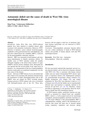
18Clin Auton Res (2014) 24:15–23Fig. 1 Poincaré plots to detect parasympathetic autonomic functionover the course of disease in a hamsters and b mice injectedsubcutaneously with WNV. Data from representative animals anddays after viral challenge are shown from a total of a five sham-infected hamsters, six WNV-infected hamsters, b four sham-infectedmice, and five WNV-infected mice. Unlabeled axes have the samevalues as listed in the first upper left graph for a hamsters and b mice.Arrows indicate loss of HRVthrough the axons into the respective neuron cell bodies ofthe DMNV and the NA. Positive cells stained with fluorogold could be visualized in coronal sections of the medullaoblongata using as references the fourth ventricle, centralcanal, and a hamster brain atlas [22] (Fig. 2a). The sametechnique was applied to hamsters injected s.c. with WNVto determine if WNV infects these cells (Fig. 2b). In a totalof six hamsters, no WNV-infected cells co-localized withfluorogold in the DMNV (0 WNV-positive cells co-localized with 193 fluorogold-positive cells), whereas, 14 % ofWNV-stained cells (11 WNV-positive cells co-localizedwith 80 fluorogold-positive cells) co-localized with fluorogold-stained cells in the NA (Table 2). These data suggestthat parasympathetic autonomic function controlled by theDMNV were not affected by WNV infection, whereas theNA may have been affected by WNV.To determine if infection of parasympathetic neuronsaffected cardiac parasympathetic function, tachycardia wasinvestigated in WNV-infected hamsters using surgicallyimplanted radio telemetry chips. These chips faithfullyreported heart rate, in addition to body temperature andmovement activity in two sham-infected control hamstersover 24 days, as illustrated with sham #102 (Fig. 3). Diurnaland nocturnal cycling was readily observed in these animalsand underscored the need to record heart rate throughout the24-h cycle. The data from three representative WNV-infected hamsters are shown in Fig. 3. Animals #903 and #293showed no evidence of tachycardia. Animal #292 showedsome evidence of possible tachycardia at about days 8–10.Nevertheless, tachycardia did not correlate with mortality,which suggested that loss of parasympathetic autonomicfunction was not the main mechanism of death.123DiscussionThe main contribution of this study is the evidence generated to show that loss of parasympathetic autonomic
Clin Auton Res (2014) 24:15–2319Table 1 Quantification of heart rate variability from Poincaré plot data in WNV-infected rodents using Kubios HRV Analysis software (version21)HamstersAnimal #Death dayDay 0ADay 4Day 8Day 12#227920, 338.4, 1311, 11#2291442, 5721, 258.0, 10BBDay 14Day 15Day 16Day 18Day 19Day 20Day 2548, 62126, 1256.8B, 7.8B8.2, 8.8B#23618101, 10766, 748.4, 9.4#2381937, 4326, 3164, 8910.8, 6.6B#2392522, 4111, 1731, 3675, 8491, 103#2401628, 4020, 3358, 6833, 3530, 3182, 120134, 14358, 7616, 17MiceAnimal #Death dayDay 1Day 3Day 5Day 7Day 89.6, 165.6, 1119, 2156, 86#4087.1, 9.8#4284.1, 9.13.6, 6.87.3, 124.3, 1026, 37#44#47882.5C, 4.33.9, 4.81.7, 6.16.9, 118.3, 125.8, 128.1, 8.012, 2421, 4736, 68#50104.2, 4.13.4, 5.73.3, 8.77.1, 124.2, 11Day 9Day 102.0C, 4.748, 90The smaller numbers of SD1 and SD2a indicated smaller heart rate variability. SD1 and SD2 are the standard deviations in the X1 (perpendiculardirection) and X2 (parallel direction) to the scatter distribution of the Poincaré plotADays after WNV subcutaneous challengeBValues considered abnormal when below two standard deviations of sham-infected hamsters, SD1, SD2 B7.8, 10.5CValues considered abnormal when below two standard deviation of sham-infected mice, SD1, SD2 B2.6, 1.8function does occur in hamsters, but is not the mechanismof death in WNV-infected rodents. Three physiologicalparameters provided this evidence, namely, HRV as represented in Poincaré plots, staining of the DMNV and NAfor WNV envelope antigen, and measurement of tachycardia. In a previous publication by us [26] demonstratingreduced HRV in WNV-infected hamsters, standard timedomain measurements (e.g., standard deviation of normalRR, root mean square of the differences between consecutive RR) and frequency domain measurements (low- andhigh-frequency power) were used. Subsequently in thisreport, we have used the Poincaré plots to visualize theHRV, and subjectively believe that it is more dependableand unambiguous in detecting reduced HRV in theseWNV-rodent models. Moreover, the parasympathetic analysis of the current study differed from the prior study inthat measurements were used when the animals weremotionless and appeared to be sleeping. This reducederroneous recordings due to movement of the electrodes orby muscle EMGs during times of activity. Clearly, someanimals had loss of HRV on the last day of death asidentified by condensed data points, but other animals didnot have condensed data points on the day of death.Moreover, mice never did develop condensed data pointsduring the course of disease. Therefore, loss of HRV couldnot have been the mechanism of WNV-induced death inrodents.Autonomic function is divided into the ‘fight-or-flight’sympathetic response and ‘rest-and-digest’ parasympathetic response. The neuron cell bodies innervating thethorax and abdominal viscera parasympathetic responseswere identified by observing stained neurons in the DMNVand NA from animals injected in the cervical trunk of thevagus nerve with fluorogold dye [6]. No WNV-stainedcells were observed in the DMNV, and a small number ofcells were observed in the NA. The parasympatheticautonomic response alone was likely to not be the mechanism of WNV-induced death, because the vast majority ofthese cell bodies were not stained for WNV envelopeantigen reflecting viral replication.This hypothesis was supported by analysis of heart rate.If the viruses were to functionally damage neurons in theDMNV and NA, the cardiac parasympathetic responsewould be reduced and leave a dominant sympatheticresponse. This typically results in tachyarrhythmia, and ifsevere enough, cardiac arrest [13]. Since hamsters manifeststrong circadian rhythm [7], single measurements of heartrates during the day may result in misidentification oftachyarrhythmias. To overcome this problem, the heart ratewas monitored continuously over the course of the experiment using radio telemetry chips. As expected, strongfluctuations of diurnal and nocturnal rhythms wereobserved prior to development of neurological disease.There was no consistent tachycardia observed, however,123
20Fig. 2 Staining of the DMNVand NA for WNV envelopeantigen. a Five hamsters wereinjected into the cervical trunkof the vagus nerve withfluorogold. After 3 days,sections of the brains in the areaof the DMNV and NA wereprepared and stained for WNVenvelope antigen. Thefluorogold-stained areas wereidentified to be the DMNV andNA using a hamster stereotaxicatlas [22]. On the left of arepresentative hamster, imageswere assembled from the samebrain section to construct ahemisphere identified to be thefluorogold staining of DMNVand NA. On the right, highermagnifications of the image ofthe DMNV (upper) and NA(lower) are shown. b Images ofthe DMNV and NA identifiedwith fluorogold were stained forWNV envelope antigen inWNV- and sham-infectedhamsters. The animals wereinjected s.c. with WNV or sham.Six days later they were injectedinto the cervical trunk of thevagus nerve with fluorogold,after which the brains wereprocessed for staining for WNVenvelope antigen and withDAPI to detect nuclear DNA(3 days after dye injection).Arrows identify WNV-positivecells123Clin Auton Res (2014) 24:15–23
Clin Auton Res (2014) 24:15–2321Table 2 Co-localization of WNV-stained neurons with fluorogoldstained neurons in the dorsal motor nucleus of vagus (DMNV) andnucleus ambiguus (NA)WNV-positive staining co-localized with fluorogoldpositive cells per total fluorogold-positive cells (%)DMNVNAWNV-infected hamstersSham-infected hamsters0/193 (0 %)A0/157 (0 %)BC11/80 (14 %)0/35 (0 %)DCriteria for killing and necropsy—no stepping when proddedAFifteen slides from six hamstersBSeven slides from two hamstersCNine slides from four hamstersFour slides from one hamsterDduring the development of fatal disease. Moreover, in arecent study, cardiac arrhythmia did not correlate withmortality [26]. Therefore, tachyarrhythmia did not consistently occur in lethal WNV disease in hamsters.An interesting occurrence can be observed in Fig. 3where the circadian rhythm of a particular hamster #903appeared to be lost beginning at day 8 after WNV challenge. Other hamsters, but not all, that recover fromWNV infection manifest this loss of circadian rhythm(data not shown), which may be attributed to loss ofhigher-order functions in areas such as the suprachiasmatic nucleus [7]. The cause of loss of circadian rhythmin WNV-infected rodents and how this might relate tosleep disorders or fatigue in WNV patients await furtherinvestigations.Another interesting observation seen qualitatively inFig. 1 and quantitatively in Table 1 is the increase in HRVin some hamsters (#238, #239) and in all mice just beforedeath. Even though cardiopulmonary coupling is largelyresponsible for high-frequency rhythms, low-frequencyrhythms controlled by higher brain structures might alsodominate during WNV infection and could conceivably beresponsible for increased HRV. Moreover, the differencesin cytokine responses that may occur between individualanimals and between species may contribute to differencesin HRV due to viral infection [9]. These issues should beinvestigated in future studies.The results of this study augmented the finding thatrespiratory insufficiency correlated strongly with the mortality of rodents infected with WNV and were the likely thepredominant mechanism of death. Gliosis, paravascularcuffing, neuronal phagia, and WNV-specific staining wereobserved in the rostral ventrolateral medulla controllingrespiratory motor functions of hamsters with respiratoryinsufficiency as measured by plethysmography, but littlestaining was observed in the DMNV or NA. These priorstudies [26, 33] and the current study suggest thatFig. 3 Heart rate (blue), body temperature (red), and movementactivity (gray) measured by radio telemetry in hamsters challengeds.c. with sham or WNV. Data from representative animals are shownfrom a total of two sham-infected hamsters, 8 WNV-infectedhamstersrespiratory insufficiency, not loss of parasympatheticautonomic function, is the primary mechanism of death inWNV-infected rodents and possibly human patients.123
22In this work we showed that WNV infection of rodentsmay or may not cause autonomic function deficits asmeasured by HRV depending on the species, hamsters andmice, yet both species succumb to infection. The lack oftachycardia and WNV infection in the dorsal motor nucleusof vagus or nucleus ambiguous controlling parasympatheticfunctions also suggest that autonomic dysfunction inrodents and possibly human patients is not likely to be thephysiological mechanism of death, despite the clinicalobservation that disease signs and symptoms may reflectsome degree of autonomic function deficits.Acknowledgments The work was supported by the Rocky Mountain Regional Centers of Excellence, National Institute of Allergy andInfectious Diseases (NIAID), National Institutes of Health (NIH;grant U54 AI-065357 to J. D. M.); Virology Branch, NIAID, NIH(grant HHSN272201000039I to J. D. M.); and the Utah AgricultureResearch 480 Station (grant UTA00424 to J. D. M.). We speciallythank Ashley Dagley and Neil Motter for expert technical assistance,Ronald M. Harper, Ph.D. (Department of Neurology, University ofCalifornia at Los Angeles) for helpful suggestions, and RamonaSkirpstunas, D.V.M., Ph.D., Diplomat of the American College ofVeterinary Pathology.Conflict of interest On behalf of all authors, the correspondingauthor states that there is no conflict of interest.Open Access This article is distributed under the terms of theCreative Commons Attribution License which permits any use, distribution, and reproduction in any medium, provided the originalauthor(s) and the source are credited.References1. Abbott SB, Stornetta RL, Coates MB, Guyenet PG (2011)Phox2b-expressing neurons of the parafacial region regulatebreathing rate, inspiration, and expiration in conscious rats.J Neurosci 31:16410–164222. Betensley AD, Jaffery SH, Collins H, Sripathi N, Alabi F (2004)Bilateral diaphragmatic paralysis and related respiratory complications in a patient with West Nile virus infection. Thorax59:268–2693. Bochorishvili G, Stornetta RL, Coates MB, Guyenet PG (2012)Pre-Botzinger complex receives glutamatergic innervation fromgalaninergic and other retrotrapezoid nucleus neurons. J CompNeurol 520:1047–10614. Bode AV, Sejvar JJ, Pape WJ, Campbell GL, Marfin AA (2006)West Nile virus disease: a descriptive study of 228 patientshospitalized in a 4-county region of Colorado in 2003. Clin InfectDis 42:1234–12405. Brennan M, Palaniswami M, Kamen P (2002) Poincare plotinterpretation using a physiological model of HRV based on anetwork of oscillators. Am J Physiol Heart Circ Physiol283:H1873–H18866. Cheng Z, Powley TL (2000) Nucleus ambiguus projections tocardiac ganglia of rat atria: an anterograde tracing study. J CompNeurol 424:588–6067. Coogan AN, Piggins HD (2003) Circadian and photic regulationof phosphorylation of ERK1/2 and Elk-1 in the suprachiasmaticnuclei of the Syrian hamster. J Neurosci 23:3085–3093123Clin Auton Res (2014) 24:15–238. Depuy SD, Kanbar R, Coates MB, Stornetta RL, Guyenet PG(2011) Control of breathing by raphe obscurus serotonergicneurons in mice. J Neurosci 31:1981–19909. Fairchild KD, Srinivasan V, Moorman JR, Gaykema RP, GoehlerLE (2011) Pathogen-induced heart rate changes associated withcholinergic nervous system activation. Am J Physiol Regul IntegrComp Physiol 300:R330–R33910. Fortuna MG, Stornetta RL, West GH, Guyenet PG (2009) Activation of the retrotrapezoid nucleus by posterior hypothalamicstimulation. J Physiol 587:5121–513811. Gross V, Tank J, Obst M, Plehm R, Blumer KJ, Diedrich A,Jordan J, Luft FC (2005) Autonomic nervous system and bloodpressure regulation in RGS2-deficient mice. Am J physiol RegulIntegr Comp Physiol 288:R1134–R114212. Grossman P, Taylor EW (2007) Toward understanding respiratory sinus arrhythmia: relations to cardiac vagal tone, evolutionand biobehavioral functions. Biol Psychol 74:263–28513. Huikuri HV, Valkama JO, Airaksinen KE, Seppanen T, KesslerKM, Takkunen JT, Myerburg RJ (1993) Frequency domainmeasures of heart rate variability before the onset of nonsustainedand sustained ventricular tachycardia in patients with coronaryartery disease. Circulation 87:1220–122814. Janssen BJ, Leenders PJ, Smits JF (2000) Short-term and longterm blood pressure and heart rate variability in the mouse. Am Jphysiol Regul Integr Comp Physiol 278:R215–R22515. Kanbar R, Stornetta RL, Cash DR, Lewis SJ, Guyenet PG (2010)Photostimulation of Phox2b medullary neurons activates cardiorespiratory function in conscious rats. Am J Respirat Crit CareMed 182:1184–119416. Lanciotti RS, Ebel GD, Deubel V, Kerst AJ, Murri S, Meyer R,Bowen M, McKinney N, Morrill WE, Crabtree MB, Kramer LD,Roehrig JT (2002) Complete genome sequences and phylogeneticanalysis of West Nile virus strains isolated from the UnitedStates, Europe, and the Middle East. Virology 298:96–10517. Lanciotti RS, Kerst AJ (2001) Nucleic acid sequence-basedamplification assays for rapid detection of West Nile and St.Louis encephalitis viruses. J Clin Microbiol 39:4506–451318. Lazarenko RM, Stornetta RL, Bayliss DA, Guyenet PG (2011)Orexin A activates retrotrapezoid neurons in mice. Respir PhysiolNeurobiol 175:283–28719. Leis AA, Stokic DS (2005) Neuromuscular manifestations ofhuman West Nile virus infection. Curr Treat Options Neurol7:15–2220. Lindsey NP, Staples JE, Lehman JA, Fischer M (2010) Surveillance for human West Nile virus disease—United States,1999–2008. MMWR Surveill Summ 59:1–1721. Mongue-Di
h. wang v. siddharthan j. d. morrey (&) department of animal, dairy, and veterinary sciences, school of veterinary medicine, institute for antiviral research, utah state university, 4700 old main hill, logan, ut 84322-4700, usa e-mail: john.morrey@usu.edu j. o. hall department of animal, dairy, and veterinary sciences, utah veterinary