Transcription
G C A TT A C GG C A TgenesArticleExploring the Genetic Landscape of Retinal Diseasesin North-Western Pakistan Reveals a High Degree ofAutozygosity and a Prevalent Founder Mutation inABCA4Atta Ur Rehman 1 , Virginie G. Peter 2,3,4 , Mathieu Quinodoz 3,5 , Abdur Rashid 6 ,Syed Akhtar Khan 7 , Andrea Superti-Furga 1 and Carlo Rivolta 3,4,8, *12345678*Division of Genetic Medicine, Lausanne University Hospital and University of Lausanne, 1011 Lausanne,Switzerland; atta.rehman@unil.ch (A.U.R.); asuperti@unil.ch (A.S.-F.)Institute of Experimental Pathology, Lausanne University Hospital and University of Lausanne,1011 Lausanne, Switzerland; virginie.peter@unil.chDepartment of Genetics and Genome Biology, University of Leicester, Leicester LE1 7RH, UK;mathieu.quinodoz@unil.chClinical Research Center, Institute of Molecular and Clinical Ophthalmology Basel (IOB), 4031 Basel,SwitzerlandDepartment of Computational Biology, University of Lausanne, 1015 Lausanne, SwitzerlandGovernment Degree College Ara Khel, FR Kohat 26000, Khyber Pakhtunkhwa, Pakistan;rasheedkhan037@gmail.comDepartment of Ophthalmology, Khalifa Gul Nawaz Hospital, Bannu 28100, Pakistan; drak8304@gmail.comDepartment of Ophthalmology, University Hospital Basel, 4031 Basel, SwitzerlandCorrespondence: carlo.rivolta@iob.chReceived: 31 October 2019; Accepted: 17 December 2019; Published: 21 December 2019 Abstract: Variants in more than 271 different genes have been linked to hereditary retinal diseases,making comprehensive genomic approaches mandatory for accurate diagnosis. We explored thegenetic landscape of retinal disorders in consanguineous families from North-Western Pakistan,harboring a population of approximately 35 million inhabitants that remains relatively isolated andhighly inbred ( 50% consanguinity). We leveraged on the high degree of consanguinity by applyinggenome-wide high-density single-nucleotide polymorphism (SNP) genotyping followed by targetedSanger sequencing of candidate gene(s) lying inside autozygous intervals. In addition, we performedwhole-exome sequencing (WES) on at least one proband per family. We identified 7 known and 4 novelvariants in a total of 10 genes (ABCA4, BBS2, CNGA1, CNGA3, CNGB3, MKKS, NMNAT1, PDE6B,RPE65, and TULP1) previously known to cause inherited retinal diseases. In spite of all familiesbeing consanguineous, compound heterozygosity was detected in one family. All homozygouspathogenic variants resided in autozygous intervals 2.0 Mb in size. Putative founder variantswere observed in the ABCA4 (NM 000350.2:c.214G A; p.Gly72Arg; ten families) and NMNAT1genes (NM 022787.3:c.25G A; p.Val9Met; two families). We conclude that geographic isolationand sociocultural tradition of intrafamilial mating in North-Western Pakistan favor both the clinicalmanifestation of rare “generic” variants and the prevalence of founder mutations.Keywords: hereditary retinal diseases; autozygosity mapping; consanguinity; Pakistan1. IntroductionInherited retinal dystrophies (IRDs) constitute a genetically heterogeneous group of rare conditionsof the eye. They are mainly characterized by the progressive loss of rod and/or cone photoreceptors,Genes 2020, 11, 12; s
Genes 2020, 11, 122 of 13resulting in complete or nearly complete blindness at the end [1]. Globally, IRDs affect approximatelyone million people, with a frequency of 1 in 3000 births. Clinically, they may range from mild andnon-progressive night blindness to more severe and degenerative phenotypes, including retinitispigmentosa (RP) and cone or cone-rod dystrophies [2]. To date, mutations in over 271 genes havebeen linked to various forms of IRDs (RetNet; https://sph.uth.edu/RETNET/; accessed on 12 December2019), and the sequencing of their coding parts has allowed the detection of pathogenic mutations inmore than 60% of the patients [3]. IRDs are inherited as an autosomal recessive, autosomal dominant,X-linked, or mitochondrial trait, with autosomal recessive being the most prominent type [1,4]. Recenttechnological advancements, such as next-generation sequencing (NGS), have significantly increasedgene discovery rates in a wide range of inherited ocular conditions [5], with a sensitivity value of 75%, when applied to a clinically focused IRDs group [6]. Since consanguinity unmasks the adverseeffects of recessive mutations through bi-parental inheritance of the same allele, it is possible toreveal the presence of disease-causing variants in consanguineous pedigrees by simply flagging largesegments of consecutive homozygous genotypes surrounding the mutations, using a technique called“autozygosity mapping” [7–10]. For a more rapid and robust analysis, scientists usually combineautozygosity mapping with NGS to maximize the acquisition of relevant genetic information [10].The combination of such information with targeted functional studies has provided significant insightsinto the molecular mechanisms of rare Mendelian diseases, including IRDs.Since children of consanguineous couples are more likely than children of non-consanguineousparents to be affected by recessive genetic anomalies [11], the incidence of rare Mendelian diseasesis higher in populations having a high degree of endogamy [12]. For example, Pakistan has one ofthe highest rates of inherited genetic diseases in the world, likely due to the fact that consanguinity ispresent in more than 50% of the population and marriages among first cousins are highly favored bythe society [13,14]. According to a recent estimate, approximately 1.12 million people in Pakistan areblind, and the vision loss burden has continued to rise in the country since 1990 [15].Although some information exists on blindness caused by cataracts or refractive errors,the prevalence of IRDs is not well documented in Pakistan at the level of the whole population.A hospital-based study in Karachi, a metropolitan city, revealed that 1 in 800 patients who visited theophthalmic outpatient department had retinal dystrophies, with RP being the most frequent type (64%),followed by Stargardt disease (14.7%) and cone dystrophies (6.7%). Unsurprisingly, more than half of thepatients from this study were born to consanguineous parents [16]. Recently, a few studies on IRDs havebeen published in Pakistan [17–20]. However, the majority of these reports were based on pedigreesfrom the Punjab and Sindh provinces, thus leaving North-Western Pakistan largely unexplored.Administratively known as Khyber Pakhtunkhwa (KP), this part of the country is predominantly aPashtuns territory and includes a heterogeneous population of approximately 35 million inhabitants.Consanguinity in KP ranges between 22% and 66%, and the rate of consanguinity was found to haveincreased over time, possibly due to the growing violence and geo-political conflicts in the region(consanguineous marriages are believed to strengthen pre-existing intra-familial relationships and thusbe advantageous in the context of civil unrest) [21–25]. To our knowledge, no comprehensive study onthe genetic spectrum of IRDs has ever been undertaken in North-Western Pakistan, possibly becauseof socio-economic and cultural limitations, a lack of infrastructure, difficult terrain, and escalatingconflicts in the region.2. Materials and Methods2.1. Enrollment of Families and Collection of SamplesOur study conforms to the standards of the Declaration of Helsinki and was approved bythe Institutional Review Boards of the Hazara University, Mansehra, Pakistan (approval code:F.No:185/HU/Zool/2018/583) and of all our respective Institutions. Informed consents were providedin written form by all families prior to their participation and were signed by all members who
Genes 2020, 11, 123 of 13were enrolled. Families with at least two or more affected persons, a history of consanguinity, anda clear autosomal recessive inheritance pattern of the disease were selected for molecular analysis.All families participating in our study were ethnically Pashtuns and were geographically located inthe KP province in North-Western Pakistan. Clinical and demographic information was obtainedvia a pre-designed questionnaire, and pedigrees were drawn and cross-checked through face-to-faceinterviews with patients and/or elder members of the family, in their native language. Clinical data wereobtained either directly from the patients’ medical reports (if available) or through consultation withthe local clinicians/ophthalmologists who examined them on our request. Due to the limited medicalinfrastructure in the region, detailed clinical investigations, such as electroretinography (ERG) orfundus images, were not available. Furthermore, most of the clinical information obtained was derivedfrom self-reported data, including, for example, difficulties in day/night vision, photophobia, diseaseonset and progression, response to medication, outcome of the Ishihara test, dark/light adaptation,ability to see/focus near and distant objects, loss of central or peripheral vision, and nystagmus. Patientswere also investigated for other parameters, such as their ability to perform routine activities, e.g.,reading, doing physical activities, and socializing with people. Electronic versions of pedigrees werecreated using the Pedigree Chart Designer (CeGaT, Tubingen, Germany). Saliva samples were collectedby using the Oragene saliva kit (OG-500, DNA Genotek, Ottawa, ON, Canada) from patients as wellas their clinically unaffected relatives, following the manufacturer’s guidelines. DNA was extractedfrom these samples following standard protocols, e.g., by following the prepIT-L2P manual (DNAGenotek, Canada). Quantitative and qualitative assessments of DNA were made using the NanoDrop 1000 Spectrophotometer (Thermo Fisher Scientific, Waltham, MA, USA) and electrophoresis on 1%agarose gels.2.2. Genotyping and Homozygosity MappingInitially, nine families were subjected to genetic analysis using genome-wide high-densitysingle-nucleotide polymorphism (SNP) arrays. For this purpose, genomic DNA of two or moreindividuals per family was genotyped by using the InfiniumCoreExome-24v1-1 array (Illumina,San Diego, CA, USA), which encompasses 550,000 genome-wide SNP markers, at the iGE3 GenomicsPlatform of the University of Geneva, Switzerland. Arrays were processed using an iScan, accordingto the manufacturer’s protocol. Genotype calls were generated using the GenomeStudio software byIllumina. PLINK was used to analyze the genotype data [26]. Following the identification of sharedautozygous intervals among two or more patients from the same family, all exons and exon–intronboundaries of candidate gene(s) inside these intervals were sequenced using the Sanger method.Additionally, families that belonged to the same geographic area and had clinically overlappingphenotypes were also investigated for the presence of shared autozygous intervals. Using this method,putative disease-causing variants were identified in four consanguineous pedigrees segregatingautosomal recessive IRDs, while the remaining unsolved families were subsequently characterized bywhole-exome sequencing (WES).2.3. Whole-Exome SequencingOverall, WES was performed for 10 pedigrees. For WES analysis, 2.0 µg of genomic DNAfrom index patients was initially processed by Novogene Co. Ltd (Hong Kong, China). Sequencinglibraries were generated using the Agilent SureSelect Human All ExonV6 kit (Agilent Technologies,Santa Clara, CA, USA), while fragmentation was carried out by hydrodynamic shearing (Covaris,Massachusetts, MA, USA). Following adapter ligation, DNA fragments were selectively enriched ina PCR reaction. Products were purified using the AMPure XP system (Beckman Coulter, Beverly,CA, USA) and quantified using an Agilent high-sensitivity DNA assay on the Agilent Bioanalyzer2100 system. Captured DNA libraries underwent paired-end sequencing on an Illumina Novaseq6000 S4 platform, resulting in sequences of 150 bases (PE150 sequencing strategy). WES data wereanalyzed using our in-house computational pipeline [27], and autozygosity mapping was done
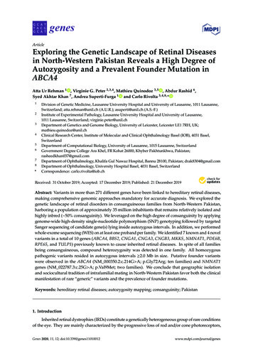
Genes 2020, 11, 124 of 13using AutoMap (unpublished). Finally, Sanger sequencing was performed to validate the potentiallypathogenic variants detected and to confirm their causality, via genotype–phenotype cosegregationwithin the families.3. Results3.1. Clinical SynopsisAs mentioned earlier, detailed clinical investigation could not be achieved for all patients. However,a summary of the clinical information of one family is shown in Figure S1. On the basis of the fewdata available, mostly based on patients’ symptoms, we could identify five major clinical IRD classes.Briefly, patients with severe early-onset blindness were tentatively categorized as individuals withLeber congenital amaurosis (LCA, two families), while patients presenting with IRD and extra-ocularsymptoms such obesity, hypogonadism, learning/developmental disabilities, post-axial polydactyly ofhands and/or feet, and renal abnormalities were examined by a local clinician who classified them assuffering from Bardet–Biedl syndrome (BBS) (two families). Patients with progressive loss of centralvision were categorized as having macular dystrophy (eleven families), while those presenting withinitial night blindness and progressive loss of peripheral vision were classified as suffering from RP(four families). Patients with RP were clinically evaluated with the help of a local ophthalmologist whoreported the presence of bilateral bone spicules and peripheral retinal vascular attenuation, throughfundus examination. Lastly, patients with a complete inability to discriminate between colors wereclassified as having achromatopsia (one family).3.2. Molecular FindingsCollectively, we identified 11 disease-causing variants in 10 IRD-associated genes, in a total of20 consanguineous IRDs pedigrees, all from North-Western Pakistan (Table 1). These variants weredetected using a genome-wide SNP array followed by Sanger sequencing (four families), WES (tenfamilies), and targeted Sanger sequencing alone (six families). Of these variants, seven were previouslyknown IRD mutations, while four variants had never been identified before. Newly detected changescomprised three protein-truncating mutations and one nonsynonymous single-nucleotide variant(SNV). While the pathogenicity of protein-truncating variants can be easily postulated, the causality ofthe nonsynonymous SNV was inferred through in silico analysis and segregation studies. In spiteof all families being consanguineous, compound heterozygosity was detected in one family (PK-E).All homozygous pathogenic variants were detected inside tractable autozygous intervals ( 2.0 Mb insize) and co-segregated with the disease in homozygosis in members from the remaining 19 families,including those who were not pre-ascertained by means of homozygosity mapping (Figure 1). Putativefounder mutations were observed in the ABCA4 (NM 000350.2:c.214G A; p.Gly72Arg; 10 families)and in the NMNAT1 genes (NM 022787.3:c.25G A; p.Val9Met; 2 families), which together accountedfor more than half of the IRD pedigrees analyzed in this study.
Genes 2020, 11, 125 of 13Table 1. Molecular findings in 20 consanguineous pedigrees from North-Western Pakistan segregating autosomal recessive inherited retinal dystrophies CA4ABCA4ABCA4ABCA4ABCA4ABCA4ABCA4ABCA4ABCA4ABCA4NM 001298.2NM 170784.2NM 031885.3NM 022787.3NM 022787.3NM 001289395.1NM 001142564.1NM 019098.4NM 000329.2NM 001145291.1NM 000350.2NM 000350.2NM 000350.2NM 000350.2NM 000350.2NM 000350.2NM 000350.2NM 000350.2NM 000350.2NM 000350.2NM 000350.2c.847C Tc.280T Cc.1438C Tc.25G Ac.25G Ac.1307A Gc.1298G Ac.1574 1575delc.550G Tc.427delc.3081T Gc.214G Ac.214G Ac.214G Ac.214G Ac.214G Ac.214G Ac.214G Ac.214G Ac.214G Ac.214G 9-Mb21-Mb4-MbNANA7-MbNA24-Mb24-MbNANANANANAWESSNP arraySNP arraySNP arraySNP SSNP-WESTSSTSSTSSTSSTSS[28]This study[29][30][30][31][18]This studyThis studyThis AF: Minor allele frequency; NA: Not available; Hom: Homozygous; Het: Heterozygous; ROH: Runs of homozygosity; WES: Whole-exome sequencing; TSS: Targeted Sanger sequencing.ACHM: Achromatopsia; BBS: Bardet–Biedl syndrome; LCA: Leber congenital amaurosis; RP: Retinitis pigmentosa; MD: Macular dystrophy.
Genes 2020, 11, 12Genes 2020, 11, 126 of 136 of 13Figure1. ervalsharboringgenesin whichFigure1. ozygousintervalsintervals asas blueblue peaks.intervalsharboringgenesin ed.
Genes 2020, 11, 127 of 133.3. Macular Dystrophy (Possibly Including Stargardt Disease and Cone-Rod Degeneration)Following SNP-based autozygosity mapping in three apparently unrelated families (PK-B, PK-D,and PK-F), we initially identified a 2.0 Mb autozygous interval on chromosome 1, which was sharedby three probands belonging to these families (Figure 1). Interestingly, the ABCA4 gene was residinginside this interval, and an approximately 100 kb haplotype flanking this gene was identical in all threepatients. WES analysis revealed a homozygous missense variant (NM 000350.2:c.214G A:p.Gly72Arg)in ABCA4. The same variant (p.Gly72Arg) was also present in a compound heterozygous state with anonsense mutation (NM 000350.2:c.3081T G:p.Tyr1027Ter) in an additional family (PK-E) belongingto the same geographic location. Both of these variants have previously been identified to causeStargardt disease [32,33]. Next, we performed targeted Sanger sequencing for p.Gly72Arg in a cohort of18 previously uncharacterized consanguineous pedigrees from the region and identified the p.Gly72Argmutation in six of them, in homozygosis. In total, p.Gly72Arg was found to cause disease in at least10 independent pedigrees from a small town in North-Western Pakistan, collectively accounting for37 patients (Figure 2). Geographically, these families belonged to Darra Adam Khel in North-WesternPakistan, an area which is mainly inhabited by the Afridi clan of Pashtuns ethnicity. Since thesefamilies were from the same geographic region and had a common ethnic affiliation, and an identicalhaplotype around ABCA4 was detected in the three patients who were investigated for it, we believethat p.Gly72Arg constitutes a founder mutation.Furthermore, WES analysis in family PK009 revealed a nonsense variant (NM 019098.4:c.15741575del:p.Phe525Ter) in the CNGB3 gene, which co-segregated with the disease in homozygosis(Figure 2). This variant has never been reported in any public databases and constitutes a loss-of-functionallele, therefore likely representing the molecular cause of disease in this family.
Genes 2020, 11, 12Genes 2020, 11, 128 of 138 of 13Figure2. 2.Segregationanalysisof ofthethemutationsdetectedin ina totalof etecteda totalof consanguineous20 tan.Probandsin each pedigreeare indicatedby arrows.to spacefrom North-WesternProbandsin each pedigreeare indicatedby Duearrows.Due limitations,to spacealllimitations,pedigrees weretrimmed.all pedigreeswere ing inin fourfour consanguineousconsanguineous ssociatedThese included:identified disease-causingdisease-causing variantsin infourfourIRD-associatedgenes.genes.These included:TULP1TULP1 (NM 001289395.1:c.1307A G:p.Lys436Arg; family PK001), PDE6B (NM 001145291.1:
Genes 2020, 11, 129 of 13c.427del:p.Ala143LeufsTer7; family PK002), CNGA1 (NM 001142564.1: c.1298G A:p.Gly433Asp; familyPK007), and RPE65 (NM 000329.2:c.550G T:p.Glu184Ter; family PK010). All genes harboring thesevariants were located inside autozygous intervals with a genomic size of 2 Mb (Figure 1). All variantswere validated by Sanger sequencing and confirmed to co-segregate with the disease in all affected familymembers (Figure 2). While variants identified in the TULP1 (NM 001289395.1:c.1307A G:p.Lys436Arg)and in the CNGA1 (NM 001142564.1: c.1298G A:p.Gly433Asp) genes were formerly known to causeRP [18,31], DNA changes in PDE6B and in RPE65 have never been reported in any public databases.Since they are both loss-of-function variants (nonsense and frame-shift variants), they can be assumedto represent bona fide mutations.3.5. Leber Congenital Amaurosis (Early-Onset Retinal Blindness)Using SNP-based autozygosity mapping in two families with early-onset visual problems (PK-L,PK-M), we identified an autozygous interval on chromosome 1 that was shared by both probandsfrom these families (Figure 1). Since NMNAT1, residing in this region, was a suitable candidate genefor LCA, we screened all exons and exon–intron boundaries of this gene, using Sanger sequencing.We found a missense variant (NM 022787.3:c.25G A:p.Val9Met) in exon 2 that co-segregated with thedisease in both families (Figure 2). The same variant (p.Val9Met) was previously reported to causeLCA in a pedigree of Pakistani descent [30]. However, we could not establish whether this previouslyidentified family had any relationship with the pedigrees analyzed in our study. Considering thegeographic proximity of these families, we suggest that p.Val9Met in NMNAT1 constitutes anotherexample of a founder mutation in North-Western Pakistan.3.6. Bardet–Biedl SyndromeAgain, autozygosity mapping identified common intervals on chromosomes 16 and 20 in twofamilies (PK006 and PK-H) with BBS (Figure 1); these intervals included the BBS2 and the MKKS genes,respectively. In affected members of family PK006, the previously reported homozygous nonsensemutation BBS2:NM 031885.3:c.1438C T:p.Arg480Ter was found [29], whereas family PK-H segregateda novel missense mutation (MKKS:NM 170784.2:c.280T C:p.Phe94Leu). Both BBS2 and MKKS variantsco-segregated with the disease in a homozygous state and were present heterozygously in healthyindividuals from both families (Figure 2). The MKKS variant was found to result in the disruptionof a highly conserved amino acid (Phe94) and was predicted to be highly deleterious by numerousonline prediction tools such as PolyPhen-2, PROVEAN, Mutation Taster, SIFT, MutationTaster2,and LRT [34–39]. Additionally, the variant was never identified in any public databases, including TheGenome Aggregation Database (gnomAD), The Exome Aggregation Consortium (ExAC), and TheHuman Gene Mutation Database (HGMD) [40–42].3.7. AchromatopsiaThrough exome sequencing in a consanguineous pedigree (PK004) with three affected childrensuffering from putative complete achromatopsia, we identified a homozygous nonsynonymoussingle-nucleotide variant (NM 001298.2:c.847C T:p.Arg283Trp) in the CNGA3 gene. Autozygositymapping revealed the CNGA3 gene to lie within a 5 Mb autozygous interval on chromosome 2 (Figure 1).The mutation is a known cause of achromatopsia [28].4. DiscussionPakistan has one of the highest prevalence of inherited genetic diseases in the world [13], likelydue to the high consanguinity rate of its population, generally exceeding 50% [11,12,14]. In this country,marriages of first cousins are highly favored, and families from Pakistan are considered a valuableresource for medical genetics research, which has led to significant scientific findings in the recentpast [13,43]. Several studies on IRDs have been conducted in Pakistan during the last few years, but themajority of them were based on pedigrees from the Punjab and Sindh provinces [17–20]. Khan et
Genes 2020, 11, 1210 of 13al. [20] showed that 90% of the mutations for non-syndromic IRDs and 100% of the mutations forsyndromic IRDs were specific to families of Pakistani origin and that mutations in 35 different geneswere found to cause non-syndromic IRDs specifically in families of Pakistani descent. In our study,we observed a different trend: out of the 11 mutations identified, only 4 were novel, whereas theremainder had previously been reported in patients from China, the UK, and Germany, and only twoof them were found in Pakistani residents [28,29,31–33]. This probably reflects the fact that we focusedour analysis on pedigrees from the North-Western part of the country, which remains largely isolatedfrom populations inhabiting central Pakistan for cultural, linguistic, and geographic reasons. Further,North-Western Pakistan is predominantly a Pashtuns territory and, in contrast to central Pakistan,these populations trace their lineage to Pashtuns living in Western Afghanistan, with whom they share,even today, linguistic and sociocultural affiliations.Our data therefore provide a first insight into the genetic landscape of IRDs in this peculiar partof the world, slightly extending the known mutational spectrum of IRDs. In particular, we havereported two founder mutations (ABCA4:c.214G A:p.Gly72Arg and NMNAT1:c.25G A:p.Val9Met)that, together, were responsible for disease in more than half of the patients in our study. At least 37patients from 10 different families were found to be affected by the mutation in ABCA4, while theNMNAT1 mutation touched at least 6 people in two unrelated families. Owing to the high rates oftraditional intra-familial marriages, strong socio-cultural and ethnic divides, as well as geographicbarriers, we predict that the number of patients/families affected by these two founder mutationsmay be even higher. Furthermore, a considerable number of variants identified in our study weredetected inside large autozygous intervals ( 10 Mb), thus reflecting recent endogamy in the population.Similarly, our data support the previous notion that the likelihood for a homozygous pathogenic variantto be found within one autozygous interval is much higher in consanguineous pedigrees [10,44–46].In summary, our study explored the genetic landscape of inherited retinal diseases inNorth-Western Pakistan, a large but relatively ignored part of the country. In addition to detecting ahigh degree of autozygosity and relevant founder mutations in the region, our data further expandthe mutational spectrum of IRD-associated genes by adding four new variants to it. These findingswill help future researchers/clinicians in their rapid screenings of patients from this region and willassist families seeking genetic counselling. We also predict that these insights on eye disorders mightapply to other rare conditions that affect individuals from this region, such as deafness, intellectualdisabilities, and developmental defects.Supplementary Materials: The following are available online at http://www.mdpi.com/2073-4425/11/1/12/s1,Figure S1: Clinical features from the proband of family PK-H (MKKS:NM 170784.2:c.280T C:p.Phe94Leu). Fundusimages of the patient’s right (A) and left (B) eye. Post-axial polydactyly of the right (C) and left (D) foot, indicatedby red arrows.Author Contributions: Conceptualization, A.U.R., C.R., and A.S.-F.; Methodology, A.U.R., A.R., and S.A.K.;Software, V.G.P. and M.Q.; Validation, A.U.R., A.R., and S.A.K., Investigation, V.G.P. and M.Q.; Writing—originaldraft preparation, A.U.R.; Writing—review and editing, V.G.P., M.Q., C.R., and A.S.-F.; Supervision, C.R. andA.S.-F. All authors have read and agreed to the published version of the manuscript.Funding: This research received funding from the Swiss National Science Foundation (Grant #176097 to CR.)Acknowledgments: We are highly indebted to all families for their volunteer participation in our study. We arealso grateful to the Swiss Confederation for the award of Swiss Government Excellence PhD Fellowship to AttaUr Rehman.Conflicts of Interest: The authors declare no conflict of interest.References1.2.Hamel, C.P. Gene discovery and prevalence in inherited retinal dystrophies. C. R. Biol. 2014, 337, 160–166.[CrossRef] [PubMed]Berger, W.; Kloeckener-Gruissem, B.; Neidhardt, J. The molecular basis of human retinal and vitreoretinaldiseases. Prog. Retin. Res 2010, 29, 335–375. [CrossRef] [PubMed]
Genes 2020, 11, 1.22.23.11 of 13Duncan, J.L.; Pierce, E.A.; Laster, A.M.; Daiger, S.P.; Birch, D.G.; Ash, J.D.; Iannaccone, A.; Flannery, J.G.;Sahel, J.A.; Zack, D.J.; et al. Inherited Retinal degenerations: Current Landscape and knowledge gaps. Transl.Vis. Sci. Technol. 2018, 7, 6. [CrossRef] [PubMed]Brown, M.D.; Voljavec, A.S.; Lott, M.T.; MacDonald, I.; Wallace, D.C. Leber’s hereditary optic neuropathy:A model for mitochondrial neurodegenerative diseases. Faseb. J. 1992, 6, 2791–2799. [CrossRef]Traboulsi, E.I. Hope and major strides for genetic diseases of the eye. J. Genet. 2009, 88, 395–397. [CrossRef][PubMed]Stone, E.M.; Andorf, J.L.; Whitmore, S.S.; DeLuca, A.P.; Giacalone, J.C.; Streb, L.M.; Braun, T.A.; Mullins, R.F.;Scheetz, T.E.; Sheffield, V.C.; et al. Clinically focused molecular investigation of 1000 cons
to the manufacturer's protocol. Genotype calls were generated using the GenomeStudio software by Illumina. PLINK was used to analyze the genotype data [26]. Following the identification of shared autozygous intervals among two or more patients from the same family, all exons and exon-intron