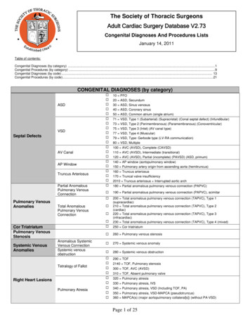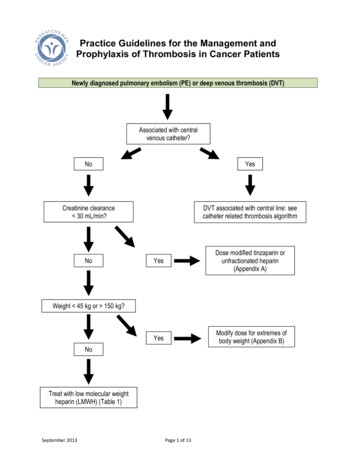
Transcription
Deep Venous Thrombosis,Pulmonary Embolus andIVC FiltersMichael Meuse, M.D.Vascular and Interventional Radiology
Pulmonary Embolism and DVT Estimated 570,000 to 650,000 cases ofsymptomatic PE each year These result in an estimated 200,000deaths per year Anticoagulation is the treatment of choicefor most, but . . .
Heparin and Coumadin Up to 26% of patients will experience acomplication related to anticoagulation As a whole, these anticoagulation relatedcomplications have an associated mortality rateof 5 to 12% 3 to 20% of patients will develop a second PEdespite therapeutic levels of anticoagulation
Heparin and Coumadin:Contraindications Hx of bleeding complication on anticoagulationKnown recent bleeding—GI, CNS, etc.Recent major trauma or surgeryThrombocytopenia—HIT or otherwiseCNS neoplasm, aneurysm, or AVM
Prevention of PE:Historical Interventions Femoral vein ligation– Hunter 1874, Homans 1934– High morbidity, recurrent PE IVC ligation– Ochsner, Debakey, O’Neal 1940’s– Operative mortaility, recurrent PE– High morbidity with venous stasis in 33%
IVC Filters:Absolute Indications Thromboembolic disease and contraindication toanticoagulation Thromboembolic disease and complicationrelated to anticoagulation Recurrent thromboembolic disease despiteanticoagulation Inability to achieve or maintain therapeuticanticoagulation
IVC Filters:Relative Indications Iliocaval DVTLarge, free-floating proximal DVTDifficulty establishing therapeutic anticoagulationMassive PE treated with thrombolysis/thrombectomyChronic PE treated with thromboendarterectomyThrombolysis for iliocaval DVT VTE with limitedcardiopulmonary reserve Recurrent PE with filter in place Poor compliance with anticoagulant medications High risk of complication of anticoagulation (eg, ataxia,frequent falls)
Prophylaxis:Is there a role for IVC filters? The only function of vena cava filters is to preventclinically significant PE by trapping venous emboli. Vena cava filters do not prevent or treat venousthrombosis. Vena cava filters are indicated when primary therapycannot be started, must be stopped, or is insufficient toprotect patients from clinically significant PE who are athigh risk. Filters placed for so-called prophylactic indications donot provide prophylaxis for development of DVT
Prophylactic Filters Usually in the setting of trauma Preoperatively in select patients Data supporting placement in setting ofmalignancy Cannot receive anticoagulation Suffers from one of the following injury patterns:– CHI with low GCS Spinal cord injury– Pelvic and long bone fractures– Multiple long bone fractures
Optional Filters Patients with accepted indications forfilters plus Well defined short period of risk Relatively young age Reasonable life expectancy Avoidance of anticoagulation for sakeof the filter
Non-Permanent Filters: Old Idea1967:
IVC filters:Adjunct to Thrombolysis Overall risk of PE low– 1% in Venous Thrombolysis Registry No proven clinical benefit– Trapped thrombus common Are we brave enough not to place a filter?– No.Mewissen et al Radiology 1999;211:39Yamagami et al Br J Radiol 2003;76:712-713
IVC Filters:Contraindications Chronically thrombosed IVC Inability to obtain direct access to IVC
Decousus et al, NEJM 1998 Only randomized, prospective study(unblinded)– 400 pts with DVT, DVT PE– 200 heparin alone, 200 heparin filter Mortality, recurrent PE at 2 yrs: nodifference
Decousus et al, NEJM 1998 DVT at 2 yrs 20% with filter, 12% without(p .05) PE during first 12 days Rx:– 1.1% with filter, no mortality– 4.8% no filter, ½ lethal– P .05
Decousus 8-Year Update Circulation Recurrent DVT– 34.1% filter group– 27.3% no filter P 0.08– Post–thrombotic syndrome 56% in both groups Symptomatic PE– 6.2% (2 fatal) in the filter group– 15.1% (5 fatal) in the no filter group P 0.01 No survival benefit with filter
IVC filtration:Historical Interventions Mobin-Uddin Filter––––1st intravascular IVC filter – 1967Placed via a surgical venotomyPercutaneous methods introduced in 1973Recurrent PE rate 3%; fatal PE rate 1%
IVC filters:Historical Interventions Stainless Steel Greenfield Filter–––––2nd intravascular IVC filter – 1972Placed via a surgical venotomyPercutaneous methods evolved during the 1980’sInserted through a 29.5 Fr sheathCrafted of medical grade 316 LStainless steel
Historical IVC Filters Stainless Steel Greenfield filter (cont.)– Shape allowed for 89-100% rate of clot trapping– Could perserve laminar flow through the IVCdespite with up to 70% of the cone filled withthrombus– Tilting reduces efficacy of the device (15 degrees)– Migration has been seen in up to 39% - caudal– Strut perforation of IVC reported
Modern IVC Filters Ideal Qualities of IVC Filter– Nonthrombogenic, biocompatible– Infinite implant lifetime and function– Secure fixation– High filtering efficiency– Ease of insertion/delivery– MR compatible– Low cost– Retrievable
Titanium Greenfield Filter -- 1988 12 Fr filter inserted through a 14 OD Frsheath Dimensions: 47mm x 38mm, 0.25 g Crafted of Beta III titanium alloy Inserted from jugular or femoral approach Not inserted over guidewire—tab release ―Modified‖ hooks from original design
Modern Greenfield Filter Insertion site thrombosis of 8.7%Recurrent PE rate of 3.3%IVC thrombosis rate of 1%Left femoral and internal jugularapproaches difficult because of catheterkinking and device tilt
Modern Greenfield IVC Filters Over-the-Wire Greenfield Filter -- 1995– 12 Fr filter inserted through a 14 OD Frsheath– Filter is designed to be deployed over a 0.035wire– System stabilizes the filter reducing theincidence of tilting– Dimensions: 49mm x 32mm– Crafted of 316 L stainless steel– Inserted from a femoral or jugular approach
Bird’s Nest Filter – 1982 14 Fr delivery Sheath Dimensions: two V-shaped stainless steelstruts connected by four 304 stainlesssteel wires each 25 cm long. Deployed in stepwise fashion ―packing‖the filter wires in between the first andsecond set of struts. Can be used in IVCs between 28 mm48mm
Simon Nitinol Filter -- 1990 Small system with deliverycatheter with a 9 Fr OD Dimensions: 45mm x 30 mm Crafted of Nitinol Deployed by unsheathing the device – notover-the-wire Inserted from jugular, femoral, orantecubital approach
VenaTech Filter – 1986 12 Fr sheath with a 14.6 Fr ODLP version uses a 7 Fr sheath with a 9 Fr ODDeployed with a pusher – not over-the-wireHas side rails with hooklets to provide stabilization andcentering Dimensions: 38mm x 30 mm Crafted of Phynox,– non-ferromagnetic alloy– MRI-compatible. Inserted via from jugular orfemoral approach
TrapEase Filter – 2000 Small system with a 6 Fr delivery catheterwith a 8 Fr OD Dimensions: vary depending on cavadiameter 50mm x 35mm. Can bedeployed in cavas up to 30mm indiameter. Crafted of laser-cut Nitinol Advertised as ―idiot proof‖ because devicecannot be deployed backwards
Non-Permanent Filters Temporary– Filter attached to tether– Must be removed Retrievable– Self-affixing to IVC wall– Permanence is optional Convertible– Self –affixing to IVC wall– Device is permanent,but filtration capability is not
Braun Tempo-filterCourtesy Dr Ian Kelly
Currently FDA approvedoptional/retrievable filters Gunter tulip filter (Cook)Celect (next gen Tulip by Cook)Optease filter (Cordis)Recovery/G-2 filter (Bard)ALN Filter (ALN)
Retrievable Filter TypesTulipRecoveryG2OptEase
Gunther Tulip Filter – 2001 8.5 Fr introducer sheath Dimensions: 45mm x 30mm Deployed with release mechanism can bereadjusted at the time of deployment Crafted of Elgiloy and MRI compatiblealloy Permanent or retrievable
Gunther Tulip Filter (cont) Migration rate very low Filter can be retrieved with a loop-snarefrom jugular Approved for 4-6 weeks. Up to 9 monthsretrieval reported.
Gunther Tulip II (Celect) Modification forextended retrieval byopening petals– 12 weeks Decreased tilt withlonger arms– Improve retrievability Early reports ofpenetration of IVCwall.
Tulip Retrieval4 weeks
Recovery Filter – 2003 9 Fr introducer sheath fromfemoral approach Delivery and releasesimilar to Simon Nitinol device Crafted of Nitinol alloy but has a dual array of sixradial arms and legs. The hooklets are designed to allow release ofthe device even after endothelialization hasoccurred Preliminary studies indicate the device can beremoved up to 134 days
Recovery Filter Removal
OptEase (Cordis) Nitinol hypotube Based on TrapEase Inserted from Jugular ,femoral of antecubitalapproach 30 mm IVC Femoral retrieval with snare Removal recommended upto 23 days– Probable 4-6 weeks
OptEase Retrieval
Filter Literature Limited data on permanent IVC filters The available data on optional devices are even morelimited than permanent filters The few published clinical reports suggest that optionalfilters, as a group, are associated with equivalentoutcomes to permanent devices There has never been a clinical study to directlycompare optional and permanent filters
Filter Literature Percutaneous removal of retrievable filters appears to bea safe procedure with few immediate or latecomplications Currently, lack of published human data on convertiblefilters The benefit of discontinuation of filtration has never beenproven Until more data are available, permanent caval filtrationshould always be considered when a filter is indicated
Comparison of IVC Filters Majority of data concerning filter use andeffectiveness is historical and has notbeen performed in consistent manner. Method of follow-up varies widely– Imaging– Clinical evaluation– Telephone interview Duration of follow-up varies widely– 2 weeks, 2 months, 2 years
Comparison of IVC Filters To standardize Filter evaluation two values arecurrently focused upon: Recurrent PE after filterplacement and Thrombosis Recurrent PE after IVC Filter– Clinically symptomatic– Infrequent for all IVC filter devices (2 to 5%)– True value of this is unknown as the rate of asymptomaticPE is still unknown as is its clinical relevance– In this setting must account for PE source: Filter, othermedical device, atypical venous source, or even cardiac– Tilt of the device should also be factored into the evaluation
Comparison of IVC Filters Thrombosis– Includes IVC and access site thrombosis– The rate of IVC thrombosis varies widely 0 to 28% Variablitiy of follow-up Screening of symptomatic vs. all patients Method of follow-up: US, CT, MR, venography– The rate of access thrombosis varies from 2 to35%, much higher for the older devices withlarger profile. Lower profile devices have veryrarely cause access thrombosis.
Is flow stagnation or turbulence linkedto thrombosis and thrombus and/or PEformation?Hemodynamics: Mobin-Uddin and TrapEase filters versus the Greenfieldfilter.Harlal A, Ojha M, Johnston KW. Vena cava filter performance based on hemodynamics and reportedthrombosis and pulmonary embolism patterns. J Vasc Interv Radiol. 2007 Jan;18(1 Pt 1):103-15.
IVC PenetrationPenetration of the IVC by filter hooks or struts is along-term complication that happens relativelyoften, though symptomatic penetration is very rare.Fang W, Hieb RA, Olson E, Carrera GF. Asymptomatic Lumbar Vertebral Erosion from InferiorVena Cava Filter Perforation. Cardiovasc Intervent Radiol. 2007 Feb 2; [Epub ahead of print]
When to Retrieve A Filter? Risk of symptomatic PE becomes low– Patient can be managed with first-line therapy– Patient no longer at risk of thromboembolicdisease Risk of anticoagulation is acceptable Risk of filter exceeds benefit Life expectancy sufficient to realize benefitfrom absence of filter
Guidelines for Retrieval Filter no longer clinically indicated No significant trapped thrombus No prior or present thromboembolic disease Prophylaxis with LMWH Negative screening LE duplex Prior or present thromboembolic disease Full anticoagulation Patient agrees to have filter removed
Filter Retrieval Rosenthal et al documented successful retrievalof forty OptEase filters, with one retrieval at 48days, and all other retrievals performed within 23days. The Jonas study showed that the OptEase filtercan be successfully retrieved up to 14 days. Hoppe et al, reported Gunther Tulip filters wereremoved at a mean of 11.1 days with 100%technical and clinical success rates.
Retrievable Vena Cava Filters inTrauma Patients: An AAST multicenterstudy 152 Gunter-Tulip (G-T), 224 Recovery (R), and 37Optease (Opt). Only 22% of filters were retrieved. The primary reason filters were not removed wasbecause of loss to follow up (31%) 115 patients in whom retrieval was attempted,retrieval failed as a result of technical issues in 15patients (10% of G-T, 14% of R, 27% of Opt) andbecause of significant residual thrombus within thefilter in 10 patients (6% of G-T, 4% of R, 46% Opt).Karmy-Jones R, Jurkovich GJ, Velmahos GC, et al. Practice patterns and outcomes of retrievablevena cava filters in trauma patients: an AAST multicenter study. J Trauma. 2007 Jan;62(1):17-24
UVA experience :From 2002 to 2006, 386 retrievable IVCfilters were deployed at our institution.Only 73 filter retrieval attempts have beenmade (18%). Of the retrieval attempts, 68were successful (93%).
Number ofAttemptedRetrievalsLongestInterval trievalsTulip253 months 4days24 (96%)1 (4%)Recovery268 months 20days26 (100%)0G273 months 9days7 (100%)0OptEase153 months 0days11 (73%)4 (27%)
UVA Experience IVC filter retrieval has been largely successful. 5 of 73 retrieval attempts were unsuccessful:– Hook and filter endothelialization (3).– Extensive thrombus which involved the filter,preventing retrieval (1).– Filter migration to the right ventricle, where the filterbecame tangled in the chordae tendinae, preventingretrieval (1).
Complications: Migration greater than 5 mm: 16%––––Tulip (8%, 2/25)Recovery (12%, 3/26)G2 (71%, 5/7)OptEase (13%, 2/15) Failed Retrievals: 7%, 5 /73 filters.–––– Tulip (4%, 1/25)Recovery (0%, 0/26)G2 (0%, 0/7)OptEase (27%, 4/15)IVC penetration: 1 (Tulip)Bacteremia: 1 (Recovery)Maldeployment: 2 (1 OptEase, 1 Tulip)Pain: 3 (2 G2, 1 Recovery)
Migration occurred with all filter types Most migrations occurred in filters withfree limb construction (G2 and Recovery)– G2 demonstrated migration in 5 of 7 cases,accounting for 5 of the 12 total migrations for all filtertypes– Leading to our institution’s discontinued use of thisfilter.
.
Other reported complications Incomplete opening Filter fracture Filter fragment embolization to thelungs IVC thrombosis
Buried Hook
Fracture of Filter
Fixed penetration of IVC wall
IVC thrombosis
Conclusions Thromboembolic disease can be due totransient risk factors Permanent filters have low but real rate ofcomplications Optional filters allow protection for limited periodof risk Physician judgment important for placement– Crucial for removal
All Optional/Retrievable filters areFDA approved permanent filters Currently no FDA approved temporary filtersavailable in the USA Retrieval is approved by FDA as an option in 5types of filter (since 2002)
Key Points of SIR Consensus report20061. The primary means of therapy and prophylaxis of VTE arepharmacologic2. No unique indications for optional vena cava filters exist that aredistinct from permanent vena cava filters3. Patients with filters in situ should be managed with pharmacologicmethods4. There are no absolute indications for discontinuation of filtrationunless the filter itself is a source of documented major morbidity thatwill be relieved by retrieval or conversion5. Discontinuation of filtration should only occur when the risk ofclinically significant PE is reduced to an acceptable level6. The quality of literature on optional vena cava filters is not sufficientto support evidence-based recommendations at this timeKaufman JA, Kinney TB, Streiff MB, Sing RF, Proctor MC, Becker D, Cipolle M, Comerota AJ, Millward SF, Rogers FB,Sacks D, Venbrux AC. Guidelines for the use of retrievable and convertible vena cava filters: report from the Society ofInterventional Radiology multidisciplinary consensus conference. J Vasc Interv Radiol. 2006 Mar;17(3):449-59.
IVC filters: Historical Interventions Stainless Steel Greenfield Filter -2nd intravascular IVC filter - 1972 -Placed via a surgical venotomy -Percutaneous methods evolved during the 1980's -Inserted through a 29.5 Fr sheath -Crafted of medical grade 316 L Stainless steel