Transcription
Contact Lens Fitting: A Guide and Methodology of Contact Lens FittingISBN: 978-1-946628-15-201
Contact Lens Fitting: A Guide and Methodology ofContact Lens FittingAliki Kantzou*Saera University, Spain*Corresponding authorAliki Kantzou, Saera University, The School of Advanced Education, Research and Accreditation, UniversidadIsabel I, Spain, Email: alikikantzou@yahoo.comPublished By :Juniper publishers Inc.United StatesDate: June 23, 2018
Index1.2.AbstractIntroduction2.1. Theme 1. Contact Lenses Correct Refractive Errorsa)Slit Lamp Examinationc)Contact Lens Fittingb)Topography2.2. Theme 2. Soft Spherical and Soft Toric LensesA.Types of Soft Contact Lensesa)Types of Presbyopia Contact Lenses for Far and Near Vision2.3. Theme 3. Contact Lenses for Presbyopia2.4. Theme 4. Extended Wear Contact Lenses2.5. Theme 5. Rigid Gas Permeable Contact Lenses2.6. Theme 6. Scleral Contact Lenses2.7. Theme 7. Hybrid Contact Lensesi.Hybrid Contact Lenses using New Technology2.8. Theme 8. Cosmetic and Prosthetic Contact Lenses2.9. Theme 9. Therapeutic Contact Lenses2.10. Theme 10. Colored Contact Lenses2.11. Theme 11. Contact Lens Fitting in Aphakic Patients2.12. Theme 12. Contact Lens Fitting in Orthokeratology2.13. Theme 13. Contact Lens Fitting in Children2.14. Theme 14. Contact Lens Fitting in Patients with Corneal Ectasiaa.Pellucid Marginal Degenerationc.Keratoconusb.Keratoglobus2.15. Contact Lenses after Keratoplasty2.16. Contact Lens Fitting after Refractive Laser Procedures2.17. PROSE Contact Lenses2.18. Contact Lenses and Scuba Diving3.4.2.19. Complications of Contact Lens WearConclusionReferences
Contact Lens Fitting: A Guide and Methodology of Contact Lens FittingAbstractPurpose: The objectives of this thesis are to provide fellow Optometrists a guideline on the importance of contact lens fitting, with afocus on its advantages and disadvantages. Topics to be discussed include:a)Types of lenses that can be prescribed to patients.c)Confronting difficulties on contact lens fitting.b)d)e)Consultation with the patient and fitting techniques.The importance of proper contact lens fitting to avoid undesirable effects such as injuries and infections.Theories and practices that Optometrists are advised to follow by accredited professors in the field.Methods: This master thesis focuses on the importance of contact lenses and on ways various problems can be prevented. Severalapproaches are discussed on how vision rehabilitation and comfort can be achieved. The guide outlined in this thesis is based on newresearch, accredited books and articles on indications for use of contact lenses and their proper fitting.Results: Herein, we discuss further that contact lenses are used not only to correct refractive errors, but also to help deter myopiaprogression in the case, for example, of the Ortho-k lenses. We discuss further that contact lenses are of utmost importance in themanagement of aphakia. They do not only help to prevent amblyopia in childhood, but also eliminate patients’ discomfort by reducinganiseikonia and anisometropia. The evaluation of the right contact lens fit is performed by observing lens centration, movement andwhether the lens has ideal position on the cornea. Finally, the use of contact lenses for vision improvement in different cases of cornealectasia is discussed as well.Conclusions: This thesis aims to serve as a guide for Optometrists outlining the proper steps to follow for safe contact lens fitting andoptimal care of patients with various conditions and ailments.04
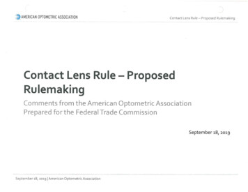
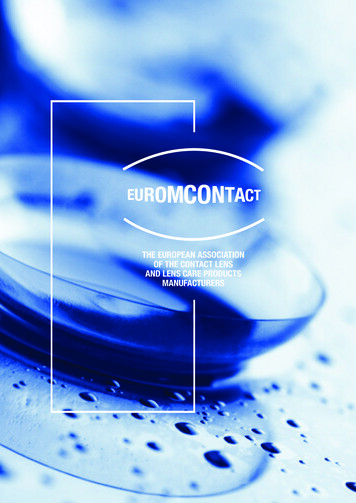
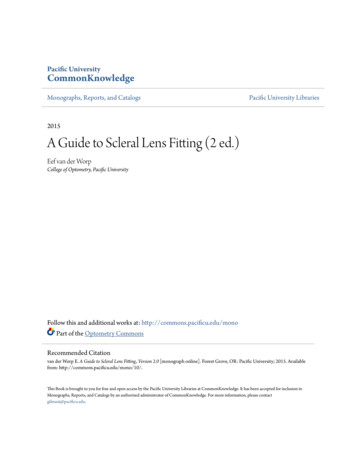
Contact Lens Fitting: A Guide and Methodology of Contact Lens FittingIntroductionVision is the ability to distinguish shapes and colors. The human eye receives an image and transfers it to the brain. Optometristsexamine the eyes to detect disease or refractive errors such as myopia, hyperopia, presbyopia and astigmatism; then the physician proceedswith an anterior and posterior segment examination to diagnose potential disease processes, prescribes eye glasses and/or contact lensesand in some cases recommends eye surgery [1]. The work of the Optometrist is not restricted only to diagnosis and refraction; each caseis treated differently and Optometrists also provide advice on disease prevention and eye health protection. For example, Optometristscounsel patients on how to use contact lenses properly in order to avoid eye injury, how to perform certain eye exercises and, in general,they provide physical and mental support. [2].In 1887, Adolf Fick created the first contact lens, made of glass, to correct irregular astigmatism. However, these lenses could not beworn for a long period of time because they were quite uncomfortable. Contact lenses became convenient for the patients when opticalinstruments were created that could measure the curvature of the cornea [3] (Figure 1).Figure 1: Photograph of two contact lenses [4].A significant number of patients with refractive errors prefer to substitute eyeglasses with contact lenses for aesthetic reasons.Furthermore, patients with high refractive errors prefer contact lenses because contact lenses improve the quality of their peripheralvision.In the mid-1900s, plastic-based contact lenses were introduced. This type of hard contact lenses could only be used for a short periodof time because of irritation of the ocular surface with long term wear. The spherical design of these contact lenses aimed to alter the shapeof the cornea in order to correct refractive errors. “In the 1970s, gas-permeable rigid contact lenses were developed that allowed muchmore oxygen to pass through to the corneal surface, thus increasing comfort and wear time.” [3].In the 1970s, soft contact lenses, known as hydrogels, were developed. Hydrogel lenses allow oxygen to enter the ocular surface, thusmaking them even more comfortable. Another advantage of these contact lenses is the fact that they cannot be easily lost due to their size.The disadvantages are that the material is more sensitive; they need to be taken care of systematically and they are not that effective incorrecting astigmatism [3].According to Davson & Perkins [2], “In 2005 hybrid lenses were developed that are gas-permeable and rigid and surrounded by a softring. These lenses provide the comfort of a soft lens with the visual sharpness of a hard lens.” (Encyclopedia Britannica) (Figure 2).Figure 2: Contact lens being inserted in the eye (Encyclopedia Britannica, Contact Lenses, 2017).05
Contact Lens Fitting: A Guide and Methodology of Contact Lens FittingI.Anatomy (Figure 3)Figure 3: The eye is our organ for sight. The eye has a number of components which include but are not limited to the cornea, iris, pupil, lens,vitreous, retina, choroid and optic nerve [5].A.B.C.D.Cornea: Clear front window of the eye that transmits light and contributes (along with the lens) to light focusing on the retina.Iris: Colored part of the eye that helps regulate the amount of light that enters the eye.Pupil: Dark aperture in the iris that determines how much light is let into the eye.Lens: Transparent structure inside the eye that focuses light rays onto the retina.E.Retina: Nerve layer that lines the back of the eye, senses light, and creates electrical impulses that travel through the optic nerveto the brain.F.Macula: Small central area in the retina that contains special light-sensitive cells and allows us to see fine details clearly.G. Optic nerve: Connects the eye to the brain and carries the electrical impulses formed in the retina to the visual cortex of thebrain.H.Vitreous: Clear, jelly-like substance that fills the middle of the eye [6].Just behind the iris and pupil lies the lens, which helps the light to focus onto the retina. Most of the eye is filled with a clear gel-likesubstance called the vitreous. The inside lining of the eye is covered by special light-sensing cells that are collectively called the retina. Theretina cells convert light into electrical impulses. Behind the eye, the optic nerve carries these impulses to the brain. The macula is a smallextra-sensitive area in the retina that is responsible for central fine-detail vision. The “color” of the eye is determined by the amount andtype of pigment in the iris.II.Eye TreatmentsA.Contact lenses and glasses: They correct common eye problems like nearsightedness, farsightedness, and astigmatism.B. LASIK (Laser-Assisted In-Situ Keratomileusis): A doctor creates a thin flap in the cornea and then uses a laser to reshape it.This procedure improves nearsightedness, excessive farsightedness and astigmatism.C.Radial Keratotomy (RK): The doctor makes a series of small incisions in the cornea to reshape it. Once a common treatment fornearsightedness, it’s rarely used today.D. Photorefractive Keratectomy (PRK): The doctor rubs surface cells off the cornea, then uses a laser to improve nearsightedness,farsightedness, or astigmatism. The cells grow back, and the eye heals the same way it would if it was scratched.E.LASEK (laser epithelial keratomileusis): It’s similar to PRK. The doctor peels back the upper layer of cornea cells and uses alaser to reshape it. The doctor may replace or remove the flap afterwards.F.G.Artificial tears: These eye drops are a lot like the natural tears. They can help treat dry or irritated eyes.Cyclosporine eye drops: This anti-inflammatory eye drop can treat dry eye caused by inflammation.H. Laser photocoagulation: A doctor uses a laser on parts of the retina with poor circulation or to treat abnormal blood vesselsdirectly. It’s often used for diabetic retinopathy but can also seal a retinal tear.I.Cataract surgery: The doctor removes the cloudy cataract and replaces the natural lens with a man-made version [7].Theme 1. Contact Lenses Correct Refractive Errorsa.Contact lenses correct refractive errors that occur in patients. The light rays travel from the cornea, pass through the lens andland on the retina. The electrical impulses formed in the retina travel via the optic nerve to the brain’s visual cortex, which turns them06
Contact Lens Fitting: A Guide and Methodology of Contact Lens Fittinginto an image. This case of no refractive error is called emmetropia. In the presence of refractive errors, the light does not focus on theretina [8].b.Myopia is a condition of blurry far vision. The patients are nearsighted, as they can see very clear in close distance. In myopia thelight rays focus in front of the retina instead of on the retina and the axial length of the eye is longer in size.c.Hyperopia is when the light rays focus on the back of the retina instead of on it. These patients are farsighted with good far visionand blurry near vision. In cases with high hyperopia, far vision will be blurry too.d.Astigmatism is the refractive error in which the light fails to focus on the retina due to the curvature of the cornea. Regularastigmatism types are:i)With-the-rule, when the meridian of the astigmatism is verticaliii)Oblique, when the meridian is between 30 - 60 and 120 - 150 ii)Without-the-rule, when it tends towards the horizontale.Irregular astigmatism is when the curvature varies in different parts of the same meridian. Lenticular astigmatism comes fromthe crystalline lens. Astigmatism can occur by itself and with other refractive errors like myopia, hyperopia and presbyopia.f.Presbyopia arises at about 40 years of age and blurry near vision occurs. The near addition, according to the patient’s age, beginsat about 0.75 D and reaches up to 3.50 D addition. This ADD power is calculated depending on the far refractive error [9].g.In younger ages, the lens changes shape easily for near vision and this is called accommodation. In presbyopia, accommodationdecreases because of the natural hardening of the lens that occurs with aging.h.Prescription for contact lenses is based on subjective refraction, binocular balance, the vertex formula and spherical equivalentcalculation. Subjective over refraction should be evaluated after contact lens fitting. This technique evaluates refractive errors thathave remained when testing patients that are wearing contact lenses. This also applies to children; retinoscopy over the contact lensaims to detect differences that occur from inappropriate vertex correction.i.Contact lens fitting may take several appointments until the proper contact lens is found. Optometrists give the patients guidelinesas to how to insert and remove the lens. Moreover, physicians give tips about the routine wash, the proper care and the storage of thecontact lenses. [10].j.The examiner must have the patient’s medical history as it has great diagnostic value. Information that should be collected incontact lenses wearers includes stability of prescription, glasses, presence of eye diseases, history of operations, environment in whichlenses will be worn, allergies and family eye diseases. A full eye examination is then performed [11].A complete eye examination includes the following:A.Slit Lamp ExaminationSlit lamp examination and biomicroscopy are used as equivalent terms. Slit lamp examination has several built-in magnification lensesthat help verify contact lens fitting. Both an anterior and a posterior segment examination should be performed [12].i)The anterior segment is located in the front of the eye and includes the eyelids, conjunctiva, cornea, limbus, sclera, iris, anteriorchamber, crystalline lens and anterior vitreous.ii) The posterior segment is tested using biomicroscopy lenses. Evaluation of the optic nerve, retina, macula and posterior vitreousis undertaken.A Goldman 3-mirror lens can be used for examination of the retina, vitreous and anterior chamber angle of the eye. Gonioscopy iscrucial in the evaluation of glaucoma as it permits direct visualization of the anterior chamber angle [13].Also, with slit lamp biomicroscopy physicians can check tear film stability by measuring the breakup time (BUT). A value of BUT below5 seconds may indicate dry eye syndrome and a poor tear film [14].Goldmann tonometry can be attached to the slit lamp to measure the eye pressure. The eye pressure is also known as intraocularpressure (IOP). Patients with high intraocular pressure could develop glaucoma with irreversible optic nerve damage and visual field loss.This eye disease usually occurs when the IOP is high, but it may occur in eyes with normal IOP as well [13].Contact lens fitting is estimated with the white light or with fluorescein light and the cobalt blue filter. The examiner observes if thereare areas of staining using fluorescein and prior contact lens wear. Afterwards, the examiner uses contact lenses to observe color patterns.This method points out where the contact lens touches or does not touch the cornea. Patients with meibomian gland dysfunction (MGD)are not good candidates for contact lenses.07
Contact Lens Fitting: A Guide and Methodology of Contact Lens FittingB.TopographyTopography images the surface of the cornea and helps Optometrists determine the types of astigmatism that the patient has. Also, itassesses the shape and power of the cornea. The topography map estimates how flat or steep the cornea is and detects the astigmatismmeridian [15]. Measurements can be conducted using Javal keratometer. The average keratometer measurements are used to select thebase curve and diameter for contact lens fitting.C.Contact Lens FittingThe examiner checks for lens movement in order to assess a flat or steep contact lens fitting. First, the patient looks straight ahead andblinks; in this way the Optometrist observes not only if the lens is in the center, but also evaluates for lens movement. Then, in the slit lampthe contact lens movement in the vertical and horizontal positions is evaluated by having the patient look up, down, right and left. An idealmovement in all positions should be about 0.2 mm to 0.4 mm. The same test is applied for toric lenses and Optometrists have to detect ifthe lens’s mark is stable. The contact lens should be centered in the middle of the cornea and the lens has to cover the entire cornea.The “push up test” is another way to estimate contact lens movement and indicates how loose or tight the lens is fitted. This methodworks when the patient looks up, blinks repeatedly, and then the Optometrist pushes the lens on the lower eye lid using his/her thumb. Inthis way, the lens movement can be assessed: when the lens has a lot of movement the result is 0% and when it falls out of the eye, there isno movement, so the result is 100% [16].Other examinations include: cover uncover test to detect any phoria or tropia, accommodation test, convergence test, subjectiverefraction, cycloplegic retinoscopy, measurement of pupil size, Schirmer tear test, measurement of base curve, keratometry, cornealthickness, corneal diameter and axial length.Theme 2. Soft Spherical and Soft Toric LensesSoft contact lenses are made of a flexible hydrophilic or modern silicone-hydrogel material and are very comfortable. The lenses havehigh oxygen permeability and there are many types in the market; some provide more oxygen than others. Soft contact lenses correctthe refractive errors of myopia, hyperopia, presbyopia and astigmatism. Soft lenses have a wide spherical and toric power range and arecomfortable for the patient. These lenses have a steep or flat base curve depending on keratometry measurements.a)Types of Soft Contact Lensesi.Daily Spherical Contact Lensesiii.Multifocal Contact Lensesii.iv.v.vi.vii.Daily Toric Contact LensesDaily Colored Contact Lens1-2 Week Spherical Contact Lenses1-2 Week Toric Contact Lenses2-Week Multifocal Contact Lensesviii. Monthly Spherical Contact Lensesix.Monthly Spherical Contact Lenses for Astigmatismxi.Monthly Multifocal Toric Contact Lensesx.xii.Monthly Multifocal Contact LensesMonthly Colored Contact Lensesxiii. 6-month up to 12-month Soft Contact lensesxiv. Extended Wear Soft Contact lensesThese lenses are very easy to adjust to and have a larger diameter than rigid gas permeable contact lenses with great stability. Softcontact lenses correct irregular astigmatism and have a large range of power but in advanced stages of keratoconus, in pellucid marginaldegeneration and in keratoglobus Optometrists can recommend other types of lenses [17]. Keratoconus, pellucid marginal degeneration(PMD) and keratoglobus are non-inflammatory eye disorders. The cornea gets thinner and the eye loses the normal round shape, whichcauses vision loss. In pellucid marginal degeneration (PMD) an inferior peripheral cornea ectasia gradually develops.Toric contact lenses are available in soft and rigid gas permeable material. Soft lenses correct astigmatism and they have a lasermark orientation at the 6 o’ clock position to check the contact lens fitting with a slit lamp examination. After 30 minutes of adaptation,Optometrists check the lens centration and lens movements with the push up test and horizontal and vertical lag. In this case, subjective08
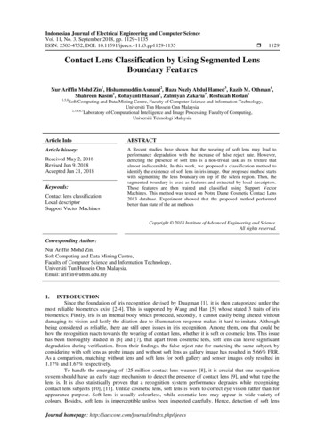
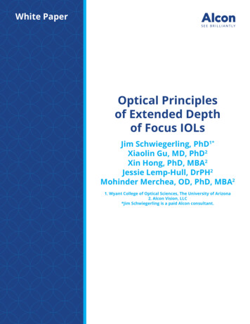
Contact Lens Fitting: A Guide and Methodology of Contact Lens Fittingrefraction is suggested to correct refractive vertical changes [18]. Prisms balance bifocal, multifocal and toric lenses with a vertical basedown prism for the contact lens position and orientation to provide the best visual acuity [19]. This design helps to keep the lens fromrotating and the meridian to stay at the correct position. Toric contact lenses correct simultaneously myopia, hyperopia, presbyopiatogether with astigmatism in different meridians and powers. Toric lenses usually have a vertical 6 o’ clock laser mark, though in themarket, there are different types of laser marks.b)Different laser marks in toric contact lenses (Figure 4)Toric lenses have a front toric design, a back toric design and a bi-toric design. The front design corrects both corneal and phakicastigmatism up to 4.50 D whereas the back design corrects higher astigmatism up to 6.00 D. Bi-toric design contact lenses have a frontand back curve and correct small amount of astigmatism of up to 2.50 D [21]. Research about tips for toric lens fitting states that whenthe lens settles down, usually after 30 minutes, then the lens rotation can be estimated [22]. This evaluation can be accomplished withthe slit lamp and the laser mark of the toric lens. According to Quinn [23], when fitting toric contact lenses, Optometrists should followthe LARS rule and “if the lens rotates left add to the axis and if it rotates right subtract from the axis”. In this way lens rotation can becorrected for up to 8 degrees [24]. In clinical practice, sometimes, this is difficult, so different applications are used to help us with thistask. For example, Optometrists can download the toric lens application of Bausch and Lomb for iPads or smartphones. This smart phoneapplication calculates the rotation and gives the correct contact lens prescription. There are many similar applications that are used inclinical practice which follow a similar principle. First, the lens prescription is inserted and then the mark is moved at the position that thelens is stable at the slit lamp. This will calculate the correct toric lens prescription.Figure 4: Various toric lens marking [20].Theme 3. Contact Lenses for PresbyopiaPresbyopia is a condition in which near vision decreases and is associated with aging. Accommodation decreases at the age of about40 years old and most people start having near vision problems. The crystalline lens gets harder after some years and loses the ability tochange shape and focus in near distance. This is called presbyopia and is a normal condition that occurs with aging. The firsts symptomsare blurry near vision; for example, patients move a book further away in order to have a clearer image. Common symptoms are headaches,fatigue and blurry vision, while better lighting is needed for reading. The addition power increases with advancing age and it depends onthe patient’s preferences and activities. The addition power for near vision in the early stages of presbyopia starts at about 0.75 D andcan reach up to 3.5 D as the patient gets older. The addition power is calculated over the far subjective refractive error and can correctboth presbyopia and astigmatism. In clinical practice, it is required to note the working distance that the patient needs. There are a varietyof materials and designs for presbyopia contact lenses. Contact lenses play a primary role in correcting presbyopia depending on thepatient’s needs. Also, presbyopia contact lenses are desired by patients because they can put aside their near glasses. Patients usually donot want to correct only presbyopia with contact lenses, they want to be able to see clearly both close and far. Several options can be testedto create a comfortable setting for both far and near correction. Material options in presbyopia are rigid gas-permeable (RGP) and softcontact lenses.A.Types of Presbyopia Contact Lenses for Far and Near Visioni)Near vision contact lenses and far vision spectaclesiii)Monovision with contact lensesii)iv)v)vi)Distance contact lenses and near vision spectaclesBifocal contact lensesMultifocal contact lensesMyopia progression with multifocal contact lenses09
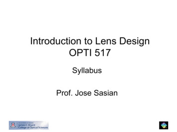
Contact Lens Fitting: A Guide and Methodology of Contact Lens Fittingvii) Contact lenses after cataract operationviii) Contact lenses in aphakiaIn the case that presbyopia is corrected binocularly, with contact lenses, the patient will see blurry in far distance and it will benecessary to use far distance glasses. Contact lenses for distance vision and reading spectacles over such contact lenses can be usedto correct presbyopia in early stages. More often than not, patients do not prefer these methods because they want to be spectacleindependent both for near and far.Another well-known technique is monovision. Using this method, the dominant eye is corrected for far and the non-dominant eye fornear vision. This solution is favorable in the early stages of presbyopia with up to 1.75 D difference in addition between the two eyes.The dominant eye evaluation technique is appropriate in other conditions such as poor visual acuity with multifocal contact lenses.Multifocal contact lenses are a good idea, but sometimes they do not provide perfect vision for both far and near vision. The dominant eyein modern design multifocal contact lenses can correct far distance with a lower addition power and the non-dominant eye can correctnear vision with a higher addition power. Multifocal contact lenses have a modern design and correct areas of far, intermediate and neardistance. Both bifocal and toric lenses have a vertical base-down prism for the contact lens position and orientation to acquire the bestvisual acuity [19]. In both bifocal and multifocal lenses, pupil diameter should be tested in normal light as well as lens centration and it ispreferable to use a flatter fit contact lens [24].Another technique of monovision, which is used by Optometrists, is applying different types of contact lenses to each eye. In one eyethe Optometrist inserts a spherical or spherical contact lens with astigmatism to correct far distance and in the other eye a multifocal lensis used to correct near vision. Also, multifocal contact lenses can correct spherical and astigmatic refractive errors providing best vision.Optometrists prefer soft contact lenses with an aspherical design.Hodd [25], mentions the following multifocal designs:a.With a distance center and near peripheryc.The concentric ring of distance and reading. (Figure 5).b.Near center with a distance peripheryFigure 5: a) The center of this contact lens corrects distance vision and the periphery provides near correction, b) This lens has a near centerand a distance peripheral correction [26].A similar monovision technique is the following: the distance center is corrected at the dominant eye and the near center is chosenat the non-dominant eye. Kassalow [27] mentions, multifocal lenses such as “AIR OPTIX AQUA Multifocal contact lenses are made from asilicone hydrogel material (lotrafilcon B) that provides both high oxygen transmissibility (with a Dk/t of 138 @ -3.00D) and a low lipiddepositing surface” and are available in LOW, MEDIUM and HIGH range addition powers for presbyopia.Optometrists should examine the patient at the beginning with a trial lens and test the patient’s visual acuity binocularly for far andnear vision. Over contact lenses refraction will be estimated in presbyopia with /- 0.25 D or /-0.50 D binocular flippers or trial lenses[25]. Perfect eye-sight in both far and near distance sometimes cannot be accomplished. Some negative aspects are difficulties in binocularfusion and dizziness, especially with monovision and high-power addition. Blurry near vision occurs in patients undergoing a cataractsurgery and in aphakia, because the crystalline lens is replaced or removed. In some occasions in cataract surgery, the physician chooses amonovision technique or a multifocal lens implantation. In such a case, patients generally cover their needs with glasses. Aphakic patientsshould wear contact lenses because they are more stable and provide better quality vision than aphakic spectacles. This is especiallyimportant in children to avoid amblyopia. Contact lenses are necessary to be used; this helps to avoid aniseikonia and anisometropia incases of monocular aphakia. Aphakia in adults and children is usually corrected with extended wear contact lenses. Otherwise, multifocalcontact lenses are a good option for patients who need prescription for near and far distance.010
Contact Lens Fitting: A Guide and Methodology of Contact Lens FittingMultifocal contact lenses are used for myopia control on a daily lens wear schedule. Follow up examination should be planned withintwo consecutive weeks of wearing them. This means that these lenses are appropriate for 8 hours up to 14 hours wear a day. These contactlenses reduce myopia progression in children and in the early teens [28].Theme 4. Extended Wear Contact LensesExtended wear soft contact lenses are worn continuously at night and day for up to 30 days followed by1 night without lens wear.This means that these lenses can be worn while sleeping and have to be removed once a week for cleaning with contact lens solution. Thiscontinuous wear increases the risk of corneal infections [29]. These lenses are suitable for patients with an active life.Patients with a history of cornea infection or inflammation are not good candidates for extended contact lens wear. Suboptimal effectsoccur more often with hydrogel than silicone hydrogel material such as, for example, vascular changes and endothelium polymegathismfrom chronic hypoxia. Kaplan [30] reports that “epithelial cells originate at the limbus and travel across the cornea before sloughing offat the ocular surface a process that might be affected by covering the cornea with a contact lens, possibly inducing hypoxia”. This studyclarifies that extended wear contact lenses decrease oxygen permeability and demonstrates that epithelial thinning leads to infections. Attimes, these lenses are required for therapeutic reasons in cases of corneal diseases and corneal dystrophies. Some examples are: Fuchs’dystrophy, bullous keratopathy, trauma and after some corneal surgeries [30].Extended wear lenses can also be used in cases of corneal scarring and injuries. These lenses correct refractive errors like myopia,hyperopia, astigmatism and presbyopia. Follow up examinations are necessary one week after the trial session, then after two weeks andfinally after six months [21].Theme 5. Rigid Gas Permeable Contact LensesRigid gas permeable contact (RGP) lenses are smaller in size. RGP lenses are fitted within the cornea and the movement of the lensdoes not cross 1mm of the limbus onto the sclera. In clinical practice, tears are provided under the lens as oxygen transmissibility occurs.RGP contact lenses are a safe option to re
2.14. Theme 14. Contact Lens Fitting in Patients with Corneal Ectasia a. Pellucid Marginal Degeneration b. Keratoglobus c. Keratoconus 2.15. Contact Lenses after Keratoplasty 2.16. Contact Lens Fitting after Refractive Laser Procedures . The disadvantages are that the material is more sensitive; they need to be taken care of systematically .