Transcription
Psychoneuroendocrinology (2015) 55, 59—71Available online at www.sciencedirect.comScienceDirectjournal homepage: www.elsevier.com/locate/psyneuenRegional volumes and spatial volumetricdistribution of gray matter in the genderdysphoric brainElseline Hoekzema a, ,1, Sebastian E.E. Schagen c,e,1,Baudewijntje P.C. Kreukels b, Dick J. Veltman d,Peggy T. Cohen-Kettenis b, Henriette Delemarre-van de Waal e,2,Julie Bakker a,faNeuroendocrinology Bakker Group, Netherlands Institute for Neuroscience, Amsterdam, The NetherlandsCenter of Expertise on Gender Dysphoria, Department of Medical Psychology, Neuroscience CampusAmsterdam, VU University Medical Center, Amsterdam, The NetherlandscDepartment of Pediatric Endocrinology, Neuroscience Campus Amsterdam, VU University Medical Center,Amsterdam, The NetherlandsdDepartment of Psychiatry, Neuroscience Campus Amsterdam, VU University Medical Center, Amsterdam,The NetherlandseDepartment of Pediatrics, Leiden University Medical Center, Leiden The NetherlandsfCentre Interdisciplinaire de Géneoprotéomique Appliquée, Université Liège, Liège, BelgiumbReceived 12 September 2014; received in revised form 29 December 2014; accepted 21 January 2015KEYWORDSMRI;Transsexual;Transsexualism;Gender identitydisorder;Voxel-basedmorphometry;Sex differences 12Abstract The sexual differentiation of the brain is primarily driven by gonadal hormonesduring fetal development. Leading theories on the etiology of gender dysphoria (GD) involvedeviations herein. To examine whether there are signs of a sex-atypical brain developmentin GD, we quantified regional neural gray matter (GM) volumes in 55 female-to-male and 38male-to-female adolescents, 44 boys and 52 girls without GD and applied both univariate andmultivariate analyses. In girls, more GM volume was observed in the left superior medial frontalcortex, while boys had more volume in the bilateral superior posterior hemispheres of thecerebellum and the hypothalamus. Regarding the GD groups, at whole-brain level they differedonly from individuals sharing their gender identity but not from their natal sex. Accordingly,using multivariate pattern recognition analyses, the GD groups could more accurately be automatically discriminated from individuals sharing their gender identity than those sharing theirnatal sex based on spatially distributed GM patterns. However, region of interest analyses indicated less GM volume in the right cerebellum and more volume in the medial frontal cortex infemale-to-males in comparison to girls without GD, while male-to-females had less volume inCorresponding author. Tel.: 31 0204444943.E-mail address: e.hoekzema@nin.knaw.nl (E. Hoekzema).Shared first yneuen.2015.01.0160306-4530/ 2015 Elsevier Ltd. All rights reserved.
60E. Hoekzema et al.the bilateral cerebellum and hypothalamus than natal boys. Deviations from the natal sex withinsexually dimorphic structures were also observed in the untreated subsamples. Our findings thusindicate that GM distribution and regional volumes in GD adolescents are largely in accordancewith their respective natal sex. However, there are subtle deviations from the natal sex in sexuallydimorphic structures, which can represent signs of a partial sex-atypical differentiation of thebrain. 2015 Elsevier Ltd. All rights reserved.1. IntroductionDuring a critical period of fetal and neonatal development,gonadal hormones are thought to primarily drive the sexualdifferentiation of the brain, sculpting brain morphology intoa gender-typical configuration. In genetic males, the testessecrete high levels of testosterone during fetal life, rendering serum levels in the adult male range. Testosterone canbe converted to 17- -estradiol by the enzyme aromatase,and animal studies have demonstrated that pre- and perinatal estradiol acts as the principal regulator of the sexualdifferentiation of the rodent brain (Bakker and Baum, 2008;McCarthy, 2008). Cerebral masculinization is precluded ingenetic females by the estrogen-binding activity of alphafetoprotein, a plasma protein secreted by the fetal liver inhigh concentrations (Bakker et al., 2006; Bakker and Baum,2008). Manipulating the exposure to sex steroid hormonesduring a critical period of prenatal and early postnatal lifesubstantially perturbs the sex-specific differentiation of thebrain (Bakker et al., 2006; Bakker and Baum, 2008).In humans, pre- and perinatal neuroendocrine factorsare also thought to primarily regulate the sexual differentiation of the brain. Interestingly, individuals (46,XY) withcomplete androgen insensitivity syndrome, who have nonfunctional androgen receptor proteins due to a mutationin the AR gene, are born phenotypically female, are sexually attracted to men and have a female gender identity(Wisniewski et al., 2000; Hines et al., 2003), suggestingthat in humans masculinization may be directed by testosterone rather than testosterone-derived estradiol. Althoughthe sexual differentiation of the brain is believed to primarily take place during prenatal development, circulatinghormones during adolescence exert activational effects andfurther potentiate neural sexual dimorphisms. In additionto gonadal hormone effects, direct effects of genes residingon the sex chromosomes may also contribute to the sexualdifferentiation of the brain (Arnold, 2004; Carruth et al.,2002).In gender dysphoria (GD) there is a persistent incongruence between a person’s natal sex and the experiencedgender identity. Leading theories on the etiology of thiscondition involve a sex-atypical cerebral programming thatdiverges from the sexual differentiation of the rest of thebody, postulated to reflect the organizational effects ofaltered levels of sex steroid hormones during a specificperiod of fetal development (Bao and Swaab, 2011; CohenKettenis and Gooren, 1999; Savic et al., 2010; Swaab, 2007).Some twin studies suggest a role for genetic factors in thedevelopment of GD, potentially involving polymorphisms ingenes encoding elements of the sex steroid signaling ormetabolic pathways (Heylens et al., 2012).Postmortem studies have observed sex-atypical volumesand neuronal numbers in hypothalamic nuclei in the brainsof individuals with GD (Garcia-Falgueras and Swaab, 2008;Kruijver et al., 2000; Zhou et al., 1995). Brain structure inindividuals with GD has also been investigated in vivo. MRIstudies examining white matter tracts using diffusion tensor imaging have shown deviations from the natal sex inwhite matter microstructure in both female-to-males (FM)and male-to-females (MF) (Rametti et al., 2011a,b). A fewneuroimaging studies have investigated gray matter (GM)in individuals with GD, observing both regional volumetric properties in line with the natal sex and others in linewith the gender identity of the GD groups (Luders et al.,2009b, 2012; Savic and Arver, 2011; Zubiaurre-Elorza et al.,2012; Simon et al., 2013). A recent study by Zubiaurre-Elorzaet al. (2012) was the first to examine GM in FM (ZubiaurreElorza et al., 2012). Interestingly, they observed signs ofsubcortical masculinization in female-to-male adults and ofcortical feminization in male-to-female adults, providingthe first insights into the developmental processes underlying GD in both sexes.In our study, we wanted to examine brain anatomy in individuals with GD during adolescence, when puberty-relatedsex steroid hormones are driving the further ‘activational’sexual differentiation of the brain rather than in adulthood, when both organizational and activational steroidhormone effects have already sculpted the brain into asex-specific configuration. Therefore, we acquired highresolution anatomical brain MRI scans of 55 female-to-maleadolescents and 38 male-to-female (MF) adolescents withGD. In addition, to allow comparisons with individuals sharing either their gender identity or their natal sex, we alsoscanned 44 boys and 52 girls without GD going through adolescence.First, we aimed to define sexually dimorphic structuresin the adolescent brain by comparing GM volume in boysand girls without GD. As the principal aim of our studywas to examine whether signs of sex-atypical cerebral programming are present in the brains of GD adolescents, wesubsequently compared their regional GM volumes to individuals sharing either their gender identity or their natalsex. To ensure that the deviations from the natal sex did notresult from hormone therapy, we repeated these analysesin the subsamples of GD participants who had never beenexposed to any form of hormonal treatment.Finally, considering the recent development of sophisticated machine learning algorithms for MRI analyses, wealso wanted to explore the data beyond the classicalmass-univariate statistical approach. Especially relevant forthe study of individuals with GD is that multivariate pattern recognition approaches provide a tool to quantify the
Regional volumes and spatial volumetric distribution of gray matter in the gender dysphoric brainpredictive value of volumetric GM patterns for group discrimination. Therefore, the final aim of our study was toexamine these data using a multivariate approach and assessthe extent to which individuals with GD can be discriminatedfrom either of the control groups based on the spatiallydistributed patterns of GM volume in the brain.2. Materials and methods2.1. SubjectsSubjects for this study were recruited by the Center ofExpertise on Gender Dysphoria of the VU University Medical Center in Amsterdam as part of a larger researchproject investigating brain development, brain functioning,growth and metabolic aspects in the clinical managementof adolescents with GD (Trial registration number ISRCTN81574253 (http://www.controlled-trials.com/isrctn/)). Alladolescents with Gender Identity Disorder were evaluatedaccording to the standard protocol at the clinic using thecriteria described in the Diagnostic and Statistical Manualof Mental Disorders-IV-TR (DSM-IV-TR) (American PsychiatricAssociation, 2000). Please note that, in this version of theDSM, ‘Gender Identity Disorder’ was still the official nameof the diagnosis. Therefore, only in the context of describing the diagnostic procedure we will use this term ratherthan the more recent ‘Gender Dysphoria’. When participants were referred to the clinic during childhood, the childand parents participated in several sessions with a mentalhealth care professional specialized in children and adolescents with Gender Identity Disorder. During these sessions,the aim was to determine whether the criteria for a GenderIdentity Disorder diagnosis in children according to DSM-IVTR were met, to evaluate the cognitive, psychological andsocial functioning of the child and functioning of the family,and, if indicated, to offer or refer for psychological treatment or counseling. The core criteria for a DSM-IV-TR GenderIdentity Disorder diagnosis are a strong and persistent crossgender identification and a persistent discomfort with his orher sex or sense of inappropriateness in the gender role ofthat sex. As a result, the child should experience a significantamount of distress or impairment in important areas of theirfunctioning. In adolescence, the individuals (re-)applied tothe clinic, where they entered the diagnostic procedure foradolescents. This implied an evaluation of the presence ofGender Identity Disorder in adolescents/adults and otherpsychopathology according to DSM-IV-TR criteria.The current analyses were performed several yearsafter data acquisition, which allowed us to ascertain thatthe sample consisted only of individuals with persistentGD. In our study, we included only participants who hadbeen followed at the center for many years and who hadindeed completed or were in the last phase of cross-sexhormone treatment at the time of the analyses rather thanrelying on participants’ declaration of intending to undergoprogressive treatment steps to change their body to fittheir gender identity. Two individuals had to be removedfrom the analyses as it became clear at some point afterthe data acquisition that their GD had not persisted intoadulthood. Our final sample consisted of 54 female-to-male61adolescents, 37 male-to-female adolescents, 44 natal maleand 52 natal female adolescents without GD.After receiving the diagnosis of Gender Identity Disorder in adolescence and meeting some additional criteriato be eligible for treatment (Kreukels and Cohen-Kettenis,2011), they were referred for hormonal treatment. Thestandard for treatment in the clinic follows the Standardsof Care of the World Professional Association for Transgender Health (WPATH, Coleman et al., 2011), a professionalorganization in the field of transgender care, and theClinical Practice Guidelines of the Endocrine Society published in 2009 (Hembree et al., 2009), comprising pubertysuppression with gonadotrophin releasing hormone (GnRH)analogues once adolescents start to display the first physical and endocrine changes of puberty (Tanner stage 2—3),followed by cross-sex hormone therapy after the age of16. From 18 years of age onward, individuals with GD canbe referred for sex reassignment surgery. After gonadectomy, GnRH analogues are no longer provided. Treatmentwith cross-sex hormones is continued after sex reassignmentsurgery. For additional information regarding the standardsfor diagnostics and treatment of adolescents with GD, see(Hembree et al., 2009; Kreukels and Cohen-Kettenis, 2011).Please see Table 1 for demographic and clinical details ofthe sample.Depending on the age of the participant, intelligence wasestimated with the Wechsler Intelligence Scale for Children(WISC-III) (Wechsler, 1991) or the Wechsler Adult IntelligenceScale (WAIS-III) (Wechsler, 1997). All participants also underwent a physical examination by a clinician to determineheight, weight, blood pressure and puberty stage accordingto Tanner (1962). Tanner staging, which defines the progressof puberty based on the development of secondary sexcharacteristics, is considered the gold standard for pubertymeasurement when performed by a trained clinician (Dorn,2006).The FM were all gynephilic, whereas the MF wereandrophilic (i.e. sexually attracted to partners of their natalsex), with the exception of one of the MF who reported to beindifferent with regard to the sex of the partner. Repeatingthe analyses without this subject did not affect our results.All GD participants had started to experience GD symptoms in childhood and met criteria for ‘early-onset’ genderdysphoria. Rather than assessing whether symptoms hadcommenced before puberty by retrospective questionnaires,we were able to ensure that our participants’ GD hadstarted in childhood as they had enrolled in our clinic atan early age and had been clinically evaluated for manyyears.No participants with any known physical inter-sex conditions, neurological disorders, cerebral damage or majormedical conditions were included in this study. To assesspsychological functioning in all groups, the Dutch translationof the Child Behavior Checklist (Achenbach and Edelbrock,1983; Verhulst et al., 1996) was used. In case of scores inthe clinical range indicating problem behavior, participantswere not included in the study.The study was approved by the Ethical Review Boardof the VU Medical Center. Written informed consent wasobtained from the subjects and from the parents orguardians of the child participants prior to their participation in the study.
62Table 1 Clinical and demographic data. GnRH: GD individuals undergoing treatment with GnRH analogues but not cross-sex hormones at the time of the MRI acquisition.Cross-sex: GD individuals undergoing cros-sex hormone therapy.Group (N)M (44)F (52)FM (54)MF (37)***Treatmentstage (N)Untreated(17)GnRH (16)Cross-sex(21)*CompletesampleUntreated(11)GnRH (14)Cross-sex(12)*CompletesampleAge (M SD)(range)Tanner (M SD)(range)IQ (M SD) (range)Duration GnRH(days) (M SD)(range)Duration cross-sex(days) (M SD)(range)Cumulative dosiscross-sex** (mg)(M SD) (range)16.42 2.75 (10.54) 4.35 1.09 (3)16.29 2.96 (11.49) 4.55 0.76 (3)15.20 2.76 (9.26) 4.12 1.15 (3)110.5 19.88 (85)105.19 16.32 (60)101.82 14.65 (50)15.88 1.54 (4.27)19.11 1.29 (4.90)3.62 1.41 (4)2.86 1.88 (4)93.31 14.71 (49)105.29 17.27 (51)611.31 435.22 (1435)1595.67 785.92 (3328)——1025.25 436.21 (1586) 9270.38 9616.43 (39,082)16.92 2.84 (10.24) 3.47 1.61 (4)100.65 16.24 (58)1170.00 816.39 (3796)1025.25 436.21 (1586) 9270.38 9616.43 (39,082)13.77 2.42 (7.99)3.45 1.13 (3)112.45 22.71 (70)15.29 0.73 (2.22)19.04 2.17 (7.11)3.71 0.99 (2)4.17 0.84 (2)93.86 10.75 (38)107.83 18.97 (58)692 247.65 (952)1619.75 398.01 (1232)——911.76 497.02 (1352) 1658.17 1133.68 (3867)16.05 2.84 (11)3.78 01.00 (3)103.92 19.02 (70)1137.32 571.89 (2141)911.76 497.02 (1352) 1658.17 1133.68 (3867)————————————Four of the MF and nine of the FM had undergone sex reassignment surgery.FM: testosterone (Sustanon), and MF: estradiol (Progynova/Meno-implant).E. Hoekzema et al.
Regional volumes and spatial volumetric distribution of gray matter in the gender dysphoric brain2.2. MRI acquisitionsThe MRI images were acquired with a 3 T Philips Intera scanner, equipped with a SENSE 6-channel head coil. To obtain ahigh-resolution anatomical brain image, a T1-weighted pulsesequence was applied using the following acquisition parameters: TR 9 ms, TE 3.5 ms, matrix size 256 256, voxelsize 1 1 1 mm, and number of slices 170.2.3. Univariate analysesMRI image processing and statistical analyses were conducted with Statistical Parametric Mapping software (SPM8;www.fil.ion.ucl.ac.uk-spm), implemented in Matlab (version7.8; www.mathworks.com).Regional GM volume was quantified using a voxelbased morphometric approach, a fully automated techniquedesigned to evaluate local volumetric properties of brainanatomy (Ashburner and Friston, 2000). We used theanalysis pipeline provided in the Gaser vbm8 toolbox(http://dbm.neuro.uni-jena.de/vbm/). With this toolbox,the anatomical images were segmented into different tissueclasses using an adaptive maximum a posteriori approachcorrected for field inhomogeneities and brought intostandard space using affine and non-linear normalization.Since we worked with a sample of adolescent participants, we created a customized sample-specific DARTELtemplate for our study. First, to obtain suitable initial agematched tissue probability maps for import into DARTEL,we generated a customized age-matched template and tissue priors using the TOM8 toolbox (https://irc.cchmc.org/software/tom.php) and used these in an affine registration.The affine registered gray matter and white matter segments were then used to create a sample-specific DARTELtemplate, which was then implemented in a DARTEL normalization to warp our data into MNI space. To preserve originaltissue volumes and allow comparisons of volumes rather thanGM concentrations, we used the GM segments that had beenmultiplied by the Jacobian determinants extracted from thenormalization step. Finally, an 8 mm full width at half maximum smoothing kernel was applied to the data. An absolutethreshold of 0.2 was applied to the analyses, and total GMvolume of the brain was included as a nuisance covariate.As there was a significant difference in IQ betweenthe groups (M SD, M: 110.50 19.88, F: 105.19 16.32,MF: 103.92 19.02, FM: 100.66 16.24. F 2.54, p 0.058),IQ was included in the between-group comparisons. Weobserved no correlations between regional GM volumes andIQ within the groups. When subgroups of the total sample were compared causing differences in age between thegroups to surface, then age was also included as a covariateof no interest.The whole-brain results are reported at a thresholdof p 0.05 FWE-corrected and an extent threshold ofk 10 voxels. Moreover, to examine whether more subtledeviations from the natal sex could be observed within sexually dimorphic brain structures, regions of interest (ROIs)were created representing these regions. ROI analyses wereperformed in Marsbar (http://marsbar.sourceforge.net/).Considering our hypothesis that individuals with GD haveundergone a sex-atypical differentiation of the brain that63diverges from their natal sex in the direction of the cerebral developmental processes typical for the other sex, theROI of larger female volume was applied to the contrasts‘F MF’ and ‘MF M’, and the ROIs of larger male volumewere applied to the contrasts ‘MF F’ and ‘FM F’. The pvalues for the ROI results are Bonferroni-corrected for thenumber of ROIs.In addition to analyses involving the complete sample, wealso separately compared the GD individuals who had neverbeen exposed to any form of hormonal therapy (excludingboth the individuals whose puberty was suppressed by meansof GnRH analogues and the GD participants who had initiated cross-sex hormone therapy at the time of the MRIacquisition) to age-matched control samples of both sexes.To minimize the contribution of puberty-related endogenoussex steroid hormones and focus primarily on organizationaleffects, the stage of pubertal development (as an index ofthe degree of puberty-related endogenous sex steroid hormones the individual had been exposed to until the time ofthe MRI acquisition) was included as a nuisance covariate inthese analyses.Furthermore, we also compared untreated FM and MFto the GD participants who had already initiated cross-sexhormone therapy at the time of MRI acquisitions.2.4. Multivariate analysesIn addition to the above-described mass-univariate statistical approach, we also performed a pattern recognitionanalysis. This is a method that stems from machine learning, and uses a multivariate approach to investigate spatiallydistributed information in MRI data — which is intrinsically multivariate — and hereby allows modeling signalpatterns in the images. In our study, we used the analysispipeline for pattern recognition analyses provided by PRoNTo(http://www.mlnl.cs.ucl.ac.uk/pronto). This pipeline canbe used to automatically search for regularities in the dataand train a classifier function that models the relationbetween spatial signal patterns and experimental factors(in our case, group) based on a training dataset (Schrouffet al., 2013). This classifier can then be used to predict thegroup a new image belongs to using the spatial distributionof the signal within the image, and compute the accuracywith which groups can be discriminated from one anotherbased on whole-brain spatial signal patterns. We selectedthe binary support vector machine (SVM) and a leave-oneout cross-validation strategy. This cross-validation strategycomputes the accuracy of classifiers by leaving one subject out at a time and predicting the group label of thissubject based on a training dataset including all remainingsubjects, which is repeated for each subject. The results ofall these runs are then used to compute the accuracy of thediscriminant function. No covariates were included in themultivariate analyses. First we trained and tested a linearSVM pattern classifier for the natal boys and girls withoutGD, to assess the accuracy with which they could be discriminated from one another based on the distribution of GMvolume. Then, we repeated these analyses for the GD groupsand individuals sharing either their gender identity or theirnatal sex. These multivariate analyses were also repeatedincluding only those MF and FM who had never been exposed
64E. Hoekzema et al.Table 2 GM group comparisons. Full results of the whole-brain comparisons between adolescent boys and girls and between theGD groups and adolescents sharing their gender identity, reported at a whole-brain statistical threshold of p 0.05 FWE-correctedand an extent threshold of 10 voxel.ComparisonBrain regionMNI coordinatesxComparisons boys and girls without GDM FL Cerebellum 42R Cerebellum414532Hypothalamus6F ML Superior Medial 8Frontal Cortex 8yz 76 46 73 37263 20 29 20 26 2226915Comparisons GD to groups sharing gender identityF MFR Superior Medial5718Frontal/R SuperiorFrontal Cortex596MF FL Cerebellum 15 48M FML Cerebellum/L 42 78FusiformFM M—Cluster size (mm3 )TP 2 0.0010.0020.0030.0010.0065.020.009243206331646.01 0.00137 59 1847545.014.804.860.0060.0220.017Comparisons GD to groups sharing natal sexM MF—MF M—F FM—FM F—to any hormonal treatment and their age-matched controls.Balanced accuracy values, which represent the fraction ofthe subjects for whom a correct class label was predictedusing the trained classifier, are provided for each of the comparisons. Permutation tests were used to examine whetherthe classifier performed above chance level.3. Results3.1. Univariate results3.1.1. Complete sampleTo identify regions of sexual dimorphism in the adolescentbrain, we first compared regional GM volumes (correctedfor total GM volume) between girls and boys without GD.These whole-brain analyses (thresholded at p 0.05 FWEcorrected and an extent threshold of k 10 voxels) renderedthree localized clusters of larger GM volumes in boys andone cluster where girls had more GM volume (see Table 2).More specifically, in girls, enlarged volume was observed ina section of the left frontal cortex extending from the superior part of the medial frontal cortex to the superior frontalcortex. For the boys, on the other hand, we observed clusters of relatively larger GM volume in the hypothalamus andin the bilateral cerebellum, covering primarily the superiorposterior lobes of the cerebellar hemispheres.Regarding the comparisons involving the GD groups, wefirst compared the adolescents with GD to individuals sharingtheir gender identity. In comparison to natal boys, femaleto-male adolescents were found to have relatively smallervolumes in a cluster extending from the left fusiform gyrusto the superior posterior cerebellum. When comparing MFto natal girls, we observed smaller GM volumes in the rightsuperior frontal and superior medial frontal cortex, andenlarged left cerebellar volume. The results of these comparisons are indicated in Table 2 and the whole-brain resultsare depicted in Fig. 1.Next, we compared the GD groups to individuals sharingtheir natal sex. For these comparisons, no supra-thresholdclusters surfaced at whole-brain level, suggesting that overall the regional GM volumes of MF and FM are largely inaccordance with those observed in their natal sex. To further evaluate the degree of masculinization or feminizationof GM in individuals with GD, we compared GM volumesspecifically within sexually dimorphic regions, by applying Regions of Interest (ROIs) representing the previouslydescribed regions of sex-typical volume (the ROIs of largervolume in females to the contrasts ‘MF M’ and ‘F FM’ andthose of larger male volume to ‘M MF’ and ‘FM F)’.For these comparisons, we detected less volume in theright superior medial frontal cortex and more volume inthe right cerebellum in FM relative to girls without GD(see Table 3). Regarding MF, when examining GM volumes
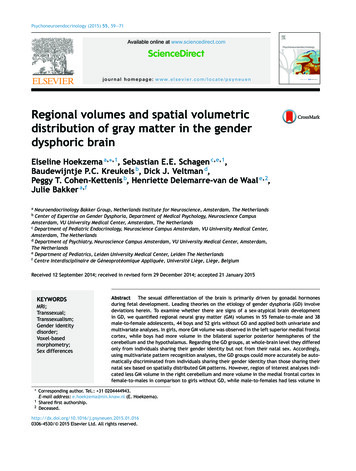
Regional volumes and spatial volumetric distribution of gray matter in the gender dysphoric brain65Fig. 1 Whole-brain results. Illustration of all the whole-brain results (see Table 2). For illustrative purposes, this figure displaysthe results at p 0.0001 (instead of p 0.05 FWE-corrected) to make them stand out more from the background in the figure.M males without GD; F females without GD; MF males-to-female individuals with GD; FM females-to-male individuals with GD.Table 3 ROI results. Full results of the Marsbar ROI analyses comparing the GD groups and their respective natalsex.RegionComplete samplesFM FL CerebellumR Cerebellum**HypothalamusF FML superiormedial frontalcortex**MF ML superiormedial frontalcortex*M MFL Cerebellum**R Cerebellum**Hypothalamus**Untreated samplesFM FL CerebellumR Cerebellum**HypothalamusF FML superiormedial frontalcortex**L superiorMF Mmedial frontalcortexM MFL Cerebellum**R CerebellumHypothalamus****trend at p 0.1.Significant at p 0.05,TCorrected .68510.0765within these sexually dimorphic regions, we observed atrend for increased left superior medial frontal volume andsignificantly less volume in the bilateral superior posteriorcerebellum and hypothalamus in comparison to boys withoutGD.As a supplementary analysis, we also volumetrically compared the two GD groups to the individuals without GD inorder to assess whether there were any alterations in GMvolume accompanying GD (independent of sex), but no significant results surfaced for this comparison.3.1.2. Untreated sampleTo examine whether these deviations from the natal sexare not driven by exposure to the hormone therapy alreadyprovided to a subsample of our GD participants, we alsorepeated these comparisons including only GD adolescentswho had not yet undergone any hormonal treatment at thetime of the MRI acquisition and age-matched controls oftheir natal sex. To minimize the contribution of ‘activational’ effects of puberty-related sex steroid hormones, thestage of pubertal development was also included in thesemodels as a nuisance covariate as an indication of the degreeto which the body had already been exposed to pubertyrelated sex steroid hormones.In line with the findings in the complete sample, weobserved no deviations from the natal sex in either the FMor MF at whole-brain level (p 0.05 FWE-corrected). However, when examining GM volumes within sexually dimorphicbrain structures using ROI analyses, we observed differencesin both GD groups with respect to their natal sex. Morespecifically, in FM, less GM volume was observed in the leftsuperior medial frontal cortex in comparison to adolescentgirls, while more GM volume
treatment steps to change their body to fit their gender identity. Two individuals had to be removed from the analyses as it became clear at some point after the data acquisition that their GD had not persisted into adulthood. Our final sample consisted of 54 female-to-male adolescents, 37 male-to-female adolescents, 44 natal male