Transcription
Voronina et al. SpringerPlus (2016) 5:1584DOI 10.1186/s40064-016-3213-2Open AccessRESEARCHDesign of a stable cell line producing arecombinant monoclonal anti‑TNFα antibodybased on a CHO cell lineE. V. Voronina1*, Y. A. Seregin1, N. A. Litvinova1, V. I. Shvets2 and R. R. ARMAPARK LLC, Bldg. 1,8 Nauchny proezd, Moscow,Russian FederationFull list of author informationis available at the end of thearticleAbstractRecombinant monoclonal antibodies (mAbs) against tumor necrosis factor alpha arewidely used in the biopharmaceutical therapy of autoimmune diseases. Currently,a large number of drugs based on these antibodies are available. Accordingly, thedevelopment of these products for the Russian market is an important goal. The aim ofthe current study is to describe the development of one such technology. CHO-DG44derived cell lines producing mAb were developed using two strategies, one based onindividual clones and the other based on cell pools. To obtain recombinant cell lineswith highly amplified genes of interest, the clones underwent dihydrofolate reductasemediated gene amplification. Using the best strategy for the selection and amplification of mAb-producing clones, we achieved the production of more than 1 g/L in smallscale, non-optimized conditions.Keywords: Monoclonal anti-TNFα antibody, Tumor necrosis factor alpha, CHO-DG44,Dihydrofolate reductase mediated gene amplificationBackgroundGenetically engineered biological agents designed from monoclonal antibodies (mAbs)have been a leading technology for pharmacotherapy in the last decade (Kalden 2002).The introduction of targeted therapy based on mAbs has promoted many important scientific achievements in rheumatology (Alonso-Ruiz et al. 2008). Specifically, recognitionof the role of tumor necrosis factor alpha (TNF-α) in the pathogenesis of inflammatoryrheumatic diseases has expanded the scope of drugs for these diseases (Alonso-Ruizet al. 2008; Schiff et al. 2005).Currently, adalimumab (otherwise known as Humira) is the representative TNF-αblocker for the treatment of rheumatoid arthritis (RA) and other common inflammatory arthropathies (Furst et al. 2007; Nasonov 2007). This drug is a completely humanantibody that determines its reduced immunogenicity, which is manifested particularlyin the reduction of allergic reactions (Furst et al. 2007; Nasonov 2007; Scott and Kingsley 2006; Wurm 2004; Chu and Robinson 2001). Worldwide, adalimumab has largelybeen successful in the treatment of inflammatory rheumatic diseases, including in multiple random clinical trials (Scott and Kingsley 2006). In the USA, adalimumab has beenapproved for the treatment of RA, psoriatic arthritis, ankylosing spondylitis, Crohn’s 2016 The Author(s). This article is distributed under the terms of the Creative Commons Attribution 4.0 International /), which permits unrestricted use, distribution, and reproduction in any medium,provided you give appropriate credit to the original author(s) and the source, provide a link to the Creative Commons license, andindicate if changes were made.
Voronina et al. SpringerPlus (2016) 5:1584disease, ulcerative colitis, and chronic plaque psoriasis, but in Russia, this drug has onlybeen registered for the treatment of RA. In addition, the annual sales volume of Humirain the USA is about 2.6 billion (Scott and Kingsley 2006), highlighting its importance asa commonly prescribed drug.Part of a strategy to develop the pharmaceutical industry in Russia includes reducingthe dependence of the Russian market on imported drugs and improving the availability of new drugs. The development of modern high-technology production of domesticdrugs based on mAbs is a challenge for the Russian medical community. Therefore, theaim of this study was to obtain a stable cell line producing a mAb for a TNF-α blockerand to characterize the purified antibody in comparison with the drug Humira (AbbVie,USA).This paper is describing the development of a cell line based on Chinese Hamster (Cricetulus griseus) Ovary (CHO) cells. Among mammalian cell lines used in biopharmaceutical production, CHO cells and their derivatives are the most ubiquitous (Urlauband Chasin 1980; Derouazi et al. 2006).For the production of therapeutic proteins, the most widely used mammalian expression system in industrial production is gene amplification using dihydrofolate reductasedeficient (DHFR ) CHO cells with DHFR-mediated gene amplification. Methotrexate(MTX) binds to and inhibits the DHFR enzyme, leading to cell death. However, DHFR CHO cells, being transfected with an expression vector containing the DHFR gene, candevelop resistance to MTX. Because the amplification unit consists a specific gene ofinterest, either co-linked to DHFR in the same expression vector or residing adjacentlyin the host chromosome, is co-amplified (Cacciatore et al. 2010).Because single-step, high-level resistance to MTX may result in cells synthesizing anMTX-resistant DHFR mutant, or in cells with altered MTX-transport properties, geneamplification is usually achieved by selection for resistance to gradually increasing concentrations of MTX in multiple steps (Cacciatore et al. 2010; Chusainow et al. 2009).However, clonal variation in foreign protein expression is significant because clones canstill acquire MTX resistance by mechanisms other than DHFR-mediated gene amplification, despite stepwise selection. Therefore, a tedious and labor-intensive effort forDHFR-mediated gene amplification is required to obtain recombinant CHO (rCHO) cellclones with a high expression level of the target gene. To do this, two strategies, onebased on individual clones and the other on parental cell pools, are commonly used.In individual clone-based selection, individual parental clones, usually isolated by thelimiting dilution method in 96-well culture plates, are independently grown in increasing concentrations of MTX. Although the final clones that are resistant to high levels ofMTX are derived clonally, they become heterogeneous with respect to the expression ofthe foreign protein (Cacciatore et al. 2010; Chusainow et al. 2009).Several CHO-derived sublines differ in the activation status of the dihydrofolatereductase (DHFR) gene, which through the inhibition of DHFR, allows the amplificationof foreign genes on genetic constructs that are introduced into the cells. In particular,the subline CHO-DG44 was developed using chemical mutagenesis. This cell line contains no active DHFR alleles (Lai et al. 2013) and is suitable for generation of stable celllines producing recombinant proteins (Kim et al. 2001; Seung and Min 2005).Page 2 of 11
Voronina et al. SpringerPlus (2016) 5:1584In this study two subcloning strategies were evaluated to produce high levels of theprotein of interest in CHO-DG44 cell line, one based on individual clones and the otherbased on cell pools.Experimental sectionPlasmid constructionThe plasmids pOptiVecTOPO and pcDNA3.3TOPO (Invitrogen, USA) were used asexpression vectors. The codon-optimized synthetic gene cDNA sequences of the heavychain (HC) and light chain (LC) mAb of Adalimumab (pUC57 HC-Adalimumab andpUC57 LC-Adalimumab, respectively) for CHO cells were ordered from Service-gene(St. Petersburg, Russia). The plasmids were modified by transferring DNA fragmentsencoding the HC and LC genes from the donor vectors to the plasmids. To do this, theplasmids were treated with the restriction enzymes BamHI, XbaI, and AgeI (Fermentas, Lithuania). Separation of the DNA fragments was performed by electrophoresis ina 1.0 % agarose gel using the Sub-Cell GT System (Bio-Rad, USA). For elution of DNAfragments from the gel, we used the QIAquick spin column kit (Qiagen, USA). Then,the generated fragments were ligated together with T4 DNA ligase (Thermo FisherScientific, Lithuania) to create the following expression vectors: pcDNA-LC Adalimumab; pOptiVec-HC Adalimumab; pOptiVec-LC Adalimumab; and pcDNA3.3-HCAdalimumab.To obtain the desired concentration of DNA for the transient transfection of CHOcells, we transformed competent XL1 E. coli cells by electroporation with the obtainedligation mixture. The contents of the isolated plasmid DNA from the resulting bacterialclones were confirmed by restriction analysis.For transfection of the highly pure (“transfection grade”) isolated plasmid DNA, weused the Plasmid Maxi kit (QIAGEN, USA). Proper assembly of the expression vectorwas verified by restriction analysis. The nucleotide sequences of both the genes and theadjoining regions were verified by sequencing.Culturing the CHO‑S and CHO‑DG44 cell linesCell cultures were carried out in 125 mL Erlenmeyer flasks in a CO2 Multitron Cellshaker-incubator (Infors HT, Switzerland) operating at a speed of 125 rpm in an atmosphere of 5 % CO2, at a temperature of 37 C and 95 % humidity. Reseeding was performed every 3–4 days to a density of 0.3–0.5 106 cells/mL. We used CD DG-44 (Lifetechnologies, USA) and PowerCHO 2CD (Lonza, Switzerland) serum-free media supplemented with 8 mM L-glutamin. Cell counts and viability analysis were performedafter staining with trypan blue (Panreac, Spain) using an automatic cell counter TC10(Bio-Rad, USA).Transfection of CHO‑DG44 and CHO‑S cell linesTransfection was performed using the following combination of expression vectors:pcDNA3.3 LC Adalimumab pOptiVec HC Adalimumab and pOptiVec LC Adalimumab pcDNA3.3 HC Adalimumab, using the lipophilic agent FreeStyle MAX (Invitrogen, USA).Page 3 of 11
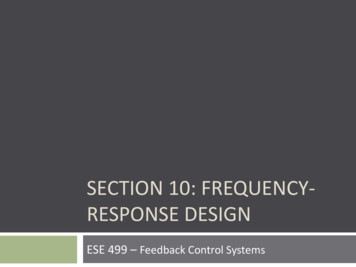
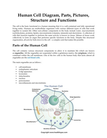
Voronina et al. SpringerPlus (2016) 5:1584One day prior to transfection, the cells were re-plated to a density of 0.5–0.6 106 cells/mL. On the day of transfection, cell density was determined, and the cellswere pelleted by centrifugation at 200g for 10 min at room temperature in an Allegra25-R centrifuge (Beckman, Germany). The supernatant was removed by decantation,and the cells were suspended in FreeStyle CHO Expression Medium, containing 8 mMalanyl-glutamine (both reagents were from Invitrogen, USA) to a final density of 1.2–1.5 106 cells/mL. Further transfection was performed in 6-well plates, according to themanufacturer’s instructions (FreeStyle CHO-DG44 Cells, Invitrogen, USA). Transfection efficiency was assessed by fluorescent microscopy of cells with the pEYFP plasmidand a blue color filter. The transfection efficiency was evaluated visually using a CKX41microscope (Olympus, Japan).According to the manufacturer’s instructions, subsequent selection of the transfectedclones was performed as shown in the schematic representation below (Fig. 1).Selection of individual clonesLimiting dilutions were used to select individual clones. After transfection for 24 h,the cells were suspended in CD OptiCHO Medium (Life technologies, USA) containing 500 μg/mL solution of G418 (Lonza, Switzerland) and 10 nM MTX at a density of10,000, 5000 or 1000 cells/well. Hundred microliter of the cell suspension was added toFig. 1 Clone selection scheme (adapted from User guide for Freedom DG44 Kit) and development ofstable cell lines for protein productionPage 4 of 11
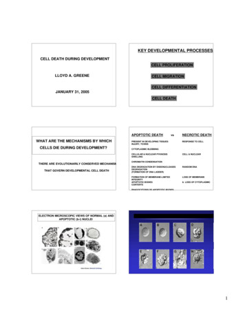
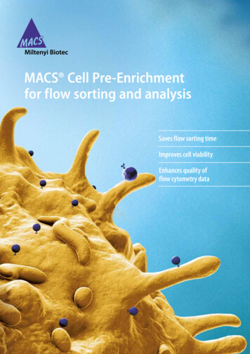
Voronina et al. SpringerPlus (2016) 5:1584Page 5 of 11every well of a 96-well plate, using 30–40 plates for each dilution. The plates were cultured in a CO2 incubator at 5 % CO2 at 37 C and 95 % humidity for 14–20 days. After12 days, the growth of cells in the wells was controlled under a microscope, registering the wells that were experiencing cell growth and division. Upon reaching 80–100 %confluence, the individual mini-pools were transferred into 24-well plates. After 5 days,the samples were analyzed for the expression level of the target antibody using the IgGELISA-BEST kit (Vector-Best, Russia). The selected pools with the highest productivitywere subcultured into 6-well plates, and then the positive pools were re-selected for celldensity and productivity by ELISA. Then the leading clones were transferred into T-75flasks, and further into 125 mL Erlenmeyer flasks in two media in parallel: CD OptiCHOMedium (Life technologies, USA) and ActiCHO SM (PAA, Austria) supplemented with8 mM alanyl-glutamine, 25 nM MTX and 500 μg/mL G418. At every step the number ofclones was reduced, basing on growth of cells, viability, and productivity (Fig. 2).For the further selection of the clones, three rounds of amplification were carried outin mini-pools at three concentrations of MTX: 25, 500 and 1000 nM. Series of batch cultures were performed for 10 amplified pools with subsequent evaluation of IgG concentration by ELISA. Then, the mini-pools were subcloned using limiting dilution in 96-wellplates as described above. From the 192 clones evaluated in 24 well plates, 60 cloneswere chosen based on the ELISA data with further reduction to 13 clones. Further batchcultures were carried out in 125 mL shaker flasks with subsequent evaluation of growthcharacteristics and productivity by ELISA, affinity chromatography, and electrophoresis(polyacrylamide gel electrophoresis under reducing conditions followed by Coomassiestaining). Based on the results of total productivity, growth characteristics, and stability,subclone 30 was selected for the further analysis (Fig. 3).bla promoterPvu I (5273)Spe I (54)Nde I (289)Amp(bla)CMVpromoterBspEI (706)Xba I (713)NarI (1275)pOptiVEC- HC adalimumabpUC oriHC adalimumab5807 bpPvu II (3837)Bam HI (2144)Not I (2162)TK polyAHpaI (2174)Apo I (3201)DHFRHindIII (2406)EMCV IRESKpn I (2627)Msc I (2760)Bgl II (2766)Fig. 2 Construct of pOptiVEC-HC adalimumab, containing the sequence of adalimumab heavy chain.HC adalimumab, synthetic gene of heavy chain mAb for adalimumab, codon optimized sequence; CMVpromoter, early gene promoter of human cytomegalovirus HCMV IE1; EMCV IRES, IRES element from theencephalomyocarditis virus (EMCV); DHFR, dihydrofolate reductase
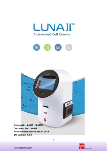
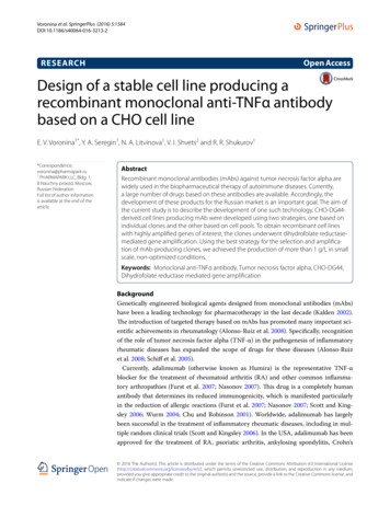
Voronina et al. SpringerPlus (2016) 5:1584Page 6 of 11Mfe I (6077)Mlu I (44)bla promoterSpe I (65)Sca I (5473)Nde I (300)CMV promoterPvu I (5363)AmpSac I (634)Kpn I (913)LC adalimumabSac II (1438)pc DNA 3.3 LC adalimumabpUC oriAge I (1451)6100 bpTK polyAf1 oriSV40 early promoterSV40 polyAXma I (2558)Sma I (2560)Nar I (2747)NeoFig. 3 Construct of pcDNA-LC adalimumab, containing the adalimumab light chain. LC adalimumab, thesynthetic gene of the light chain mAb for adalimumab, codon optimized sequence; CMV promoter, the earlygene promoter of human cytomegalovirus HCMV IE1; Neo neomycin phosphotransferaseAlternative protocol: amplification of the total pool of transiently transfected cellsOne day after transfection the cells were transferred to a selection medium, OptiCHO, containing 500 nM MTX and 500 μg/mL G418. Every 2–3 days, the medium waschanged by centrifugation at 200 g for 5 min and resuspension in the fresh medium.After 40 days, when the viability was over 90 %, the pool was transferred into a mediumcontaining an increased concentration of MTX (1000 nM). The amplification processwas performed in a similar manner as described above. The last stages of amplificationof the common pool were carried out at a concentration of 2.5 μM and 5 μM MTX,correspondingly. Upon reaching a cell viability of greater than 90 %, the total pool wassubjected to limiting dilutions to select individual clones. From the initial 40 subclones,20 subclones were selected on the basis of their specific productivity and overall productivity in the production batch. The resulting subclones were evaluated for expressionstability with or without the MTX selective agent during 60 generations. For the bestclones, production batches were carried out in the different media. Based on the totalproductivity, growth characteristics, and stability, subclone 26 was selected for furtherinvestigation.Colorimetric assay for tumor necrosis factor using WEHI 164 cellsNeutralizing activities of adalimumab against human TNF-α were measured on themouse WEHI 164 cell line treated with actinomycin D according to the methoddescribed previously (Austgulen et al. 1986; Khabar et al. 1995). Briefly, WHEI 164 cellswere seeded in triplicate at 1 104 cells/well into a 96-well plate and cultured in RPMI1640 medium supplemented with 10 % (v/v) FBS for 20 h. Then, serially diluted antibodies (final concentration: 0.5–50 ng/mL) in the medium containing 2 µg/mL actinomycin D were added to the cell culture together with 0.1 ng/mL of human TNF-α. Thecells were incubated for an additional 20 h at 37 C and cell viability was analyzed using
Voronina et al. SpringerPlus (2016) 5:1584a colorimetric MTT-based Cell Growth Determination kit (Sigma, St. Louis, MO). TheED50 value was calculated by complex sigmoid non-linear regression analysis usingSigma plot software (Systat software, Inc. Richmond, CA).Determination of the concentration of monoclonal anti‑TNFα antibodyConcentration of anti-TNF-α mAb was measured by ELISA using IgG-ELISA-BEST kit(Vector-Best, Russia), calibrated for Humira (AbbVie, USA). SDS-PAGE electrophoresisunder reducing conditions was performed according to the standard procedure in a 12 %Laemmli gel followed by staining with Coomassie-R250 to check the ratio of heavy andlight chains in the supernatant.Results and discussionAt the initial stage, genes encoding heavy (HC) and light (LC) chains of Adalimumabmonoclonal antibody (mAb) with optimized codon composition were synthesized. Genesequences were obtained from publicly available sources (DrugBank and patent databases). To confirm the sequence of monoclonal anti-TNFα antibody, a commercial drug,Humira, was subjected to peptide mapping using mass spectrometry-grade endoproteinases Chymotrypsin, Asp-N, Glu-C (Thermo Scientific, USA).For extracellular secretion of the antibody into the culture supernatant, the sequencesof the LC and HC were linked to signal peptides taken from human albumin gene.It has been shown that various genetic engineering approaches may be used for creating genetic constructs expressing mABs in CHO cells (Steven et al. 2013; Ng et al.2010). In this paper, were used a traditional approach based on the pOptiVecTOPO andpcDNA3.3TOPO plasmid vectors as carriers of the individual genes of the HC and LCantibody chains.It is known that one disadvantage of this approach is the possible survival of nonexpressing clones during selection (Steven et al. 2013; Bardor et al. 2012; Schlatter et al.2005). Another drawback is the lack of control over the ratio of expression of HC andLC. The LC is required to facilitate the folding and release of HC from binding immunoglobulin protein to form a complete IgG monomer. Each gene is under the control ofits own promoter and is transcribed separately. According to the literature, the expression of the LC in excess has a positive effect on the quality of the mAbs, and the ratioof LC:HC expression can affect mAb qualities such as glycosylation and aggregation(Schlatter et al. 2005; Lee et al. 2009). On the other hand, an excess of HC can causeER stress and proteasome overloading, creating a burden on the cell machinery that caninhibit cell proliferation (Lee et al. 2009).Thus, CHO-DG44-derived cell lines producing mAb were developed using twostrategies, one based on individual clones and the other based on cell pools. To obtainrecombinant cell lines with highly amplified genes of interest, the clones underwentdihydrofolate reductase (DHFR)-mediated gene amplification.According to the first strategy, individual clones were selected by limiting dilutions,followed by amplification of each separate clone.At the first step, the combination of selected genetic constructs was transfected intoCHO-DG44 cells. Twenty-four hours after transfection, selection of cells was performed. After 14–20 days of culture in 96-well plates, cell growth was analyzed usingPage 7 of 11
Voronina et al. SpringerPlus (2016) 5:1584Page 8 of 11light microscopy. Growth of cell colonies was found in 10 % of the wells at a dilution of100 cells/well (1000 cells/mL), in 25 % of the wells at a dilution of 500 cells/well (5000cells/mL), and in 60 % of the wells at a dilution of 1000 cells/well (10,000 cells/mL). Atthis stage, 310 clones were selected for further analysis.After an initial screening of the clones in 96-well plates using ELISA, were selected 68clones that had the highest expression level.For a more precise analysis of the productivity for the best growth characteristics, theclones were transferred to 24-well plates. Then, the leading 40 clones were cultured inT75 flasks and transferred into 125 mL flasks. For each clone, growth characteristicswere controlled and productivity was measured after 4 days of batch cultivation. For 10the most productive mini-pools, 3 rounds of amplification were performed, at 25, 500and 1000 nM MTX. After amplification, subcloning was carried out with three prospective mini-pools using limiting dilutions. As a result, 13 clones were selected with a specific productivity of 9–73 pcd [pg cell ( 1) day ( 1)] (Barnes et al. 2007) (Fig. 4). Specificproductivity was calculated using the following formula:pcd nln CDCD0pn p0·,nCDn CD0where P0 and Pn are the concentrations of the antibody in the supernatant at time zeroand the last day of cultivation, respectively, CD0 and CDn—cell densities on the corresponding days and n—culture duration in days.In total 13 leading clones were selected from 3 positive mini-pools. They were undergone an investigation of the production stability in continuous cultivation for 2 months.The drop in productivity was found to be 30–70 % during this time (Fig. 4). According tothe stability and growth characteristics, we selected clone #30 for further analysis. Whenthis clone was cultured for 8 days in fed-batch, the maximum cell density was 18 106cells/mL and productivity was more than 1 one numberFig. 4 Stability of the antibody production for the best subclones selected from the individual parentalclones. Specific productivity (pcd) was indicated before and after continuous cultivation for 60 generation
Voronina et al. SpringerPlus (2016) 5:1584Page 9 of 11All clones developed using this strategy was found to have propensity to aggregate.As known, for batch suspension cultures, cell aggregation hinders accurate cell counting, monitoring and control of the cellular environment. Transport of nutrients to, andproducts from, the cells may be impaired, cluster formation dramatically influences thegrowth behavior of the cells, cells within aggregates show a strongly reduced specificproliferation rate, apart from, shear forces exerted on large aggregates cause a considerably higher specific death rate than those exerted on single cells, reducing the specificgrowth rate up to 50 %. This issue was resolved using dextran sulfate based anti-clumping agent (Lonza, Switzerland). But when the protein was subjected to purification onMabSelect column (GE Healthcare, USA), significant protein aggregation and loss wasobserved. Moreover, all clones grew slowly (doubling time 40–50 h), and the best one,#30, reached the maximum cell density at 12.3 106 cells/mL. As a result, fed-batch inshaker flasks gave the effective productivity of only 0.4 g/L (this value indicates the yieldafter the first purification step).According to the second strategy, initially we subjected a pool of transiently transfected cells to amplification. Transfection was performed in a similar manner asdescribed above. The cells were cultured in a selective medium for 40 days, initially containing 25 nM MTX and further increased to 5000 nM. When cell viability reached morethan 90 % for the generally amplified pool, we evaluated productivity in a fed-batch.Amplified pool was subcloned using limiting dilutions as described above. From 40subclones, 20 leading ones were selected and assessed in terms of overall productivity.The best 7 subclones were evaluated for stability with or without selective agent during60 generations. Productivity per cell using the second selection strategy was significantlylower (Fig. 5). On the other hand, all clones generated using the second strategy performed much better in terms of cell growth. They didn’t aggregate, their doubling wastypical for CHO cells (23–35 h). For the leading clone, #26, maximum cell density in afed-batch process was 62.7 106 cells/mL, and process duration was 14 days. Moreover, no product aggregation was detected at purification. Consequently, the 2223263839clone numberFig. 5 Stability test for antibody pcd of best subclones selected on the basis amplification of the total pool oftransiently transfected cells after 4 days of cultivation during 60 generation
Voronina et al. SpringerPlus (2016) 5:1584productivity after the first purification step reached 1.3 g/L. Certainly, such data as1–1.3 g/L is a little low, however these are non-optimised conditions, wherein for oneof the producer has been shown a high productivity and stability of expression of prolonged cultivation. Further work is needed to optimize the culture conditions for a further increase in productivity of clone-producing.It’s important to note, the proteins obtained by both methods is identical in respect toits capacity to neutralize TNF-activity indeed. We carried out an in vitro assay to compare the binding capacity to soluble recombinant human (rh)TNFα and in vitro study tocompare the inhibition the binding of rhTNFα to TNFα receptors. These assays showedthe protein has similar activity for both cases.Thus, despite the lower specific productivity of the clones generated according to thesecond strategy, they were able to reach much higher cell density that caused betteroverall productivity.Further investigation is needed to understand the issues associated with the clonesdeveloped using the first strategy. Current data suggest that intensive amplificationcaused an excess of heavy chain that leaded to overload of translational machinery,insufficient folding and ER stress. In this case it seems unlikely to find any culture conditions to make these cell lines working without aggregation issues. On the other hand, thesecond strategy was not as effective to attain high HC expression, LC was in excess, andno stress emerged.This hypothesis was indirectly confirmed by measuring HC/LC ratio in the supernatants by SDS-PAGE. All clones from the second selection produce an excess of LC, whilethe clones from the first selection had almost 1:1 ratio (data not shown).ConclusionsTwo independent strategies were evaluated for selection and amplification of antibodyproducing clones originated from CHO-DG44 cell line. Completely different parameterswere reached for the resulting clones, both in terms of productivity and culture characteristics. The best strategy implemented in our study generated the cloned producingmore than 1 g/L of the target antibody in shaker flasks in non-optimized conditions.The results indicate that maximum specific productivity per cell should not be the onlygoal of cell line development. Intensive amplification may cause undesirable effects oncell growth and protein quality that countervail the benefits of high pcd. Thus, rationalbalance should be kept between specific productivity and maintained phenotype.Authors’ contributionsEVV, YAS, NAL carried out the genetic studies, selected clones and participated in discussing results. EVV also carriedout the immunoassays and following optimization, participated in the design of the study and performed the statisticalanalysis. YAS gave valuable impact into discussion of the study and its results conceived of the study, and participated inits design and coordination and helped to draft the manuscript. VIS also helped to draft the manuscript. All authors readand approved the final manuscript.Author details1PHARMAPARK LLC, Bldg. 1, 8 Nauchny proezd, Moscow, Russian Federation. 2 M.V. Lomonosov Moscow State Academyof Fine Chemical Technology, Moscow, Russian Federation.AcknowledgementsWe acknowledge our colleagues Nataliya Lobanova, Irina Savinova and Ravil Khamitov from Pharmapark LLC for valuableimpact into discussion of the study and its results.Competing interestsThe authors declare that they have no competing interests.Page 10 of 11
Voronina et al. SpringerPlus (2016) 5:1584Received: 23 May 2016 Accepted: 5 September 2016ReferencesAlonso-Ruiz A, Pijoan JI, Ansuategui E et al (2008) Tumor necrosis factor alpha drugs in rheumatoid arthritis: systematicreview and metaanalysis of efficacy and safety. BMC Musculoskelet Disord 9(1):52Austgulen R, Espevik T, Hammerstrøm J, Nissen-Meyer J (1986) Role of monocyte cytotoxic factor in cytolysis of actinomycin D-treated WEHI 164 cells mediated by freshly isolated human adherent mononuclear blood cells. Cancer Res46(9):4566–4570Bardor M, Feng H, Mariati Tong YW et al (2012) IRES-mediated Tricistronic vectors for enhancing generation of highmonoclonal antibody expressing CHO cell lines. J Biotechnol 157(1):130–139Barnes LM, Bentley CM, Moy N, Dickson AJ (2007) Molecular analysis of successful cell line selection in transfectedGS-NS0 myeloma cells. Biotechnol Bioeng 96(2):337–348Cacciatore Jonathan J, Chasin Lawrence A, Leonard Edward F (2010) Gene amplification and vector engineering toachieve rapid and high-level therapeutic protein production using the Dhfr-based CHO cell selection system.Biotechnol Adv 28(6):673–681Chu L, Robinson DK (2001) Industrial choices for protein production by large-scale cell culture. Curr Opin Biotechnol12:180–187Chusainow Janet, Yang Yuan Sheng et al (2009) Study of monoclonal antibody-producing CHO cell lines: what makes astable high producer? J Biotechnol Bioeng 102(4):1182–1196Derouazi M et al (2006) Genetic characterization of CHO production host DG44 and derivative recombinant cell lines.Biochem Biophys Res Commun 340(4):1069–1077Furst DE, Bree
Selection of individual clones Limiting dilutions were used to select individual clones. After transfection for 24 h, the cells were suspended in CD OptiCHO Medium (Life technologies, USA) contain-ing 500 μg/mL solution of G418 (Lonza, Switzerland) and 10 nM MTX at a density of 10,000, 5000 or 1000 cells/well.